Ultrasound (US) was once predominantly used for diagnosis but not anymore. It has the capability to noninvasively deliver energy to the deep tissues and also enable spatiotemporal control. Unlike high-intensity focused ultrasound, which primarily ablates tumors through localized heat, nonthermal US converts acoustic energy into mechanical, cavitation, and physicochemical effects. The bioeffects that are induced under nonthermal US provide a mechanistic basis for cancer therapies. Here, we present a mechanism-guided framework that connects acoustic parameters and nonthermal bioeffects with therapeutic modalities. Nonthermal US-based cancer treatments are classified into six representative modalities: histotripsy, ultrasound-targeted microbubble destruction, sonogenetics, sonomechanical therapy (SMT), sonodynamic therapy (SDT), and sonopiezoelectric therapy (SPT). Through a review of recent advances in these modalities, we highlight emerging opportunities and future directions with a focus on standardizing acoustic dosimetry, biosafety assessment, and material optimization. These insights collectively establish a foundation for creating more reliable and clinically relevant nonthermal US-based therapies for precision oncology.
Phytochlorins, a class of plant-derived tetrapyrroles, show great potential as sonosensitizers in sonodynamic therapy (SDT). The development of new phytochlorin-based sonosensitizers has significantly improved SDT, yet the absence of specialized sonodynamic systems limits their clinical translation. Herein, a dedicated ultrasound system along with a detailed step-by-step sonodynamic process from in vitro to in vivo is developed to activate phytochlorin-based sonosensitizers. Compared to standing-wave ultrasound, free-field ultrasound maintains stable acoustic pressure amplitudes and minimizes mechanical damage to cell membranes. In vitro experiments demonstrate that free-field ultrasound effectively activates naturally occurring phytochlorin, reducing the cavitation threshold for reactive oxygen species production and triggering immunogenic cell death. Furthermore, the intravenously injectable phytochlorin-based sonosensitizer (C34) enhances sonodynamic efficiency by reducing interfacial tension. Driven by in vivo free-field ultrasound, C34 effectively inhibits tumor growth in an orthotopic murine breast cancer model and elicits an immune response, preventing tumor metastasis. The reliable protocol provided by the free-field ultrasound system facilitates the activation of phytochlorin-based sonosensitizers while simultaneously stimulating the immune system, highlighting the potential of immune-sonodynamic therapy.
Breast cancer is one of the most common malignant tumors in women all over the world. Mastectomy is the most effective treatment, but there are serious problems such as high tumor recurrence rate and side effects of chemotherapy. Therefore, there is an urgent need for a therapeutic strategy that can effectively promote postoperative wound healing and inhibit local tumor recurrence. In this study, a 3D printing scaffold based on carbon dots-curcumin nano-drug release (CCNPs) was developed as a local drug delivery platform (named CCNACA using CCNPs, Sodium alginate, Nanoclay and Caffeic Acid grafted Chitosan as raw materials), which has the ability to visualize drug release. The 14-day drug release test in vitro showed that the tumor inhibition rate of CCNACA scaffolds on breast cancer cells (MCF-7) was 73.77 +/- 1.68 %. And the CCNACA scaffolds had good longterm antibacterial (Escherichia coli and Staphylococcus aureus) activity. Animal experiments have shown that implanting CCNACA scaffolds into surgical defects can inhibit postoperative residual cancer cells, reduce inflammation, promote angiogenesis, and repair tissue defects caused by surgery. In summary, the local drug delivery system of this manuscript has great potential in wound healing and prevention of tumor recurrence after breast cancer surgery.
The transplantation of islet beta cells offers an alternative to heterotopic islet transplantation for treating type 1 diabetes mellitus (T1DM). However, the use of systemic immunosuppressive drugs in islet transplantation poses significant risks to the body. To address this issue, we constructed an encapsulated hybrid scaffold loaded with islet beta cells. This article focuses on the preparation of the encapsulated structure using 3D printing, which incorporates porcine pancreas decellularized extracellular matrix (dECM) to the core scaffold. The improved decellularization method successfully preserved a substantial proportion of protein (such as Collagen I and Laminins) architecture and glycosaminoglycans in the dECM hydrogel, while effectively removing most of the DNA. The inclusion of dECM enhanced the physical and chemical properties of the scaffold, resulting in a porosity of 83.62% ± 1.09% and a tensile stress of 1.85 ± 0.16 MPa. In teams of biological activity, dECM demonstrated enhanced proliferation, differentiation, and expression of transcription factors such as Ki67, PDX1, and NKX6.1, leading to improved insulin secretion function in MIN-6 pancreatic beta cells. In the glucose-stimulated insulin secretion experiment on day 21, the maximum insulin secretion from the encapsulated structure reached 1.96 ± 0.08 mIU ml-1, representing a 44% increase compared to the control group. Furthermore, conventional capsule scaffolds leaverage the compatibility of natural biomaterials with macrophages to mitigate immune rejection. Here, incorporating curcumin into the capsule scaffold significantly reduced the secretion of pro-inflammatory cytokine (IL-1β, IL-6, TNF-α, IFN-γ) secretion by RAW264.7 macrophages and T cells in T1DM mice. This approach protected pancreatic islet cells against immune cell infiltration mediated by inflammatory factors and prevented insulitis. Overall, the encapsulated scaffold developed in this study shows promise as a natural platform for clinical treatment of T1DM.
The release of antibiotics or anions by traditional bacteriostatic agents led to the development of bacterial drug resistance and environmental pollution. Ionic liquids (ILs) have become important choices for antibacterial agents because of their excellent physical, chemical and biological properties. In this paper, the bioactivities of 1-vinyl-3-butylimidazolium chloride ([VBIM]Cl, IL) and poly (1-vinyl-3-butylimidazolium chloride) (P[VBIM]Cl, PIL) were evaluated, and the potential antibacterial material was used to synthesize hydrogels. Using the colony formation assay and the Oxford cup method, antibacterial effect of IL and PIL were tested. Cell-Counting-Kit-8 (CCK-8) experiments were used to study the IC 50 (half maximal inhibitory concentration) values of IL and showed 1.47 mg/mL, 0.35 mg/mL and 0.33 mg/mL at 24 h, 48 h and 72 h, respectively. The IC 50 value of PIL were 12.15 μg/mL, 12.06 μg/mL and 11.76 μg/mL at 24 h, 48 h and 72 h, respectively. The PIL is further crosslinked with polyvinyl alcohol (PVA) to form a novel hydrogel through freeze-thaw cycles. The newly fabricated hydrogel exhibited a high water content, excellent water absorption properties and outstanding mechanical performance. Using the colony formation assay and the inhibition zone assay, the hydrogels exhibited favorable antibacterial effects (against E.coli and S.aureus) such that nearly 100% of the bacteria were killed in liquid medium while cultivating with H4 (synthesized by 0.5 g PIL and 1g PVA). In addition, the cytotoxicity of PIL was significantly reduced through hydrogen bond crosslinking. H4 showed the highest antibacterial activity and a good biocompatibility. The results indicated that the PVA&PIL hydrogels had great potential for wound dressing.
Therapeutic cancer vaccines fail to produce satisfactory outcomes against solid tumors since vaccine-induced anti-tumor immunity is significantly hampered by immunosuppression. Generating an in situ cancer vaccine targeting immunological cold tumor microenvironment (TME) appears attractive. Here, a type of free-field based whole-body ultrasound (US)-driven nanovaccines are constructed, named G5-CHC-R, by conjugating the sonosensitizer, Chenghai chlorin (CHC) and the immunomodulator, resiquimod (R848) on top of a super small-sized dendrimeric nanoscaffold. Once entering tumors, R848 can be cleaved from a hypoxia-sensitive linker, thus modifying the TME via converting macrophage phenotypes. The animals bearing orthotopic pancreatic cancer with intestinal metastasis and breast cancer with lung metastasis are treated with G5-CHC-R under a free-field based whole-body US system. Benefit from the deep penetration capacity and highly spatiotemporal selectiveness, G5-CHC-R triggered by US represented a superior alternative for noninvasive irradiation of deep-seated tumors and magnification of local immune responses via driving mass release of tumor antigens and "cold-warm-hot" three-state transformation of TME. In addition to irradiating primary tumors, a robust adaptive anti-tumor immunity is potentiated, leading to successful induction of systemic tumor suppression. The sono-nanovaccines with good biocompatibility posed wide applicability to a broad spectrum of tumors, revealing immeasurable potential for translational research in oncology.
Radiotherapy is a common local treatment for breast cancer, and while it is effective in targeting tumor cells, it inevitably causes significant side effects. These include excessive production of reactive oxygen species (ROS), repeated inflammatory, and severe skin ulceration, all of which can hinder the wound healing process. As a result, there is a pressing need for multifunctional medical dressings that can support wound repair following radiotherapy. In this study, we introduced a novel double-network interpenetrating hydrogel (GEMC), which combined gelatin grafted dopamine (GEDA), acrylamide, nano-clay (NC), and curcumin loaded nanoparticles (CCNPs). Unlike traditional single-function hydrogels, the GEMC hydrogel offered a combination of antioxidant properties, tissue adhesion, and real time drug tracking, effectively addressing the multifaceted challenges of wound healing after radiotherapy. The GEMC hydrogel exhibited impressive antioxidant activity and superior mechanical properties, which collectively improve the support and protection of wounded surfaces. Furthermore, GEMC promoted skin regeneration, angiogenesis and reduced inflammatory in a mouse model of radiotherapy-induced skin ulceration. These results highlight the hydrogel's potential to accelerate would healing and enhance the effectiveness of post-radiotherapy wound care, providing a promising new approach to improving the quality of skin recovery following radiotherapy.
Photodynamic therapy (PDT), performed with low-fluence rates, can improve antitumor responses and prevent adverse effects. However, photosensitizers (PSs) for low-fluence PDT treatment are rarely reported. Herein, we exploited an amphiphilic chlorin-based PS, named DYSP-C34, which has a variety of beneficial biological properties, such as improved water solubility, better cellular permeability, specific localization and enhanced phototoxicity under low light dose irradiation. In addition, DYSP-C34 could effectively accumulate in a mouse subcutaneous xenograft tumor and exhibit substantial tumor regression after irradiation with an extremely low light fluence (6 J/cm2). Meanwhile, the excellent phototoxicity could stimulate the host immune system and lead to a strong inhibition of tumor growth synergistically. These results indicated the potential value of DYSP-C34 as a chlorin-type PS for low-fluence PDT application.
Exploring facile and effective therapeutic modalities for synergistically controlling primary tumor and metastasis remains a pressing clinical need. Sonodynamic therapy (SDT) offers the possibility of noninvasively eradicating local solid tumors, but lacks antimetastatic activity because of its limited ability in generating systemic antitumor effect. Here, we exploited a previously unidentified ultrasound-driven “molecular machine,” DYSP-C34 (C34 for short), with multiple attractive features, emerging from preferential tumor accumulation, potent ultrasound-triggered cytotoxicity, and intrinsic immune-boosting capacity. Driven by the ultrasound, C34 functioned not only as a tumor cell killing reagent but also as an immune booster that could potentiate robust adaptive antitumor immunity by directly stimulating dendritic cells, resulting in the eradication of the primary solid tumor along with the inhibition of metastasis. This molecular machine, C34, rendered great promise to achieve systemic treatment against cancer via unimolecule-mediated SDT.
Photodynamic therapy (PDT) has garnered immense research interest. PDT can directly kill the cells via a combination of photosensitizer, light, and molecular oxygen. It has emerged as a promising therapeutic option for cancer treatment owing to its advantages such as minimized systemic toxicity, minimal invasiveness, high therapeutic efficacy, and potential for developing antitumor immunity. The novel photosensitizer 32-(4-methoxyphenyl)-152-aspartyl-chlorin e6 (DYSP-C34) was synthesized by introducing a 32-aryl substitution and amino acid substituent of the Chenghai chlorin (CHC). Briefly, 32-(4-methoxyphenyl) substitution was achieved via olefin metathesis reactions. The aspartic acid side chain was introduced regioselectively at C-152, followed by hydrolysis to yield the target DYSP-C34. CHC with the same chemical structure as chlorin e6 was prepared from chlorophyll a, which was extracted from Spirulina powders derived from Chenghai Lake in the Yunnan province of China. This strategy successfully endowed the resultant photosensitizer with better cellular permeability and increased water solubility. In addition, the photodynamic antitumor effects of PDT largely depend on the dose of photosensitizer used, time between photosensitizer administration and light exposure, and possibly other still poorly known variables. Determination of optimal conditions for PDT requires a coordinated interdisciplinary effort. Therefore, the pharmacokinetics and tissue distribution of DYSP-C34 in vivo are critical for the efficacy and safety of PDT. Herein, a high performance liquid chromatography-ultraviolet (HPLC-UV) detection method was established for the determination of the new photosensitizer DYSP-C34 in rat plasma. The sample preparation involved a protein-precipitation and liquid-liquid extraction method. Methanol was used to precipitate proteins and chloroform was used to extract chlorins. Then, DYSP-C34 was separated on a Unitary C18 column (250 mm×4.6 mm, 5 μm) with a mobile phase comprising methanol and 5 mmol/L tetrabutylammonium phosphate buffer solution (70∶30, v/v). The flow rate was 1.0 mL/min with UV detection using a wavelength of 400 nm at 40 ℃. Results showed that DYSP-C34 and chlorin e6 trimethyl ester (IS) were well separated under these conditions. The method was sensitive and sufficiently precise with a good linear relationship (determination coefficient (r2)=0.9941) over the range of 1-200 μg/mL in rat plasma. At three spiked levels (8, 40, and 120 μg/mL), the average recoveries were 74.39%, 69.71%, and 65.89%, respectively. The intra-day and inter-day relative standard deviations (RSDs) were lower than 5%. The precision met the requirements of biological sample determination. Furthermore, DYSP-C34 was stable in rat plasma under various storage conditions at room temperature, three freeze-thaw cycles, and long-term cryopreservation. The validated method was successfully applied to the pharmacokinetic study of DYSP-C34 after intravenous injection of a single dose in rat plasma. The pharmacokinetic parameters after intravenous injection of DYSP-C34 (16 mg/kg) were calculated. The plasma half-life (t1/2z) was 6.98 h, the area under the plasma concentration-time curve AUC(0-∞) was 1025.01 h·mg/L and the mean retention time MRT(0-∞) was 9.19 h. In addition, the results of DYSP-C34 distribution in tumor-bearing mice showed that DYSP-C34 could accumulate in tumor tissues, with higher concentrations in liver and kidney tissues, and lower concentrations in heart, spleen, and lung tissues. In summary, a specific, simple, and accurate HPLC-UV method was developed and validated for the determination of DYSP-C34 in rat plasma and tumor-bearing mouse tissues. The pharmacokinetics of DYSP-C34 after intravenous administration in rats and the tissue distribution characteristics of tumor-bearing mice were clarified for the first time. It is significant for clinical rational drug use and pharmacodynamic research. Therefore, choosing an appropriate time for light treatment time can achieve the best photodynamic effect. The results of pharmacokinetics and tissue distribution of DYSP-C34 provide vital guidance for subsequent pharmacodynamic research and further clinical trials in terms of dosage, light time, light toxicity and side effects.
Photodynamic therapy (PDT) has garnered immense research interest. PDT can directly kill the cells via a combination of photosensitizer, light, and molecular oxygen. It has emerged as a promising therapeutic option for cancer treatment owing to its advantages such as minimized systemic toxicity, minimal invasiveness, high therapeutic efficacy, and potential for developing antitumor immunity. The novel photosensitizer 32-(4-methoxyphenyl)-152-aspartyl-chlorin e6 (DYSP-C34) was synthesized by introducing a 32-aryl substitution and amino acid substituent of the Chenghai chlorin (CHC). Briefly, 32-(4-methoxyphenyl) substitution was achieved via olefin metathesis reactions. The aspartic acid side chain was introduced regioselectively at C-152, followed by hydrolysis to yield the target DYSP-C34. CHC with the same chemical structure as chlorin e6 was prepared from chlorophyll a, which was extracted from Spirulina powders derived from Chenghai Lake in the Yunnan province of China. This strategy successfully endowed the resultant photosensitizer with better cellular permeability and increased water solubility. In addition, the photodynamic antitumor effects of PDT largely depend on the dose of photosensitizer used, time between photosensitizer administration and light exposure, and possibly other still poorly known variables. Determination of optimal conditions for PDT requires a coordinated interdisciplinary effort. Therefore, the pharmacokinetics and tissue distribution of DYSP-C34 in vivo are critical for the efficacy and safety of PDT. Herein, a high performance liquid chromatography-ultraviolet (HPLC-UV) detection method was established for the determination of the new photosensitizer DYSP-C34 in rat plasma. The sample preparation involved a protein-precipitation and liquid-liquid extraction method. Methanol was used to precipitate proteins and chloroform was used to extract chlorins. Then, DYSP-C34 was separated on a Unitary C18 column (250 mm×4.6 mm, 5 μm) with a mobile phase comprising methanol and 5 mmol/L tetrabutylammonium phosphate buffer solution (70∶30, v/v). The flow rate was 1.0 mL/min with UV detection using a wavelength of 400 nm at 40 ℃. Results showed that DYSP-C34 and chlorin e6 trimethyl ester (IS) were well separated under these conditions. The method was sensitive and sufficiently precise with a good linear relationship (determination coefficient (r2)=0.9941) over the range of 1-200 μg/mL in rat plasma. At three spiked levels (8, 40, and 120 μg/mL), the average recoveries were 74.39%, 69.71%, and 65.89%, respectively. The intra-day and inter-day relative standard deviations (RSDs) were lower than 5%. The precision met the requirements of biological sample determination. Furthermore, DYSP-C34 was stable in rat plasma under various storage conditions at room temperature, three freeze-thaw cycles, and long-term cryopreservation. The validated method was successfully applied to the pharmacokinetic study of DYSP-C34 after intravenous injection of a single dose in rat plasma. The pharmacokinetic parameters after intravenous injection of DYSP-C34 (16 mg/kg) were calculated. The plasma half-life (t1/2z) was 6.98 h, the area under the plasma concentration-time curve AUC(0-∞) was 1025.01 h·mg/L and the mean retention time MRT(0-∞) was 9.19 h. In addition, the results of DYSP-C34 distribution in tumor-bearing mice showed that DYSP-C34 could accumulate in tumor tissues, with higher concentrations in liver and kidney tissues, and lower concentrations in heart, spleen, and lung tissues. In summary, a specific, simple, and accurate HPLC-UV method was developed and validated for the determination of DYSP-C34 in rat plasma and tumor-bearing mouse tissues. The pharmacokinetics of DYSP-C34 after intravenous administration in rats and the tissue distribution characteristics of tumor-bearing mice were clarified for the first time. It is significant for clinical rational drug use and pharmacodynamic research. Therefore, choosing an appropriate time for light treatment time can achieve the best photodynamic effect. The results of pharmacokinetics and tissue distribution of DYSP-C34 provide vital guidance for subsequent pharmacodynamic research and further clinical trials in terms of dosage, light time, light toxicity and side effects.
7-Hydroxyneolamellarin A (7-OH-Neo A, 1), a natural marine product derived from sponge Dendrilla nigra, was first synthesized with 10% overall yield under the instruction of convergent synthetic strategy. We found that 7-OH-Neo A could attenuate the accumulation of hypoxia-inducible factor-1α (HIF-1α) protein and inhibit vascular epidermal growth factor (VEGF) transcriptional activity, showing well inhibitory effect on HIF-1 signaling pathway. Meantime, 7-OH-Neo A had the well anti-tumor activities, such as inhibiting tumor angiogenesis, proliferation, migration and invasion. More importantly, 7-OH-Neo A exhibited profound anti-tumor effect in mice breast cancer model by suppressing the accumulation of HIF-1α in tumor tissue. Mechanism study demonstrated that 7-OH-Neo A might target the protein with the ability of stabilizing HIF-1α in hypoxia. Due to the excellent water solubility, superior anti-tumor activity and good biocompatibility, 7-OH-Neo A shows the promising potential for being exploited as an anti-tumor agent in near future.
Photodynamic therapy (PDT) is a non-invasive and innovative therapeutic approach which has been increasingly applied in clinical cancer therapy. As the central element of PDT, the development of novel photosensitizers (PSs) with longer absorption wavelength, proper lipophilic/hydrophilic profiles, target tissue selectivity, and higher photo−/lowest dark-cytotoxicity is a challenging task. Previously, we designed and synthesized a series of novel long-wavelength chlorin e6 (Ce6)-based PSs via introducing aromatic groups to the vinyl of Ce6 skeleton. The new formed compounds with π-extension system exhibited improved photodynamic effects and spectral characteristics. Among these π-conjugated chlorin PSs, (E)-32-(4-methoxyphenyl)-chlorin e6, named A15, was expected to be a potent antitumor candidate as a PDT agent due to its good photobiological properties. Herein, in this work, we evaluated the effectiveness of A15 in cancer PDT. In vitro, a novel rare earth probe, ATTA-Eu3+ was applied to detect the singlet oxygen (1O2) production of A15 in solution and human hepatoma HepG2 cells, respectively. Moreover, A15 exhibited strong phototoxicity and weak dark cytotoxity to HepG2 cells. In H22 tumor bearing mice, A15 showed excellent tumor accumulation ability via i.v. administration and induced tumor regression, followed by laser treatment. These results indicated that A15 is a potential novel π–extension chlorin-type PS for PDT applications.
Targeting tumor acidic microenvironment, a cis-aconitate linked pyropheophorbide a dimer 3 was designed and prepared The observed fluorescence quenching of 3 verified our FRET based molecular design. Acid-dependent cleavage in aqueous solution, singlet oxygen generation, and cytotoxiciry against HepG2 cell lines of dimer 3 were investigated. The dimer demonstrated different levels of fluorescence recovery when incubated in acidic aqueous environment as well as effective phototoxicity against HepG2 cells.
Bimodal fluorescence-magnetic resonance imaging (MRI) technique has shown great utilities in bioassays because it combines the advantages of both optical imaging and MRI to provide more sufficient information over any modality alone. In this work, on the basis of a MnO2 nanosheet-Ru(II) complex nanoarchitecture, a bimodal phosphorescence-MRI nanoprobe for glutathione (GSH) has been constructed. The nanoprobe, Ru(BPY)3@MnO2, was constructed by integrating MnO2 nanosheets with a phosphorescent Ru(II) complex [Ru(BPY)3](PF6)2 (BPY = 2,2'-bipyridine), which resulted in complete phosphorescence quenching of the Ru(II) complex, accompanied by very low longitudinal and transverse relaxivity. Upon exposure to GSH, the reduction of MnO2 nanosheets by GSH triggers a recovery of phosphorescence and simultaneously produces a number of Mn2+ ions, a perfect MRI contrast agent. The as-prepared nanoprobe showed good water dispersion and biocompatibility and a rapid, selective, and sensitive response toward GSH in the phosphorescence and MR detection modes. The practicability of the nanoprobe was proved by time-gated luminescence assay of GSH in human serum, phosphorescent imaging of endogenous GSH in living cells, zebrafish, and tumor-bearing mice, as well as the MRI of GSH in tumor-bearing mice. The research outcomes suggested the potential of Ru(BPY)3@MnO2 for the bimodal phosphorescence-MRI sensing of GSH in vitro and in vivo.
A visible-light-excitable Eu3+ complex-based luminescent probe, [Eu(pdap)(3)(DPBT)], has been proposed for time-gated luminescence imaging of singlet oxygen in the mitochondria of living cells, as well as in tumor tissues and laboratory animals. Extension of the excitation window to the visible-light region makes the probe more favorable for practical usage.
In the development of new photosensitizers jar cancer therapy, increasing the capacity of intracellular reactive oxygen species (ROS) production is an important strategy. In this paper, artesunate as a ROS generation group was regioselectively introduced to the chlorin e(6) scaffold to obtain four conjugates of chlorin e(6) and artesunate. By irradiation with 440 nm, 630 nm and 660 nm of light source the four conjugates exhibited significantly improved phototoxicity against HepG2 cells compared with chlorin e(6) and artesunate. Irradiation with 440 nm or 660 nm light source gave higher phototoxicity as well as intracellular ROS level.
In order to increase the photodynamic effect of Chlorin e6, four Chlorin e6-artesunate conjugates were designed and synthesized. Among them, three conjugates (3, 6, 9) contained single artesunate side chain at 152, 173 and 131 of Chlorin e6, respectively, and one conjugate (11) contained three artesunate side chains. In the in vitro evaluation of photodynamic effect, the four conjugates showed more potent phototoxicity against HepG2 cells than Chlorin e6. The introduction of artesunate side chain significantly increased the intracellular ROS production, although the production of singlet oxygen was not improved. Compound 11 exhibited much more potent phototoxicity than the other conjugates because the three artesunate side chains greatly enhanced the ROS production and cellular uptake. The results demonstrated that the conjugation of Chlorin e6 and artesunate could accomplish synergistic effects of chemo-phototherapy, and finally enhanced their antiproliferative effects.
32-Aryl substitution generated the maximum wavelength redshift and increased the in vitro phototoxicity against HepG2 cells simultaneously.
A possible biosynthetic pathway of bis-alpha-substituent pyrrolidine alkaloids was proposed. Based on the proposed biosynthetic pathway, six pyrrolozoxazine alkaloids and one N-alkyl-5-hydroxymethyl-pyr-role-2-carbaldehyde alkaloid were synthesized. In a mixture of acetic acid and triethylamine, condensation of n-fructose and D-amino acids produced pyrrolozoxazine alkaloids. Replacing D-amino acids with tyramine can afford pyrrolezanthine a N-alkyl-5-hydroxymethyl-pyr-role-2-carbaldehyde alkaloid. (C) 2016 Published by Elsevier Ltd.
 Weijie Zhao (赵伟杰)合作论文数School of Chemical Engineering, Dalian University of Technology16
 Yueqing Li (李悦青)合作论文数School of Chemical Engineering, Dalian University of Technology13
 Shisheng Wang (王世盛)合作论文数School of Chemical Engineering, Dalian University of Technology9
 Lei Cao合作论文数8
 Xiuhan Guo (郭修晗)合作论文数School of Chemical Engineering, Dalian University of Technology8
 Guangzhe Li (李广哲)合作论文数School of Chemical Engineering, Dalian University of Technology8
 Kun Shao (邵堃)合作论文数School of Chemical Engineering, Dalian University of Technology5Yi Dong合作论文数4Ya Su合作论文数3
 Zhi Cao (曹直)合作论文数Institute of Coal Chemistry, Chinese Academy of Sciences;University of Chinese Academy of Sciences3Yang Wang合作论文数3
 Bo Song (宋波)合作论文数School of Chemical, Dalian University of Technology3
 Yuen Yee Cheng合作论文数Institute for Biomedical Materials and Devices, University of Technology Sydney;School of Mathematical and Physical Sciences, Faculty of Science, University of Technology Sydney3Yueqi Lu合作论文数2Hailin Ma合作论文数2Yaqian Liu合作论文数2
 Jie Xu合作论文数2Bo Pan合作论文数2Xu Wang合作论文数2
 Yi Nie (聂毅)合作论文数Institute of Process Engineering, Chinese Academy of Sciences2
 Jingli Yuan (袁景利)合作论文数School of Chemistry, Dalian University of Technology2
 Rui Cai (蔡蕊)合作论文数Instrumental Analysis Center, School of Chemical Engineering, Dalian University of Technology2
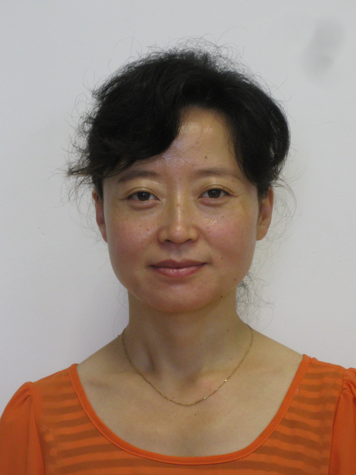 Yuming Sun (孙玉明)合作论文数School of Chemical Engineering, Dalian University of Technology2
 Xiaojun Peng (彭孝军)合作论文数School of Chemical Engineering, Dalian University Of Technology2Kedong Song合作论文数2Zejia Yang合作论文数1Qing Li合作论文数1Guanyi Zhang合作论文数1Yuxuan Zhang合作论文数1Hao Wang合作论文数1Jingdong Zhang合作论文数1Xin Wang合作论文数1Tianjiao Han合作论文数1Yanchun Guan合作论文数1Xiaodan Qin合作论文数1Wenjing Shi合作论文数1Jiaqi Liu合作论文数1Wenfang LI (李文芳)合作论文数1Chengdong Wang合作论文数1Xi Zhang合作论文数1Jianlong Su合作论文数1Huan Fang合作论文数1Yujie Shao合作论文数1Xueyan Hu (胡雪岩)合作论文数1Hua Ma合作论文数1Zhixin Tang合作论文数1Bowen SHI (石博文)合作论文数1YiMing Zhong (钟怡鸣)合作论文数1




