Neuroinflammation is a core pathological mechanism in neurodegenerative diseases. Although natural many compounds, derived from traditional Chinese medicine have shown promise in modulating neuroinflammation, conventional evaluation methods remain inefficient and fail to meet modern drug development needs. This study aimed to develop a neuroinflammation-on-a-chip for efficient and accurate evaluation of the anti-neuroinflammatory activity of such compounds. By integrating gelatin methacryloyl (GelMA) hydrogel with a microchamber array structure into a multi-channel concentration-gradient microfluidic chip, we constructed a functional neuroinflammation-on-a-chip suitable for high-throughput drug screening. Preliminary results demonstrated that the chip can successfully model lipopolysaccharide (LPS)-induced neuroinflammation and test the anti-inflammatory effects of curcumin (Cur) and resveratrol (RSV). Relative to traditional approaches, the chip offers the advantages of low sample consumption, rapid detection, and high data reliability. This study provides a novel tool for the efficient evaluation of anti-neuroinflammatory activity of traditional Chinese medicine active compounds and offers an innovative platform for research on neuroinflammation-related diseases.
Extracellular vesicles have shown great potential in treating ultraviolet (UV)-induced skin photoaging. However, effective delivery of these bioactive agents to achieve long-term therapeutic effects remains a significant challenge. In this study, we have developed recombinant human collagen (RHC)-based microcarriers loaded with native exosomes as well as Collagen type I alpha 1 chain (COL1A1) mRNA-encapsulating nanovesicles (Emvs) to treat skin photoaging. The microcarriers mitigated UV-induced cellular senescence in HaCaT cells, as evidenced by reduced oxidative stress, decreased apoptosis, and attenuation of senescence-associated markers. In the animal model, the microcarriers not only attenuated wrinkle formation, but also promoted type I collagen deposition in UV-damaged skin. These results together with the demonstrated biocompatibility highlight the potential of the microcarrier delivery system in photoaging treatment.
Detecting double-stranded DNA sequences typically requires enzymes to unwind and amplify the target. A new study by Yan et al. challenges this view by combining gamma peptide nucleic acid with multicomponent deoxyribozyme in droplets, achieving single-molecule sensitivity without protein enzymes or amplification, enabling concise, economical, and highly sensitive genetic diagnosis.
As a chronic, immune‐mediated condition, psoriasis manifests through the dysregulated proliferation of keratinocytes and the recruitment of assorted inflammatory cells into the skin. Immunosuppressants and photothermal therapy are both effective strategies. However, their applications are often challenged by systemic side effects or low efficiency of topical therapies. To address this, we developed a synergistic treatment strategy using a near‐infrared (NIR)‐responsive separable microneedle patch for NIR‐triggered photothermal activation and on‐demand immunosuppressant delivery. Gold nanorods (AuNRs), serving as both photothermal agents and drug nanocarriers, were loaded with an immunosuppressant and encapsulated into detachable microneedle tips. Upon NIR irradiation, the AuNRs induce localized hyperthermia, which provides direct thermal stimulation to psoriatic lesions and accelerates the release of the immunosuppressant at the target site. In a psoriasis mouse model, the system demonstrated significant therapeutic efficacy, effectively alleviating symptoms. This microneedle system synergistically combines controlled drug release and photothermal treatment, presenting a versatile and promising approach for managing psoriasis effectively, with significant potential for broader biomedical use.
Organ-on-a-chip technology holds great value for modeling intestinal tumors. However, how to more accurately replicate the tumor microenvironment and implement high-throughput drug screening on-chip remains to be further explored. In this study, we developed a biomimetic intestinal tumor-on-a-chip platform that simulates the 3D structure of intestinal crypts and villi, and incorporates a herringbone fluidic mixer. Compared to conventional chips, this design significantly improves the dynamic interplay between tumor cells and their surrounding microenvironment. Inside the chip, patient-derived primary epithelial and tumor cells successfully form a 3D tumor model. Furthermore, the platform enables monitoring of dynamic drug response in tumor cells derived from individual patients. Preliminary findings indicate that mitophagy could be a key contributor to the development of chemoresistance. This biomimetic intestinal tumor-on-a-chip platform effectively reproduces the complex characteristics of intestinal tumors through its high-fidelity 3D structure and dynamic microenvironment.
Macromolecular therapeutics demonstrate significant advantages and potential for treating diverse diseases. However, their intrinsic physicochemical properties often hinder the selection of suitable carriers for efficient intracellular delivery. Here, we develop coacervates formed via liquid-liquid phase separation of oligopeptides and DNA as macromolecular carriers for cellular membrane translocation. These materials enable the efficient recruitment and release of biomacromolecules, including proteins and enzymes. Notably, our experimental data suggest that their uptake may not be entirely identical to classical endocytic pathways and instead involves cholesterol-dependent lipid raft interactions, indicating a mechanism that may be distinct from canonical clathrin-mediated endocytosis. Upon reaching the cytoplasm, the DNA component of the coacervates is degraded by intracellular DNA-processing enzymes, leading to coacervate disassembly and subsequent release of the therapeutic macromolecules. Together, these coacervates establish a generalizable platform for intracellular delivery of macromolecular therapeutics, integrating membrane translocation with programmable cytosolic release.
Activated molecular pathways with elevated chemokine secretion are critical in driving pathogenesis of the mucosal inflammation in gastroesophageal reflux disease (GERD). The therapeutic efficacy of acid-suppressants is often constrained in GERD patients due to the high prevalence of non-acid reflux. Curcumin, a natural polyphenol with robust anti-inflammatory and antioxidant effects, offers promising therapeutic potential. In this study, leveraging the favorable biocompatibility and bioactive property of gelatin methacryloyl (GelMA), we developed GelMA microspheres as microcarriers encapsulating curcumin nanoparticles (Cur) for local treatment of GERD. The further incorporation of magnetic nanoparticles (MNPs) facilitates esophageal localization of the microspheres under an external magnetic field. Additionally, Cur-MNPs@GelMA microspheres sustain drug release in simulated gastric fluid, ensuring effective local concentrations. In vitro and in vivo investigations corroborated that Cur-MNPs@GelMA microspheres markedly inhibited the generation of inflammatory cytokines and reactive oxygen species (ROS). Mechanistically, the microspheres can restore mitochondrial function and inhibit NF-kappa B pathway. Furthermore, the multifunctional microspheres can effectively ameliorate histopathological damage and suppress inflammation in a mouse model of GERD. These findings endorse the therapeutic potential of Cur-MNPs@GelMA microspheres as a novel clinical intervention for GERD.
The mechanisms governing pH regulation in membrane-less organelles (MLOs) are essentially different from those relying on membrane proteins, yet it remains poorly understood due to the difficulty in directly controlling the conditions across the MLOs interface. Here, we develop a coacervate-based, in vitro model to investigate how liquid-liquid phase separation (LLPS) could contribute to pH regulation in MLOs. We construct peptide-based coacervate droplets using microfluidics and find that charged polymers within the coacervates help create uneven H+/OH- distributions, resulting in pH-regionalized microenvironments similar to the nucleolus. More interestingly, such a pH difference weakens or even disappears following the destruction of LLPS, a phenomenon observed in both nucleolus and coacervate droplets. Based on these findings, we demonstrate the ability to finely tune the local pH of the coacervate droplets over a wide range by incorporating enzymes, which can drive and control cascade reactions, and perform basic molecular biology operations such as polymerase chain reaction (PCR) and in vitro transcription and translation reaction (IVTT). This study highlights the role of LLPS in pH modulation within MLOs and provides insights into the potential of coacervates as protocells for broader applications. This study shows that liquid–liquid phase separation can help create and regulate pH differences across cell-like coacervate droplets, which can mimic simple cellular functions by organizing enzyme reactions and supporting basic biological processes.
Milk is an essential nutritional source for human health, yet developing efficient and sustainable indoor dairy production systems to produce milk-associated components remains a major challenge. Here, we propose a biomimetic mammary gland-on-a-chip system that recapitulates both the structural features and lactation mechanism of the mammary gland for producing milk-associated bioactive components. The chip is designed with a medium channel providing circulation, an intermediate porous hydrogel layer for substance diffusion, and highly specialized microchambers for culturing mammary epithelial cells. The mammary gland-on-a-chip enables tight-junction formation and active secretion of milk bioactive components such as lactotransferrin and triglycerides. More importantly, by expanding the microchambers and parallelizing the chips, increased collection of partial milklike secretions containing selected bioactive components was achieved. Our work represents the successful development of a robust microphysiological system as a potential in vitro platform for mammary gland research and is promising in addressing future food and environmental crises.
Phlorizin (PHZ) is a compound naturally found in apples, hawthorns and other plants, which shows potential in treating diabetes and its complications. However, its intestinal stability and precise targeted release ability remain challenging. Herein, we proposed a PHZ-integrated magnetic pollen micromotor for the treatment of diabetes combined with intestinal inflammation, which can well solve these problems and achieve the goal of effective drug release. We used porous pollen to load PHZ and then wrapped it with a calcium alginate (CaAlg) shell. Additionally, Fe3O4@SiO2 nanoparticles were incorporated to enhance the movement and retention of the micromotors in the intestine through magnetic control. Based on these characteristics, the PHZ-integrated magnetic pollen micromotors exhibited promising efficacy in the treatment of diabetes combined with intestinal inflammation, including reducing blood glucose and achieving anti-inflammatory and antioxidant effects. These results demonstrated that the proposed magnetic pollen micromotor delivery system has great potential for clinical gastrointestinal drug delivery.
Bone defect poses a major challenge for both patients and clinicians. Stem cell therapy based on multifunctional cell delivery microcarriers is a prospective approach for bone repair. However, the realization of efficient and precise stem cell delivery remains to be developed. Here, a bio-inspired magnetic Janus micromotor (MJM) stem cells delivery carrier is proposed for bone repair. The MJM carrier consists of an alginate shell and a solid/aqueous Janus core with different properties for the delivery of stem cells and vascular endothelial growth factor (VEGF). The solid core consists of photopolymerized silk fibroin methacrylate (SFMA) along with magnetic Fe3O4@MgSiO3, which effectively improves the drug release efficiency of VEGF. The aqueous core provides a favorable microenvironment for stem cell proliferation and delivery, which avoids mechanical damage and improves the cell implantation rate. The magnetic Fe3O4@MgSiO3 nanoparticles enable rapid collection of MJM and accurate localization of MJM to the bone defect area with the assistance of a magnet. Based on these features, it is verified that the MJM stem cell microcarriers have favorable angiogenic and osteogenic properties for bone repair. These characteristics indicate that MJM is an effective carrier for stem cell delivery, and is expected to be a viable option for clinically relevant diseases therapy.
In recent years,macromolecules such as antibodies,peptides,and nucleic acids have captured a significant share of the pharma-ceutical market.Compared to small-molecule drugs,macro-molecules exhibit superior target specificity due to their structural complexity and precise biological mechanisms,includ-ing antigen-antibody recognition,ligand-receptor interactions,and complementary base pairing[1,2].
Smart contact lenses (SCLs), an innovative evolution of conventional contact lenses, have recently attracted increasing attention for their substantial potential for use in the healthcare field. With advancements in materials science and medical technology, SCLs have integrated electronic information technology with biomedical engineering to enable the incorporation of various medical functionalities. Recent developments have focused on applying SCLs to provide intelligent, efficient, and personalized healthcare solutions in the surveillance, diagnosis, and treatment of chronic ocular surface inflammation, glaucoma, and diabetes complications.
Long-term exposure to ultraviolet radiation compromises skin structural integrity and results in disruption of normal physiological functions. Stem cells have gained attention in anti-photoaging, while controlling the tissue mechanical microenvironment of cell delivery sites is crucial for regulating cell fate and achieving optimal therapeutic performances. Here, we introduce a mechanically regulated human recombinant collagen (RHC) microcarrier generated through microfluidics, which is capable of modulating stem cell differentiation to treat photoaged skin. By controlling the cross-linking parameters, the mechanical properties of microcarriers could precisely tuned to optimize the stem cell differentiation. The microcarriers are surface functionalized with fibronectin (Fn)-platelet derived growth factor-BB (PDGF-BB) to facilitate adipose derived mesenchymal stem cells (Ad-MSCs) loading. In in vivo experiments, subcutaneous injection of stem cell loaded RHC microcarriers significantly reduced skin wrinkles after ultraviolet-injury, effectively promoted collagen synthesis, and increased vascular density. These encouraging results indicate that the present mechanically regulated microcarriers have great potential to deliver stem cells and regulate their differentiation for anti-photoaging treatments.
Live-cell therapy has emerged as a revolutionary treatment modality, providing a novel therapeutic avenue for intractable diseases. However, a major challenge in live-cell therapy is to maintain live-cell viability and efficacy during the treatment. Microcarriers are crucial for enhancing cell retention, viability, and functions by providing a protective scaffold and creating a supportive environment for live-cell proliferation and metabolism. For microcarrier construction, the microfluidic technology demonstrates excellent characteristics in terms of controllability over microcarrier size and morphology as well as potential for high-throughput production. To date, multiple live-cell delivery microcarrier types (e.g., microspheres, microfibers, and microneedles) are prepared via microfluidic liquid templates to meet different therapeutic needs. In this review, recent developments in microfluidics-based microcarriers for live-cell delivery are presented. It is focused on categorizing the structural design of microfluidic-derived cell-laden microcarriers, and summarizing various therapeutic applications. Finally, an outlook is provided on the future challenges and opportunities in this field.
Hydroxypropyl cellulose (HPC), a cellulose derivative with biocompatibility, edibility, and exceptional solubility in many polar solvents, holds significant potential for biomedical applications. Within a specific concentration range, HPC undergoes self-assembly to form cholesteric liquid crystals, which display distinct structural colors. These colors result from the interaction between incident light and the periodic nano-architecture of HPC, providing long-lasting visual effects that can be dynamically adjusted by factors such as concentration, temperature, and functional additives. This review includes the mechanisms underlying the genesis of structural colors and the regulation of HPCs while summarizing advanced techniques for fabricating HPC-based materials with diverse configurations. Furthermore, through representative examples, we highlight the multifaceted applications of these materials in sensors, bionic skins, drug delivery, and anti-counterfeiting labels. We also propose strategies to address current research and application challenges with the goal of exploring the potential of structural color HPCs for scientific breakthroughs and societal well-being. We hope this review catalyzes HPC-based structural color materials' advancement and future biomedical applications.
Optical barcodes are versatile information carriers widely applied for encryption, commercial anti-counterfeiting, and biomedical fields. Hydroxypropyl cellulose (HPC), as a natural derivative, exhibits excellent biocompatibility and can self-assemble into cholesteric liquid crystals (CLCs) with structure color. However, the high viscosity of HPC CLCs is a huge hurdle for material processing and thus limits their applications. In this study, a high-speed revolving microfluidic platform is developed for emulsifying high-viscosity methacrylate functionalized HPC (HPC-MA) solution to form droplets. HPC-MA molecules in the droplets can self-assemble into CLCs by water evaporation, and the resultant CLCs droplets can be cross-linked to form structural color barcode particles. The prepared HPC-MA CLCs barcoded particles exhibit well-defined and adjustable encoding information while maintaining excellent biocompatibility. Furthermore, the prepared barcode particles also demonstrate great potential in 3D cell culture and multiplex immunoassays. This work introduces an efficient way to continuously produce HPC-MA CLCs barcode particles with finely tunable size and uniformity. Such barcode particles are promising for widespread applications in bioanalysis and biodiagnostics.
Hydrogel-based patches have demonstrated their values in diabetic wounds repair, particularly those intelligent dressings with continuous repair promoting and monitoring capabilities. Here, we propose a type of dual physiological responsive structural color particles for wound repair. The particles are composed of a hyaluronic acid methacryloyl (HAMA)-sodium alginate (Alg) inverse opal scaffold, filled with oxidized dextran (ODex)/quaternized chitosan (QCS) hydrogel. The photo-polymerized HAMA and ionically cross-linked Ca-Alg constitute to the dual-network hydrogel with stable structural color. Furthermore, the ODex/QCS hydrogel, combined with glucose oxidase (GOX), exhibits pH/glucose dual responsiveness. Moreover, antimmicrobial peptide (AMP) plus vascular endothelial growth factor (VEGF) are comprised within the GOX-doped ODex/QCS hydrogel. In the high-glucose wound environment, GOX catalyzes glucose to generate acidic products, triggering rapid release of AMP and VEGF. Importantly, this process also leads to structural color changes of the particles, offering significant potential for wound monitoring. It has been demonstrated that such particles greatly promote the healing progress of diabetic wound in vivo. These results indicate that the present dual responsive particles would find valuable applications in diabetic wounds repair and the associated areas.
Organ-on-a-chip is emerging as a vital platform for in vitro modeling of biological systems. However, its application in the gut–islets axis and assessing regulators of endocrine hormone secretion has yet to be explored. Here, we developed an organ-on-a-chip platform featuring a microfluidic chip with scaffolds of a closed-packed porous structure to recapitulate the characteristics of the gut–islets axis for bile acid (BA) evaluation. The scaffolds were fabricated by negative replication of assembled droplet templates, enabling intestinal L-cells and pancreatic β-cells to form uniform spheroids. The scaffolds were embedded within a well-designed cascading microfluidic chip capable of generating a concentration gradient. Through this, the assessment of different concentrations of BAs in promoting GLP-1 and insulin secretion was achieved, with results consistent with previous studies, indicating the high accuracy of our platform. This novel system holds promise for evaluating other drugs or signaling molecules involved in glucose homeostasis, offering a new avenue for metabolic drug discovery.
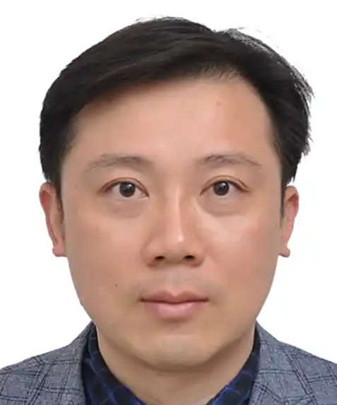 Yuanjin Zhao (赵远锦)合作论文数Medical School of Nanjing University140
 Yunru Yu合作论文数32
 Zhongze Gu (顾忠泽)合作论文数School of Biological Science and Medical Engineering, Southeast University;State Key Laboratory of Bioelectronics, Southeast University26Zhuohao Zhang合作论文数20
 Yuetong Wang (王月桐)合作论文数School of Food Science and Pharmaceutical Engineering, Nanjing Normal University19Qiao Wang合作论文数18
 Wenzhao Li合作论文数16Yao Cheng合作论文数15
 Chaoyu Yang合作论文数Univ Sci & Technol China, Dept Precis Machinery & Precis Instrumentat, Hefei 230026, Anhui, Peoples R China15
 Lingyu Sun (孙凌玉)合作论文数School of Transportation Science and Engineering, Beihang University15Chong Wang合作论文数14
 Fu Fanfan合作论文数14
 Jie Wang (王洁)合作论文数College of Artificial Intelligence, Nanjing Agricultural University14Huan Wang合作论文数13
 Fangfu Ye (叶方富)合作论文数Institute of Physics, Chinese Academy of Sciences;Wenzhou Institute, University of Chinese Academy of Sciences13Xinyuan Yang合作论文数11
 Hanxu Chen合作论文数11Zhuoyue Chen合作论文数11Changmin Shao合作论文数10
 Feika Bian合作论文数Dept Clin Lab, Nanjing Univ10
 Xiaocheng Wang合作论文数Zhejiang Lab for Regenerative Medicine, Vision and Brain Health), Wenzhou Institute, University of Chinese Academy of Sciences10Xiaoxuan Zhang合作论文数10Yu Wang合作论文数9
 Dagan Zhang合作论文数Dept Rheumatol & Immunol, Nanjing Univ9
 Hongbo Zhang合作论文数Pharmaceutical Sciences Laboratory, Department of Pharmaceutical Science, Åbo Akademi University;Shanghai Jiao Tong University9Guopu Chen合作论文数Nanjing Univ, Dept Gen Surg, Jinling Hosp, Sch Med, 305 East Zhongshan Rd, Nanjing 210002, Jiangsu, Peoples R China8Weijian Sun (孙维建)合作论文数8Hui Zhang合作论文数7
 Ze Zhao (赵泽)合作论文数School of Resource and Environmental Sciences, Wuhan University7
 Keqing Shi合作论文数7
 Gaizhen Kuang合作论文数6Junjie Chi合作论文数6Jiali Wang合作论文数6Lu Fan合作论文数6Yuxiao Liu合作论文数6Qingfei Zhang合作论文数6
 Weixia Zhang合作论文数Tsinghua Univ, Dept Chem, Key Lab Organ Optoelect & Mol Engn, Beijing 100084, Peoples R China6
) Li Wang (王立)合作论文数College of Chemical and Biological Engineering, Zhejiang University6
Li Wang (王立)合作论文数College of Chemical and Biological Engineering, Zhejiang University6 Fei Rong (戎非)合作论文数School of Biological Science & Medical Engineering, Southeast University6Xiao-ya DING (丁小雅)合作论文数5Cheng Zhao合作论文数5Qihui Fan合作论文数5
 Filppula Anne M合作论文数Department of Clinical Pharmacology, University of Helsinki5
 Ding Haibo合作论文数School of Biological Science and Medical Engineering, Southeast University5
 Liu Cihui合作论文数5
 Jiahui Guo合作论文数5
 David A. Weitz合作论文数Experimental Soft Condensed Matter Group, Department of Physics, School of Engineering and Applied Sciences, Harvard University;BASF Advanced Research Initiative, Harvard University;Materials Research Science and Engineering Center, Harvard University5Ping Duan合作论文数4
 Xin Shou合作论文数4




