Background: Systemic autoimmune rheumatic diseases-associated interstitial lung disease (SARD-ILD) presents with varied disease courses, emphasizing the need for reliable predictors of progression. The prognostic utility of bronchoalveolar lavage (BAL) in SARD-ILD remains underexplored. The objective of this study was to evaluate the role of BAL fluid lymphocyte count in predicting disease progression in patients with SARD-ILD. Methods: This observational study included patients with SARD-ILD undergoing BAL as part of their diagnostic workup. Disease progression was defined as either Forced vital capacity (FVC) decrease >10%, two out of the following three criteria within two years: FVC decrease of 5-10%, worsening symptoms, increased fibrosis on imaging, or any of the following: escalation of treatment, Interstitial lung disease (ILD) exacerbation, lung transplantation, or disease-specific mortality. Logistic regression identified predictors of progression. Time-to-progression was assessed using Kaplan-Meier survival curves. The optimal BAL lymphocyte threshold for predicting progression was identified using the Youden Index and the Wilcoxon method. Results: We identified 89 patients, of whom 30 (33.7%) had progressive disease. Progressors had a significantly higher BAL lymphocyte count compared to non-progressors (31.6 ± 24.8% vs. 14.3 ± 16.5%, p < 0.001). BAL lymphocyte proportion was significantly and independently associated with disease progression (odds ratio, 1.05; 95% confidence interval 1.02-1.07; p < 0.01). A lymphocyte count above 9 percent was associated with a markedly increased risk of disease progression (odds ratio, 13.14; 95% confidence interval, 4.20-51.98; p < 0.01). Conclusions: BAL lymphocyte count was associated with a higher likelihood of progression in SARD-ILD. BAL assessment may help identify patients at increased risk of disease progression. However, these findings should be considered exploratory and require validation in larger prospective studies and across individual SARD-ILD subtypes.
BACKGROUND:ABO blood group has been associated with SARS-CoV-2 susceptibility. Little data exist regarding the impact of ABO and Rhesus (Rh[D]) type on breakthrough infections and antibody responses following SARS-CoV-2 booster vaccination. METHODS:This multicenter, population-based cohort study includes individuals ≥ 18 years who received a booster vaccination against SARS-CoV-2. Antibody levels against SARS-CoV-2 receptor binding domain (RBD) and neutralizing antibodies against wild-type (WT) virus and Omicron variant were assessed at baseline, after 4 weeks and after 6 months. At 6 months follow-up, self-reported ABO and Rh(D) type, time to, and severity of breakthrough infections were collected. RESULTS:In total, 3066 participants (mean age 49 [35-59], 62% female) were included in multivariable regression models, showing no association of anti-RBD and neutralizing antibodies against wild-type and Omicron variant with ABO or Rh(D) 4 weeks after booster vaccination. Time-dependent Cox regression analysis showed significantly lower rates of breakthrough infections in patients with AB (hazard ratio [HR] 0.67 [0.50-0.90]; p = 0.008) compared to O, even after adjustment for relevant covariates and a trend for blood group B (HR 0.82 [0.66-1.01]; p = 0.06) compared to O; there were no associations between Rh(D) and breakthrough infection rates or between observed (asymptomatic to moderate) COVID-19 symptom severity and ABO or Rh(D) blood group. CONCLUSIONS:ABO or Rh(D) blood group was not associated with COVID-19 severity or antibody responses following SARS-CoV-2 vaccination. Breakthrough infections were least common in blood groups AB and B. Although our data do not support previously discussed protective effects of anti-A/B/Rh(D) agglutinins, differences in ABO group appear relevant for Omicron transmission.
OBJECTIVES:Methotrexate (MTX) is the first-line treatment for rheumatoid arthritis (RA), yet its immunological mechanisms remain incompletely understood. This study aimed to define the time-resolved cellular and molecular changes induced by MTX. METHODS:In a prospective longitudinal study, newly diagnosed patients with RA initiating MTX were systematically profiled over time. We applied an integrated single-cell multiomics approach combining high-parameter immunophenotyping and single-cell RNA sequencing of PBMCs (peripheral blood mononuclear cells) obtained from 6 patients at baseline and at sequential time points during therapy. Functional in vitro assays were conducted to evaluate direct effects of MTX. Key findings were validated in an independent cohort of 22 patients with RA, stratified by clinical response, and integrated with external transcriptomic datasets. RESULTS:MTX-induced rapid immune remodelling that was detectable as early as 3 weeks after treatment initiation and preceded clinical improvement. The earliest and most pronounced alterations occurred in T follicular helper (Tfh) cells and plasmablasts, which declined selectively in clinical responders but remained largely unchanged in nonresponders across independent cohorts and external datasets, underscoring their potential as early biomarkers of treatment response. Functional plasmablast differentiation assays further linked MTX treatment to impaired development. Transcriptomic network analyses identified novel regulatory hubs associated with MTX exposure, including LMNA, SCO2, GVQW3, and members of the GIMAP family. To promote data accessibility, we developed an interactive single-cell browser for community-driven exploration. CONCLUSIONS:Together, these findings reveal an unexpectedly rapid and coordinated immune reconfiguration after MTX initiation and position early alterations in Tfh cells and plasmablasts as biomarkers of treatment response.
Throughout history, herbal medicines and natural products have played a crucial role as therapeutics for humans, yet their molecular mechanisms of action often remain elusive. Here, we investigate whether primulagenin A (PGA) from the traditionally used herbal substance Primula root acts via the nuclear receptor RORγ, a key regulator of pro-inflammatory Th17 cells, which are linked to autoimmune diseases like psoriasis. Full-length luciferase assays revealed a high potency (IC50 = 119 nmol/L) and efficacy (I max = 87%) of PGA as an inverse agonist of RORγ. To ensure sufficient supply, we established methods to isolate and synthesize PGA. Its binding to the human RORγ ligand binding domain was confirmed by nano differential scanning fluorimetry, and a structure-activity relationship was proposed by docking and site-directed mutagenesis. qPCR revealed PGA-mediated downregulation of RORγ target gene expression. Furthermore, PGA inhibited murine and human Th17 differentiation in a concentration-dependent manner and reduced the proportion of IL-17A-producing Th17 cells, as assessed by flow cytometry. In this work, we identify PGA as a new, potent, and efficacious inverse agonist of RORγ, with potential for modulating immune responses in inflammatory and autoimmune diseases.
OBJECTIVE:Idiopathic inflammatory myopathies (IIM) are autoimmune disorders characterised by muscle inflammation, high creatine kinase (CK) levels and disability. Despite immunomodulating therapies, patients often experience progressive disease, resulting in refractory conditions or dependence on glucocorticoids (GC). This study addresses the efficacy and safety of immunoadsorption (IAS) in refractory IIM. METHODS:A retrospective, monocentric study was conducted on patients with highly active IIM subjected to IAS between January 2000 and September 2021. Inclusion criteria were adult patients with confirmed IIM unresponsive to standard therapies, including GC and at least one disease-modifying antirheumatic drug. Primary end point was defined as minor improvement, requiring a ≥ 20% reduction in both daily GC dosage and CK levels at week 12, alongside patient comparative self-assessment (PCSA) rated as 'no change' or 'better'. Secondary endpoints included moderate (≥40%) and major (≥60%) improvements at weeks 4, 8 and 12. Relapse rates were assessed over a 3-month period following the last IAS procedure. RESULTS:The study included 23 refractory IIM patients treated with IAS. Primary end point was reached by 52.2% (n = 12) patients. Moderate improvement was observed in 8.7% (n = 2), 26.1% (n = 6) and 34.8% (n = 8) and major improvement in 8.7% (n = 2), 17.4% (n = 4) and 30.4% (n = 7) at weeks 4, 8 and 12, respectively. No unexpected adverse events were reported, with low relapse rates within 3 months post-IAS (18%). CONCLUSION:IAS may be an effective adjunctive therapy in patients refractory to GC and conventional treatments, leading to rapid CK level reductions, decreased GC usage and improvements in PCSA and muscle function.
Rheumatoid arthritis (RA) is characterized by immune dysregulation, including alterations in peripheral blood mononuclear cell (PBMC) populations and aberrant cytokine signaling. Methotrexate (MTX) is the preferred first-line treatment for RA, yet its precise mechanisms of action remain incompletely understood. This study employed a multi-omics strategy-combining single-cell RNA sequencing (scRNA-seq) and immunophenotyping-to identify key effector peripheral immune cells and their cellular responses in RA patients over 12 weeks of MTX treatment. In our study, MTX was associated with significant immune modulation, including the restoration of naive T and B cells and reductions in T cell memory subsets with these effects detectable as early as three weeks post-treatment. Plasmablast levels also emerged as a potential biomarker for early therapeutic response, reflecting MTX's impact on immune homeostasis. Transcriptional analysis revealed modulation of key pathways, including TNF-alpha; signaling, B cell receptor signaling, and T cell receptor-mediated apoptosis. Network analysis identified critical regulatory hubs, such as EGR1, JAK2, and SOCS1, in monocytes and CD4 memory T cells, highlighting these cell types as key mediators of MTX's effects. In conclusion, these findings advance our understanding of MTX's effects on immune cell dynamics at different stages of treatment, showing for the first time the early cellular changes leading to immune modulation in RA. Altogether, our results provide the foundation for further mechanistic investigations into MTX. ### Competing Interest Statement MaB and TP received funding from Lilly. MiB received grants from AlphaSigma. DA received grants and consulting fees from AbbVie, Amgen,Lilly, Merck, Novartis, Pfizer, Roche and Sandozand.
Autoimmune diseases like rheumatoid arthritis (RA) are characterized by a systemic inflammation caused by autoreactive immune cells. Epigenomic modulation of these cells offers a strategy to reprogram pathogenic pathways without altering the genome, potentially restoring immune balance. Epigenetic inhibitors are already utilized in oncology but often exhibit adverse effects due to lack of selectivity and cytotoxic concentrations. Applying these drugs to treat autoimmune diseases necessitates more selective inhibitors and the use of tolerable concentrations. In this study, we screened a library of 25 compounds with varying degrees of target selectivity and different concentrations. Spectral cytometry enabled the analysis of cell-subset distribution and activation, followed by bulk RNA-sequencing for transcriptomic profiling. We could demonstrate cell-subset specific and concentration-dependent immune modulation in PBMCs. Transcriptomic analysis showed that inhibitors of histone acetylation-modulating enzymes significantly altered gene expression, particularly in immune regulation pathways relevant to autoimmune diseases. Comparative analysis between in-vitro treated healthy controls and RA patients demonstrated both shared and selective drug effects, with some inhibitors like Ricolinostat overlapping with established RA drug pathways. Our findings highlight the potential of epigenetic inhibitors, especially those targeting histone acetylation, to modulate immune responses in a target-selective manner. ### Competing Interest Statement MaB and TP received funding from Lilly. MiB received grants from AlphaSigma. DA received grants and consulting fees from AbbVie, Amgen,Lilly, Merck, Novartis, Pfizer, Roche and Sandozand.
Viral infections, including respiratory diseases such as Coronavirus disease 2019 (COVID-19), are hypothesized to contribute to the onset of autoimmune disorders. Although elevated levels of autoantibodies have been observed following COVID-19, the role of specific autoantibodies linked to autoimmune diseases and their correlation with disease severity remains poorly defined. In this study, we used a comprehensive autoantibody panel to assess the autoantibody production across different cohorts of COVID-19 patients, categorized by disease severity. We also compared patients with severe COVID-19 to a control group with other severe, non-COVID-related diseases. Our findings indicate that the severity of COVID-19 corresponds to the overall production of specific autoantibodies, which are particularly associated with COVID-19. This association might predispose to an increased risk for the development of autoimmune conditions after a severe course of COVID-19.
Regulatory T (Treg) cells are essential for maintaining immune homeostasis, with Foxp3 acting as the master transcription factor governing their differentiation and function. The acquisition of effector signatures in Treg cells is closely tied to the surrounding tissue-specific immune environment and typically occurs alongside Foxp3 expression. In this study, we investigated the transcriptomic and functional consequences of Treg-mediated regulation in a Th2-driven disease setting. The application of both in vitro systems and in vivo disease models allowed us to mimic Th2-mediated environments. We could demonstrate Th2-driven loss of Foxp3 expression in Treg cells in vitro and in vivo. Transcriptomic analysis revealed a maintained Treg signature despite the loss of active Foxp3 expression. Functional characterization of Tregs both in vitro and in vivo uncovered a preserved suppressive capacity even in the absence of Foxp3. Our findings unveil that, despite loss of Foxp3, a preserved Treg signature remains intact enabling the regulation of Th2-mediated diseases. The persistence of this regulatory transcriptome highlights the importance for developing Treg-cell therapy strategies in cancer and autoimmune diseases independent of Foxp3 expression.
Background Interstitial lung disease (ILD) is a common manifestation of idiopathic inflammatory myopathies (IIM) and a substantial contributor to hospitalisation, increased morbidity, and mortality. In-vivo evidence of ongoing tissue remodelling in IIM-ILD is scarce. We aimed to evaluate fibroblast activation in lungs of IIM-patients and control individuals using Ga-68-labelled inhibitor of Fibroblast-Activation-Protein (FAPi) based positronic emission tomography and computed tomography imaging (PET/CT). Methods In this prospective observational pilot study, consecutive patients with IIM and participants without rheumatic conditions or ILD serving as a control group were recruited at the Medical University of Vienna, Austria, and underwent FAPi PET/CT imaging. Standard-of-care procedures including clinical examination, assessment of severity of dyspnoea, high-resolution computed tomography (HR-CT), and pulmonary function testing (PFT) were performed on all patients with IIM at baseline and for patients with IIM-ILD at follow-up of 12 months. Baseline pulmonary FAPi-uptake was assessed by the maximum (SUVmax) and mean (SUVmean) standardized uptake values (SUV) over the whole lung (wl). SUV was corrected for blood pool background activity and target-to-background ratios (TBR) were calculated. We compared pulmonary FAPi-uptake between patients with IIM-ILD and those without ILD, as well as controls, and correlated baseline FAP-uptake with standard diagnostic tools such as HR-CT and PFT. For predictive implications, we investigated whether patients with IIM and progressive ILD exhibited higher baseline FAPi-uptake compared to those with stable ILD. Metrics are reported as mean with standard deviation (+/- SD). Findings Between November 16, 2021 and October 10, 2022, a total of 32 patients were enrolled in the study. Three participants from the control group were excluded due to cardiopulmonary disease. In individuals with IIM-ILD (n = 14), wlTBR(max) and wlTBR(mean) were significantly increased as compared with both non-ILD-IIM patients (n = 5) and the control group (n = 16): wlTBR(max): 2.06 +/- 1.04 vs. 1.04 +/- 0.22 (p = 0.019) and 1.08 +/- 0.19 (p = 0.0012) and wlTBR(mean): 0.45 +/- 0.19 vs. 0.26 +/- 0.06 (p = 0.025) and 0.27 +/- 0.07 (p = 0.0024). Similar values were observed in wlTBR(max) or wlTBR(mean) between non-ILD IIM patients and the control group. Patients with progressive ILD displayed significantly enhanced wlTBR(max) and wlTBR(mean) values at baseline compared to patients with stable ILD: wlTBR(max): 1.30 +/- 0.31 vs. 2.63 +/- 1.04 (p = 0.0084) and wlTBR(mean): 0.32 +/- 0.08 vs. 0.55 +/- 0.19 (p = 0.021). Strong correlations were found between FAPi-uptake and disease extent on HR-CT (wlTBRmax: R = 0.42, p = 0.07; wlTBRmean: R = 0.56, p = 0.013) and severity of respiratory symptoms determined by the New York Heart Association (NYHA) classification tool (wlTBRmax: R = 0.52, p = 0.022; wlTBRmean: R = 0.59, p = 0.0073). Further, pulmonary FAPi-uptake showed inverse correlation with forced vital capacity (FVC) (wlTBRmax: R = -0.56, p = 0.012; wlTBRmean: R = -0.64, p = 0.0033) and diffusing capacity of the lungs for carbon monoxide (DLCO) (wlTBRmax: R = -0.52, p = 0.028; wlTBRmean: R = -0.68, p = 0.0017). Interpretation Our study demonstrates higher fibroblast activation in patients with IIM-ILD compared to non-ILD patients and controls. Intensity of pulmonary FAPi accumulation was associated with progression of ILD. Considering that this study was carried out on a small population, FAPi PET/CT may serve as a useful non-invasive tool for risk stratification of lung disease in IIM. Copyright (c) 2024 The Authors. Published by Elsevier Ltd.
Due to optimised treatment strategies and the availability of new therapies during the last decades, formerly devastating chronic inflammatory diseases such as rheumatoid arthritis or systemic sclerosis (SSc) have become less menacing. However, in many patients, even state-of-the-art treatment cannot induce remission. Moreover, the risk for flares strongly increases once anti-inflammatory therapy is tapered or withdrawn, suggesting that underlying pathological processes remain active even in the absence of overt inflammation. It has become evident that tissues have the ability to remember past encounters with pathogens, wounds and other irritants, and to react more strongly and/or persistently to the next occurrence. This priming of the tissue bears a paramount role in defence from microbes, but on the other hand drives inflammatory pathologies (the Dr Jekyll and Mr Hyde aspect of tissue adaptation). Emerging evidence suggests that long-lived tissue-resident cells, such as fibroblasts, macrophages, long-lived plasma cells and tissue-resident memory T cells, determine inflammatory tissue priming in an interplay with infiltrating immune cells of lymphoid and myeloid origin, and with systemically acting factors such as cytokines, extracellular vesicles and antibodies. Here, we review the current state of science on inflammatory tissue priming, focusing on tissue-resident and tissue-occupying cells in arthritis and SSc, and reflect on the most promising treatment options targeting the maladapted tissue response during these diseases.
Background High content imaging-based functional precision medicine approaches have been developed and successfully applied in the field of haemato-oncology. For rheumatoid arthritis (RA), treatment selection is still based on a trial-and-error principle, and biomarkers for patient stratification and drug response prediction are needed. Methods A high content, high throughput microscopy-based phenotyping pipeline for peripheral blood mononuclear cells (PBMCs) was developed, allowing for the quantification of cell type frequencies, cell type specific morphology and intercellular interactions from patients with RA (n = 65) and healthy controls (HC, n = 33). Samples were exposed to a curated set of RA-specific small molecules, biologicals and reference stimuli for 24 h to assess ex vivo drug effects. Data on ex vivo PBMC phenotypes were integrated with information on patients’ in vivo medication and disease activity. Findings The unbiased data from in total 6.9e8 individual cells were collected and allowed for the identification of PBMC phenotypes specific to disease activity as well as in vivo and ex vivo treatment. The arrayed ex vivo drug perturbation enabled the systematic characterization of drug effects, clustering by mode of action and uncovered morphologic alterations associated with biologic disease-modifying anti-rheumatic drug (DMARD) treatment. Individual in vivo treatment regimens translated into altered immune cell abundances in patients with a comedication of conventional synthetic DMARDs when compared to HCs. Global integration of PBMC characteristics led to clustering of patients according to disease activity and correlation with clinical data. Interpretation The application of the developed screening tool demonstrates a technical proof-of-concept for feasibility of a functional precision medicine approach to the ex vivo immunophenotypic characterisation of patients with RA. Funding This work was supported by the Austrian Academy of Sciences, the Medical University of Vienna and a grant (RMG2235 to L.X.H.) from the European Alliance of Associations for Rheumatology (EULAR).
Fundamental insight gained over the last decades led to the discovery of cytokines as pivotal drivers of inflammatory diseases such as rheumatoid arthritis, psoriasis/psoriasis arthritis, inflammatory bowel diseases, atopic dermatitis and spondylarthritis. A deeper understanding of the pro-inflammatory and anti-inflammatory effects of various cytokines has prompted new cytokine-targeting therapies, which revolutionised the treatment options in the last years for patients with inflammatory disorders. Disease-associated immune responses typically involve a complex interplay of multiple cytokines. Therefore, blockade of one single cytokine does not necessarily lead to a persistent remission in all patients with inflammatory disorders and fostered new therapeutic strategies targeting intracellular pathways shared by multiple cytokines. By inhibiting JAK-STAT signalling pathways common to families of cytokines, JAK-inhibitors (JAKinibs) have created a new paradigm for the treatment of inflammatory diseases. Multiple agents have been approved for various disorders and more are being investigated for several new indications. Second-generation selective JAKinibs have been devised with the aim to achieve an increased selectivity and a possible reduced risk of side effects. In the current review, we will summarise the current body of evidence of pan versus selective JAKinibs and the most recent insights on new side effects and indications, including COVID-19.
Background: The pathogenesis of synovitis in systemic sclerosis (SSc) is unknown. Due to the lack of evidence-based therapies, SSc patients with synovitis are treated as rheumatoid arthritis (RA) patients, however with unknown effect. Previously, we showed that SSc synovitis differs from RA synovitis with a prevalent pauci-immune pathotype. Objectives: To decipher the histological, cellular, and transcriptional differences between SSc and RA synovitis, specifically in synovial fibroblasts (SF). Methods: We collected ultrasound-guided synovial biopsies from 11 SSc and 7 RA patients. All samples were analysed histologically, including Krenn synovitis score (0-9) and pathotype characterisation. Additionally, 7 SSc and 6 RA samples were dissociated and processed for single-cell RNA-sequencing (20,705 cells in SSc and 31,681 in RA). Due to the pauci-immune nature of the synovitis, we focused our analysis on the SF population (4,638 SF in SSc, 6,097 in RA). We performed differential gene expression analysis, over-representation pathway analyses and calculated signature expression scores between SSc and RA SF. Pathway analyses included publicly available datasets and bulk RNA sequencing data from SF stimulated in vitro with TNF-α, IFN-α/b, IFN-γ, and IL-6 from internal experiments and published data1 (Figure 1A). Results: The Krenn synovitis score was lower in SSc than in RA (2.1 ± 1.4 vs. 4.2 ± 1.0, p = 0.01). There was no difference in fibrosis (Elastica van Gieson staining) and vascularisation (CD31 staining) between SSc and RA synovitis. We confirmed a pauci-immune pathotype characteristic of SSc synovitis (91% vs. 43% of RA biopsies, p = 0.04). Consistently, neutrophils were more prevalent in RA as compared to SSc synovitis (p < 0.01). On the single-cell level, we identified seven SF cell states defined by the expression of the marker genes PRG4, CHI3L2, COMP, CXCL12, CXCL14, MFAP5, and POSTN consistent with the current literature in RA2. The proportion of SF clusters differed between both diseases with a higher enrichment of MFAP5+ (Fold-change (FC) = 3.81, false discovery rate (FDR) < 0.001) and COMP+ SF in SSc (FC = 2.28, FDR < 0.001), and POSTN+ (FC = 3.12, FDR < 0.001) and CXCL12+ SF in RA (FC = 1.70, FDR < 0.001). We identified 675 significant differentially expressed genes (FDR < 0.01, |log2FC| > 0.25) and several differentially activated signalling pathways between SSc and RA SF. In particular, SSc SF were enriched in IFN-α and IFN-γ (both FDR < 0.018) and complement and coagulation pathways (FDR < 0.008), especially in genes of the alternative complement pathway (Figure 1B). RA SF showed a strong enrichment in TNF-α signaling (FDR < 6.3e-12) and inflammatory response genes (FDR < 0.018). Using the transcriptional signature of in vitro stimulated SF as gene sets, we confirmed the IFN response in SSc SF, whereas TNF-α and IL-6 response signatures were more dominant in RA SF (Figure 1C). Interestingly, IFN-α/β and IFN-γ stimulation of SF in vitro led to an activation pattern of alternative complement pathway genes similar to that observed in SSc SF (Figure 1D). IFN and complement signature scores were significantly higher in sublining CXCL12+ SF in SSc compared to RA (both p < 2.22e-16). To identify the drivers of the IFN signature in SSc SF, we assessed the expression of the different IFN genes in synovial cell populations. Only IFNG was expressed, mainly by T cells, which is consistent with the preferential expression of IFN response genes in the sublining synovium. Conclusion: This first in-depth characterization of the SSc synovium revealed hallmark tissue, cellular and molecular changes between SSc and RA synovitis. We found a strong enrichment of the IFN signalling pathway in SSc SF, possibly leading to a dysregulation of the alternative complement pathway. Our findings provide the basis for the development of specific targeted therapies for synovitis in SSc. REFERENCES: [1] Tsuchiya, Haruka, et al. Annals of the Rheumatic Diseases 80.4 (2021): 440-450.[2] Zhang, Fan, et al. Nature (2023): 1-9. Acknowledgements: This project is funded by FOREUM – Foundation for Research in Rheumatology. We thank our patient research partners Joëlle Messmer & Andreas Eisenring. Disclosure of Interests: Celina Geiss: None declared, Miranda Houtman: None declared, Raphael Micheroli: None declared, Yannis Djeffal: None declared, Mojca Frank Bertoncelj: None declared, Sam G. Edalat: None declared, Kristina Buerki: None declared, Chantal Pauli: None declared, Michael Bonelli GSK, Galapagos, Thomas Karonitsch: None declared, Oliver Distler 4P-Pharma, Abbvie, Acceleron, Alcimed, Altavant, Amgen, AnaMar, Argenx, Arxx, AstraZeneca, Blade, Bayer, Boehringer Ingelheim, Corbus, CSL Behring, Galderma, Galapagos, Glenmark, Gossamer, Horizon, Janssen, Kymera, Lupin, Medscape, Merck, Miltenyi Biotec, Mitsubishi Tanabe, Novartis, Orion, Prometheus, Redxpharma, Roivant, Topadur and UCB, Co-founder of CITUS AG, 4P-Pharma, Abbvie, Acceleron, Alcimed, Altavant, Amgen, AnaMar, Argenx, Arxx, AstraZeneca, Blade, Bayer, Boehringer Ingelheim, Corbus, CSL Behring, Galderma, Galapagos, Glenmark, Gossamer, Horizon, Janssen, Kymera, Lupin, Medscape, Merck, Miltenyi Biotec, Mitsubishi Tanabe, Novartis, Orion, Prometheus, Redxpharma, Roivant, Topadur and UCB, 4P-Pharma, Abbvie, Acceleron, Alcimed, Altavant, Amgen, AnaMar, Argenx, Arxx, AstraZeneca, Blade, Bayer, Boehringer Ingelheim, Corbus, CSL Behring, Galderma, Galapagos, Glenmark, Gossamer, Horizon, Janssen, Kymera, Lupin, Medscape, Merck, Miltenyi Biotec, Mitsubishi Tanabe, Novartis, Orion, Prometheus, Redxpharma, Roivant, Topadur and UCB, Caroline Ospelt: None declared, Muriel Elhai: None declared.Figure 1Abbreviations: log2FC = log2 fold-change, RA = rheumatoid arthritis, SF = synovial fibroblasts, SSc = systemic sclerosis, stim = stimulation. Panel A created with BioRender.
 Josef Smolen合作论文数Universitätsklinik für Innere Medizin III, Medizinische Universität Wien73
 Clemens Scheinecker合作论文数Medical University of Vienna Oberarzt;Division of Rheumatology, Medical University of Vienna57
 Daniel Aletaha合作论文数48Daniel Mrak合作论文数25Birgit Niederreiter合作论文数25
 Helga Radner合作论文数Department of Internal Medicine III, Medical University of Vienna23Thomas Karonitsch合作论文数Department of Internal Medicine 3, Medical University of Vienna22
 Stephan Blüml合作论文数Med Univ Vienna, Dept Rheumatol, Vienna, Austria21Elisabeth Simader合作论文数18
 Gunter Steiner合作论文数18Hans P. Kiener合作论文数18Lisa Goeschl合作论文数16
 Helmuth Haslacher合作论文数Dept Lab Med, Med Univ Vienna16
 Antonia Puchner合作论文数16
 Victoria Saferding合作论文数16
 Thomas Perkmann合作论文数Department of Laboratory Medicine, Medical University of Vienna16
 Daniela Sieghart合作论文数15
 Wilfried Ellmeier合作论文数Institute of Immunology, Center for Pathophysiology, Infectiology and Immunology, Medical University of Vienna15
 Leonhard X. Heinz合作论文数Institute of Immunology;Medical University Vienna;Institute of Immunology, Medical University Vienna15Kastriot Kastrati合作论文数14
 Peter Mandl合作论文数University of Vienna14
 Kiyoshi Hirahara合作论文数14Silvia Hayer合作论文数Department of Internal Medicine III, University of Vienna14A. Savitskaya合作论文数13Felix Kartnig合作论文数Austrian Acad Sci, CeMM Res Ctr Mol Med, Vienna, Austria13
 Carl Walter Steiner合作论文数13
 Teresa Preglej合作论文数Institute of Immunology, Medical University of Vienna12
 Philipp Hofer合作论文数12Stefan Winkler合作论文数12
 John J. O'Shea合作论文数National Institute of Arthritis and Musculoskeletal and Skin Diseases, National Institutes of Health12K. Von Dalwigk合作论文数12
 Tosevska Anela合作论文数Research Platform Active Ageing, University of Vienna11Göschl Lisa合作论文数11
 Kurt Redlich合作论文数10L. Goeschl合作论文数9Andreas Kerschbaumer合作论文数9
 Han-Yu Shih合作论文数National Eye Institute, National Institutes of Health9Yuka Kanno合作论文数9Thomas Deimel合作论文数8Gessl Irina合作论文数8
 Marije I Koenders合作论文数Department of Rheumatic Diseases, Radboud University;Department of Experimental Rheumatology, Radboud University Medical Center8Renate Kain合作论文数Department of Nephrology;Department of Clinical Pathology;University Vienna;Department of Clinical Pathology, University Vienna8Hladik Anastasiya合作论文数7
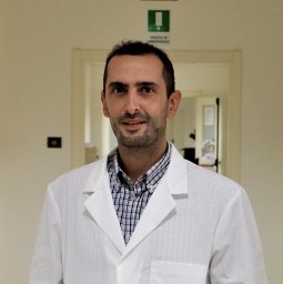 Giuseppe Sciume合作论文数Department of Molecular Medicine-Istituto Pasteur-Fondazione Cenci Bolognetti, Sapienza University of Rome7
 Karin Stiasny合作论文数Medical University of Vienna7Arian Laurence合作论文数7Renate Thalhammer合作论文数6A. Rapp合作论文数6Blueml Stephan合作论文数6




