The RNA-dependent protein kinase PKR regulates responses to viral infection and has emerging roles in memory formation. Inhibition of PKR enhances long-term memory in mice and reverses cognitive decline in models of aging and Alzheimer's disease. However, existing PKR inhibitors have poor selectivity and pharmacokinetic properties, limiting therapeutic development. Here, we describe the transformation of dabrafenib, an FDA-approved oncogenic BRAF inhibitor, into a selective PKR inhibitor. Dabrafenib was identified by screening as a promising PKR lead with similar potency against BRAF and PKR. Guided by X-ray cocrystal structures, we introduced modifications that removed BRAF while retaining PKR inhibition. This optimization yielded OICR-403184, which shows markedly reduced BRAF activity, improved PKR selectivity (IC50 > 10,000 nM against BRAF vs IC50 = 263 nM against PKR in vitro), and minimal activity against related eIF2α kinases in cells. These findings establish OICR-403184 as a promising chemical starting point for further PKR inhibitor optimization.
Persistent activation of the integrated stress response (ISR) is a central driver of cognitive decline in both neurodevelopmental and neurodegenerative disorders. However, the cell type-specific mechanisms underlying these deficits remain poorly understood. By integrating single-cell RNA-seq and single-cell assay for transposase-accessible chromatin sequencing, we generated a brain ISR atlas using Ppp1r15bR658C mice, a clinically relevant model of intellectual disability characterized by selective and persistent ISR activation. We find that distinct brain cell types differentially engage transcriptional and chromatin remodeling programs. Notably, selective deletion of the major ISR downstream effector ATF4 in GABAergic neurons, but not in glutamatergic neurons, exacerbates ISR-mediated cognitive decline in Ppp1r15bR658C mice, demonstrating that different neuronal subtypes rely on distinct ISR effectors. We define a molecular single-cell signature of persistent ISR activation that serves as a metric of ISR-mediated cellular vulnerability and as a biomarker for cognitive dysfunction across human cognitive disorders. These findings demonstrate that cell type-specific responses drive cognitive dysfunction during persistent ISR activation.
The integrated stress response (ISR) plays a crucial role in cognition via bidirectional modulation of the two major forms of synaptic plasticity, long-term potentiation, and long-term depression (LTD). Specifically, inhibition of the ISR blocks metabotropic glutamate receptor-dependent LTD (mGluR-LTD), whereas its activation facilitates this form of synaptic depression. However, the contribution of activating transcription factor 4 (ATF4), the best studied downstream effector of the ISR, to mGluR-LTD remains unknown. Here, we show that pharmacological activation of group I mGluRs in mouse hippocampal slices increases ATF4 protein levels without altering its transcription and concurrently downregulates the expression of oxidative phosphorylation (OXPHOS) proteins. Selective deletion of ATF4 in excitatory neurons impairs mGluR-LTD and prevents the downregulation of OXPHOS proteins. Notably, administration of a small molecule inhibitor of OXPHOS rescues the impaired mGluR-LTD in ATF4-depleted hippocampal slices, indicating that ATF4 regulates this type of synaptic plasticity by modulating mitochondrial function. Moreover, ATF4 deletion in excitatory neurons disrupts object-place learning, an mGluR-LTD-dependent behavior paradigm. Together, these findings reveal a role of ATF4 as a key mediator of protein synthesis-regulated synaptic depression and related behaviors.
The integrated stress response (ISR) is essential for cellular homeostasis and cognitive function. We investigated how persistent ISR activation affects cognitive performance by studying the PPP1R15BR658C genetic variant associated with intellectual disability. To model this condition, we generated a mouse line with the pathogenic allele inserted. This variant destabilized the PPP1R15B•PP1 phosphatase complex, causing persistent ISR activation, impaired protein synthesis, and long-term memory deficits. We demonstrated that the cognitive and synaptic impairments in Ppp1r15bR658C mice arise directly from ISR activation. Furthermore, we characterized DP71L, a viral ortholog of PPP1R15B, which acted as a potent pan-ISR inhibitor. DP71L reversed the cognitive and synaptic deficits across mouse models of Down syndrome, Alzheimer's disease, and aging, and enhanced synaptic plasticity and memory in healthy mice.
Childhood absence epilepsy (CAE) is a common genetic epilepsy with a frequently complex polygenic etiology, for which a genetically tractable animal model is lacking. Current anti-seizure medications fail to address the significant neurocognitive and social comorbidities of CAE, highlighting a critical unmet need for comprehensive therapies and a better understanding of the underlying mechanisms. To address this, we developed a digenic mouse model (Cacng2stargazer/+;Cacna1a+/-, hereafter DiGstg+Ca1A) that mimics human polygenicity. The double mutant mice exhibit both absence seizures and altered social novelty preference. We found that ethosuximide, a first-line anti-CAE medication targeting thalamic low-threshold T-type calcium currents, suppressed seizures in DiGstg+Ca1A mice but failed to rescue their social deficit. Similarly, deletion of Cacna1g, which encodes the T-type Ca2+ channel CaV3.1, prevented seizure generation but did not ameliorate the social deficit. These findings reveal a dissociation between the T-type current that mediates seizures and the mechanism underlying the social deficits. We further demonstrate that in Cacng2stargazer/+ mice, selective deletion of one copy of Cacna1a in stargazin-enriched parvalbumin (PV) interneurons induced absence seizures and impaired social behavior. Remarkably, chemogenetic activation of cortical and thalamic PV interneurons using DREADDs not only suppressed seizures but also rescued the impaired social behavior. Together, our results suggest that selective modulation of PV interneuron activity in polygenic absence epilepsy could serve as a promising therapeutic strategy to address both absence seizures, and the often treatment-resistant neurocognitive comorbidities observed in CAE.
Alterations in the gut microbiome are associated with neurodevelopmental disorders, but causal mechanisms and therapeutic strategies remain undefined. Here, we demonstrate that human infant microbiomes isolated during the first six months of life drive behavioral impairments in mice and that microbiota-based interventions restore mice to normal behavior. Early-life microbiomes from twelve infants who later exhibited cognitive deficits at 2 years old (low-scoring) transferred adverse metabolic, brain, and behavioral phenotypes to mice, in contrast to microbiomes from twenty-three cognitively typical or high-scoring infants. Deficits in mice were rescued by fecal microbiota transplant from high-scoring infants or a rationally designed consortium that promoted amino acid levels. We confirmed lower fecal amino acid concentrations in low-scoring infants and replicated the association between early-life microbiome composition and cognitive outcomes in a second geographically independent infant cohort. Altogether, we discovered an early-life microbiome-mediated metabolic state causally linked to cognitive deficits and amenable to microbial intervention.
The ternary complex, composed of eIF2, GTP and initiator methionyl-tRNA, delivers the first amino acid to the ribosome to initiate protein synthesis. Eukaryotic initiation factor 2B (eIF2B) catalyzes GDP to GTP exchange on eIF2, thereby setting the ternary complex level. Stress-induced phosphorylation converts eIF2 from the substrate of eIF2B into an inhibitor (eIF2-P). This conversion reduces ternary complex levels and induces the integrated stress response (ISR). Here we chart an allosteric axis running through eIF2B, revealing the importance of an α-helix in its β-subunit, the 'latch-helix', that hooks onto the α-subunit to induce eIF2B activity. eIF2-P binding promotes latch-helix unhooking, opening eIF2B, which inhibits its activity. Convergently evolved viral proteins stabilize this latch-helix-binding active state of eIF2B. Using these insights, we generated ISR-activating compounds that stabilize eIF2B in its inhibited, unlatched state. Our study thus highlights how long-range eIF2B allostery can be pharmacologically manipulated to sustain or attenuate the ISR.
Genetic variants altering brain function have been considered the primary cause of many neurological disorders. Therefore, current therapeutics target the brain directly. However, an often-overlooked source of genetic variation comes from microbes inhabiting our bodies, which can profoundly modulate host brain function via metabolic and immune pathways. As a result of the dynamic interplay and co-evolution between the host and microbes, the concept of the hologenome-the collective genomes of host and microbes-has emerged. Intriguingly, recent studies suggest a paradigm shift in our understanding of disease etiology-neurological symptoms in some genetic diseases may have distinct hologenomic origins and may even originate outside the brain, potentially evading brain-targeting therapies. This perspective will summarize recent findings on the hologenome's role in neurological disorders and recommend a roadmap for future research. The proposed approach aims to provide new insights into human/animal biology, enhancing the rigor and reproducibility of both preclinical and clinical research.
The gut-microbiome-brain axis is a bidirectional communication system influencing host physiology and overall fitness. While "bottom-up" effects-where gut microbes influence brain function and behavior-are well established, direct evidence for "top-down" modulation-where the brain shapes the gut microbial ecology-remains elusive. Here, we show that the selective expression of the bacterial ion channel mNaChBac in glutamatergic lateral habenula (LHb) neurons increases their bursting activity and leads to endophentypes associated with depression. Importantly, this chronic activation leads to changes in the gut microbiome composition over time, as reflected by shifts in alpha-diversity and alterations in specific microbial taxa. These findings provide direct evidence that the persistent activation of the brain acts as a selective pressure that affects the stability and diversity of the gut microbiome, providing a new dimension by which gut-microbiome-brain interactions may cooperate to modulate host physiology and health.
The integrated stress response (ISR) is a highly conserved network essential for maintaining cellular homeostasis and cognitive function. Here, we investigated how persistent ISR activation impacts cognitive performance, primarily focusing on a PPP1R15B R658C genetic variant associated with intellectual disability. By generating a novel mouse model that mimics this human condition, we revealed that this variant destabilizes the PPP1R15B•PP1 phosphatase complex, resulting in chronic ISR activation, impaired protein synthesis, and deficits in long-term memory. Importantly, we found that the cognitive and synaptic deficits in Ppp1r15b R658C mice are directly due to ISR activation. Leveraging insights from evolutionary biology, we characterized DP71L, a viral orthologue of PPP1R15B, through detailed molecular and structural analyses, uncovering its mechanism of action as a potent pan-ISR inhibitor. Remarkably, we found that DP71L not only buffers cognitive decline associated with a wide array of conditions-including Down syndrome, Alzheimer's disease and aging-but also enhances long-term synaptic plasticity and memory in healthy mice. These findings highlight the promise of utilizing evolutionary insight to inform innovative therapeutic strategies.
IntroductionDespite the rise in psychiatric disorders worldwide, the underlying brain circuits responsible for these devastating conditions remain elusive. The lateral habenula (LHb) has emerged as a key brain structure in depression studies due to its hyperactive state in both patients and animal models. While this aligns with known roles in driving aversive states and regulating serotonin release, it is still unclear how acute and transient activity changes in the LHb can influence higher order cognitive processes such as learning, memory, and behavioral adaptation. Given the importance of these processes to psychiatric conditions, understanding how LHb activity impacts cognitive function allows novel insights into the neurobiological mechanisms of disorders like depression.MethodsTowards this goal, we used chemogenetic activation to temporarily excite glutamatergic neurons in the mouse LHb and assessed impacts on associative memory.Results and DiscussionSurprisingly, we found that transient activation of LHb impaired long-term memory, without affecting anxiety or depression-like behaviors. Specifically, post-training activation of LHb glutamatergic neurons disrupted object recognition and reward-based associative long-term memory, while sparing fear associated long-term memory. The memory impairment was restricted to a critical temporal window post-training/conditioning that corresponded with the consolidation stage of long-term memory. Strikingly, pairing LHb glutamatergic neuronal activation with systemic ketamine administration rescued the long-term memory deficits, indicating that LHb glutamatergic neurons modulate consolidation of associative memories via a NMDA-mediated mechanism. Together, these findings support a novel role for LHb glutamatergic neuronal activity in the consolidation of associative long-term memories.
Persistent activation of the integrated stress response (ISR) is a major driver of cognitive decline in both neurodevelopmental and neurodegenerative disorders. Using a new mouse model (Ppp1r15bR658C mice) that mimics the persistent ISR activation and cognitive decline observed in humans, we generated the first single-cell ISR atlas of the brain. By integrating single-cell RNA-seq and single-cell ATAC-seq with proteomics, we discovered that distinct brain cell types respond differently to persistent ISR activation and elicit cell-type-specific ISR programs. Interestingly, chromatin accessibility analyses revealed that the ISR downstream factor ATF4 is a key ISR effector in GABAergic neurons, while AP-1 (JUNB) is implicated in glutamatergic neurons. More importantly, selective deletion of ATF4 in GABAergic neurons, but not in glutamatergic neurons, impacts ISR-mediated cognitive decline in Ppp1r15bR658C mice, demonstrating that different neuronal subtypes rely on unique ISR downstream effectors to regulate mnemonic processes. Furthermore, we defined a comprehensive molecular signature of persistent ISR activation, which we showed could serve as a biomarker for cognitive dysfunction across neurodevelopmental, neurodegenerative disorders and normal aging. This multi-omic framework provides a key platform for exploring and validating new scientific hypotheses, significantly advancing our understanding of ISR-related brain disorders. ### Competing Interest Statement K.T., L.C.R., S.W.D.,D.N.I., B.Y., D.H., P.W., S.T., and M.C.-M. are employees and hold shares in Altos Labs, Inc.
The integrated stress response (ISR) modulates protein homeostasis in response to both intracellular and extracellular signals. The four kinases involved in the ISR all phosphorylate the same target, the alpha subunit of eukaryotic initiation factor 2 (eIF2α), to integrate various stress signals, thereby regulating cell fate. The activation of the ISR reprograms the proteome by inhibiting general protein synthesis while increasing the translation of specific mRNAs. In the brain, the ISR regulates the type of synaptic plasticity necessary for forming long-term memory. More importantly, the activation of the ISR has emerged as a causal mechanism underlying cognitive decline associated with a wide range of neurological disorders, prompting several pharmaceutical companies to target the ISR to promote brain health. However, whether the ISR acts at specific localities within neurons, including synapses, remains unclear. Here, we examined the presence, activity, and spatial arrangement of the ISR branch driven by the double-stranded RNA-dependent protein kinase (PKR) (PKR-eIF2α axis) in synapses and assessed the role of PKR in maintaining synaptic proteostasis over time. Our findings demonstrate that both PKR and eIF2α are localized at synapses, where a dynamic PKR-eIF2α axis regulates synaptic size and the abundance of synaptic proteins in an age-dependent manner. Moreover, PKR deficiency leads to an increase in protein synthesis in synapse-enriched fractions. Thus, the PKR branch of the ISR serves as a new regulator of synaptic structural plasticity.
Autism spectrum disorder (ASD) is characterized by the presence of restricted/repetitive behaviors and social communication deficits. Because effective treatments for ASD remain elusive, novel therapeutic strategies are necessary. Preclinical studies show that L. reuteri selectively reversed social deficits in several models for ASD. Here, in a double-blind, randomized, placebo-controlled trial, we tested the effect of L. reuteri (a product containing a combination of strains ATCC-PTA-6475 and DSM-17938) in children with ASD. The treatment does not alter overall autism severity, restricted/repetitive behaviors, the microbiome composition, or the immune profile. However, L. reuteri combination yields significant improvements in social functioning that generalized across different measures. Interestingly, ATCC-PTA-6475, but not the parental strain of DSM-17938, reverses the social deficits in a preclinical mouse model for ASD. Collectively, our findings show that L. reuteri enhances social behavior in children with ASD, thereby warranting larger trials in which strain-specific effects should also be investigated.
The integrated stress response (ISR), a pivotal protein homeostasis network, plays a critical role in the formation of long-term memory (LTM). The precise mechanism by which the ISR controls LTM is not well understood. Here, we report insights into how the ISR modulates the mnemonic process by using targeted deletion of the activating transcription factor 4 (ATF4), a key downstream effector of the ISR, in various neuronal and non-neuronal cell types. We found that the removal of ATF4 from forebrain excitatory neurons (but not from inhibitory neurons, cholinergic neurons, or astrocytes) enhances LTM formation. Furthermore, the deletion of ATF4 in excitatory neurons lowers the threshold for the induction of long-term potentiation, a cellular model for LTM. Transcriptomic and proteomic analyses revealed that ATF4 deletion in excitatory neurons leads to upregulation of components of oxidative phosphorylation pathways, which are critical for ATP production. Thus, we conclude that ATF4 functions as a memory repressor selectively within excitatory neurons.
Recurrent seizures lead to accumulation of the activity-dependent transcription factor ∆FosB in hippocampal dentate granule cells in both mouse models of epilepsy and mouse models of Alzheimer’s disease (AD), which is also associated with increased incidence of seizures. In patients with AD and related mouse models, the degree of ∆FosB accumulation corresponds with increasing severity of cognitive deficits. We previously found that ∆FosB impairs spatial memory in mice by epigenetically regulating expression of target genes such as calbindin that are involved in synaptic plasticity. However, the suppression of calbindin in conditions of neuronal hyperexcitability has been demonstrated to provide neuroprotection to dentate granule cells, indicating that ∆FosB may act over long timescales to coordinate neuroprotective pathways. To test this hypothesis, we used viral-mediated expression of ∆JunD to interfere with ∆FosB signaling over the course of several months in transgenic mice expressing mutant human amyloid precursor protein (APP), which exhibit spontaneous seizures and develop AD-related neuropathology and cognitive deficits. Our results demonstrate that persistent ∆FosB activity acts through discrete modes of hippocampal target gene regulation to modulate neuronal excitability, limit recurrent seizure activity, and provide neuroprotection to hippocampal dentate granule cells in APP mice.
Evolution of antibiotic resistance is a world health crisis, fueled by new mutations. Drugs to slow mutagenesis could, as cotherapies, prolong the shelf-life of antibiotics, yet evolution-slowing drugs and drug targets have been underexplored and ineffective. Here, we used a network-based strategy to identify drugs that block hubs of fluoroquinolone antibiotic-induced mutagenesis. We identify a U.S. Food and Drug Administration– and European Medicines Agency–approved drug, dequalinium chloride (DEQ), that inhibits activation of the Escherichia coli general stress response, which promotes ciprofloxacin-induced (stress-induced) mutagenic DNA break repair. We uncover the step in the pathway inhibited: activation of the upstream “stringent” starvation stress response, and find that DEQ slows evolution without favoring proliferation of DEQ-resistant mutants. Furthermore, we demonstrate stress-induced mutagenesis during mouse infections and its inhibition by DEQ. Our work provides a proof-of-concept strategy for drugs to slow evolution in bacteria and generally.
Dysregulation of protein synthesis is one of the key mechanisms underlying autism spectrum disorder (ASD). However, the role of a major pathway controlling protein synthesis, the integrated stress response (ISR), in ASD remains poorly understood. Here, we demonstrate that the main arm of the ISR, eIF2a phosphorylation (p-eIF2a), is suppressed in excitatory, but not inhibitory, neurons in a mouse model of fragile X syndrome (FXS; Fmr1-/y). We further show that the decrease in p-eIF2a is mediated via activation of mTORC1. Genetic reduction of p-eIF2a only in excitatory neurons is sufficient to increase general protein synthesis and cause autism-like behavior. In Fmr1-/y mice, restoration of p-eIF2a solely in excitatory neurons reverses elevated protein synthesis and rescues autism-related phenotypes. Thus, we reveal a previously unknown causal relationship between excitatory neuron-specific translational control via the ISR pathway, general protein synthesis, and core phenotypes reminiscent of autism in a mouse model of FXS.
Epilepsy is a neurological disorder that poses a major threat to public health. Hyperactivation of mTOR complex 1 (mTORC1) is believed to lead to abnormal network rhythmicity associated with epilepsy, and its inhibition is proposed to provide some therapeutic benefit. However, mTOR complex 2 (mTORC2) is also activated in the epileptic brain, and little is known about its role in seizures. Here we discover that genetic deletion of mTORC2 from forebrain neurons is protective against kainic acid-induced behavioral and EEG seizures. Furthermore, inhibition of mTORC2 with a specific antisense oligonucleotide robustly suppresses seizures in several pharmacological and genetic mouse models of epilepsy. Finally, we identify a target of mTORC2, Nav1.2, which has been implicated in epilepsy and neuronal excitability. Our findings, which are generalizable to several models of human seizures, raise the possibility that inhibition of mTORC2 may serve as a broader therapeutic strategy against epilepsy.
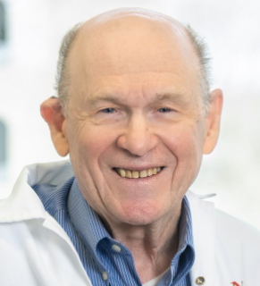 Nahum Sonenberg合作论文数Department of Biochemistry, School of Biomedical Sciences, McGill University;Rosalind and Morris Goodman Cancer Institute, Faculty of Medicine and Health Sciences, McGill University30
 Zhu Ping Jun合作论文数15Wei Huang合作论文数13
 Juan Cristina合作论文数12
 Buffington Shelly A合作论文数Department of Neuroscience, Baylor College of Medicine11
 Peter Walter合作论文数School of Medicine, University of California, San Francisco;Altos Labs11
 Sean W Dooling合作论文数Memory & Brain Res Ctr, Baylor Coll Med9
 Rodney Colina合作论文数Centro de Investigaciones Nucleares, Facultad de Ciencias9Loredana Stoica合作论文数Departments of Neuroscience and;Center for Addiction, Learning and Memory (CALM),, Baylor College of Medicine;Center for Addiction, Learning and Memory (CALM),, Baylor College of Medicine8Sylviane Billaudel合作论文数8
 Jean-Claude Lacaille合作论文数Département de Neurosciences, Faculté de Médecine, Université de Montréal8
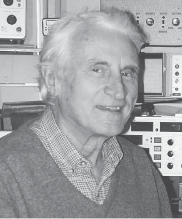 Kresimir Krnjevic合作论文数McGill University8Virginie Ferre合作论文数7Hongyi ZHOU (周虹屹)合作论文数6
 Sgritta Martina合作论文数6
 Randal J. Kaufman合作论文数Center for Genetic Disorders and Aging Research, Sanford Burnham Prebys;Designated Cancer Center, NCI;Department of Biological Chemistry, University of Michigan6
 Andon Placzek合作论文数Roskamp Institute, University of South Florida6
 Yuri Svitkin合作论文数6
 Gonzalo Viana Di Prisco合作论文数5Sanjeev Khatiwada合作论文数5
 Lucas Charles Reineke合作论文数Department of Molecular Virology and Microbiology, Baylor College of Medicine5
 Karim Nader合作论文数The Nader Lab, Department of Psychology, McGill University5
 Ola Segnestam Larsson合作论文数Center for Genomics and Bioinformatics, Karolinska Institutet5
 Barbara Herdy合作论文数Department of Biochemistry, McGill University5
 Michael Bidinosti合作论文数5Yvan Martineau合作论文数5Jacqunae Mays合作论文数4
 Jennifer Johnson合作论文数4
 Tommy Alain合作论文数Childrens Hosp Eastern Ontario, Res Inst, Ottawa, ON K1H 8L1, Canada4Wayne Sossin合作论文数Department of Neurology & Neurosurgery, Faculty of Medicine and Health Sciences, McGill University4
 Ivan Topisirovic合作论文数Department of Biochemistry, Faculty of Medicine and Health Sciences, McGill University4
 Arkady Khoutorsky合作论文数Faculty of Dentistry, McGill University4
 Claudio Cuello合作论文数Department of Pharmacology & Therapeutics, School of Biomedical Sciences, Faculty of Medicine and Health Sciences, McGill University4Ryan J. O. Dowling合作论文数4Liwei Rong合作论文数4
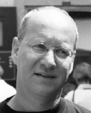 Kobi Rosenblum合作论文数Department of Neurobiology;Weizmann Institute of Science;Department of Neurobiology, Weizmann Institute of Science4Serge Monpoeho合作论文数4
 Mikhail Kochukov合作论文数3Linyan Meng合作论文数Department of Molecular and Human Genetics, Baylor College of Medicine;Baylor Genetics;Department of Molecular and Human Genetics, Baylor College of Medicine3Chien-Ju Chen合作论文数Univ Calif San Diego, Dept Bioengn, La Jolla, CA 92093 USA3
 Yuwei Liu合作论文数Dept Biochem & Biophys, Univ Calif San Francisco3
 Wayne S. Sossin合作论文数Department of Psychology, Faculty of Science, McGill University3
 Jerry Pelletier合作论文数Department of Biochemistry, School of Biomedical Sciences, McGill University;Rosalind and Morris Goodman Cancer Centre, McGill University3
 John A. Dani合作论文数Department of Microbiology, Perelman School of Medicine, University of Pennsylvania3
 Gkogkas Christos合作论文数3
 Delphine Gobert合作论文数3
 Sidrauski Carmela合作论文数Howard Hughes Medical Institute, University of California3
 Vijendra Sharma合作论文数Sagol Department of Neurobiology3




