Der Schock wird im Kindesalter in seiner Schwere und seinem zeitlichen Verlauf häufig unterschätzt. Er stellt einen bedrohlichen Zustand dar, bei dem aufgrund des Kreislaufversagens der Sauerstoffbedarf wichtiger Organe nicht mehr gedeckt werden kann. Dies geht bei inadäquater oder spät eingeleiteter Therapie – insbesondere im Fall von Kindern – mit einer hohen Letalität einher; sekundäre Organschäden bis zum Multiorganversagen können resultieren. Die verlängerte Kapillarfüllungszeit (CRT) ist ein sehr spezifisches und frühes Zeichen der Zentralisation, während die Blutdruckwerte im Kindesalter trotz schweren Schocks durch Erhöhung des peripheren Widerstands lange noch normal sein können. Nach seiner Ursache wird der Schock in hypovolämisch, distributiv, kardiogen und obstruktiv eingeteilt. Er muss zügig behandelt werden; hierbei erfordern die einzelnen Schockformen jeweils eine differenzierte Therapie. Bei unklarer Ätiologie bestehen die initialen Maßnahmen aus Volumen- und Katecholamingaben.
Der Schock wird im Kindesalter in seiner Schwere und seinem zeitlichen Verlauf haufig unterschatzt. Er stellt einen bedrohlichen Zustand dar, bei dem aufgrund des Kreislaufversagens der Sauerstoffbedarf wichtiger Organe nicht mehr gedeckt werden kann. Dies geht bei inadaquater oder spat eingeleiteter Therapie – insbesondere im Fall von Kindern – mit einer hohen Letalitat einher; sekundare Organschaden bis zum Multiorganversagen konnen resultieren. Die verlangerte Kapillarfullungszeit (CRT) ist ein sehr spezifisches und fruhes Zeichen der Zentralisation, wahrend die Blutdruckwerte im Kindesalter trotz schweren Schocks durch Erhohung des peripheren Widerstands lange noch normal sein konnen. Nach seiner Ursache wird der Schock in hypovolamisch, distributiv, kardiogen und obstruktiv eingeteilt. Er muss zugig behandelt werden; hierbei erfordern die einzelnen Schockformen jeweils eine differenzierte Therapie. Bei unklarer Atiologie bestehen die initialen Masnahmen aus Volumen- und Katecholamingaben.
As there are limited data about the clinical practice of catheter ablation in asymptomatic children and adolescents with ventricular preexcitation on ECG, we performed the multicenter “CASPED” (Catheter ablation in ASymptomatic PEDiatric patients with Ventricular Preexcitation) study.
Ein dreijähriger Junge mit bekannter Adipositas permagna wird nach einem Reanimationsereignis auf die pädiatrische Intensivstation verlegt. Es imponieren wiederholt Phasen einer Hypocalcämie getriggerten reversiblen QTc-Zeit-Verlängerung mit resultierenden Torsade-de-pointes-artigen ventrikulären Tachykardien. In der weiteren Abklärung ergibt die Konstellation aus Hypocalcämie, Hyperphosphatämie, Parathormonerhöhung, symmetrischen kalzifikationsverdächtigen Hyperdensitäten bifrontal und im Bereich der Stammganglien sowie die vorbekannte Adipositas permagna die Verdachtsdiagnose eines Pseudohypoparathyreoidismus Typ 1a (Albright-Osteodystrophie), die molekulargenetisch bestätigt werden kann. Die Therapie der Endorganresistenzen verschiedener Hormone erfolgt symptomatisch.
Objectives: There is limited data on catheter ablation of accessory pathways in asymptomatic children and adolescents with a Wolff-Parkinson-White ECG pattern. We therefore performed a multicenter (n = 193) retrospective study ("CASPED").
Haemostatic disorders can lead to bleeds or thrombosis. Acquired platelet dysfunction is the most common cause of haemorrhagic diathesis and is induced by pharmacological agents in >80%. Apart from antiplatelet drugs, non steroidal anti-inflammatory agents and serotonin reuptake inhibitors etc. can also impair platelet function and cause or aggravate haemorrhages. About 40% of intensive care unit patients suffer from thrombocytopenia which, at plate, let counts <100.000/mu l, is responsible for manifest bleeding. Coagulation disorders are significantly less frequent. Congenital platelet disorders and hereditary deficiencies of haemostatic plasma components are extremely rare, except for von Willebrand disease, representing a congenital or acquired disorder. Venous thromboembolism is considered a multicausal and multifactorial disease. Virchow's triad, including alterations in blood flow (stasis), vessel wall (trauma, inflammation) and blood composition (with subsequent hypercoagulability), is still an up-to-date concept of patho-genesis. Hypercoagulability can result from acquired and/or congenital risks, specifically when genetic thrombophilic defects are present in combination. For example, distinct gain-of-function mutations have been identified in the genes of coagulation factors II and V. Hereditary deficiencies of antithrombin, protein C, or protein S are associated with a high risk of venous thromboembolism (VTE), but are infrequent (<1%). Arterial thrombotic occlusion is mediated by platelets and can cause myocardial infarction, stroke, or peripheral ischaemia. Recently, genetically determined platelet receptor variants are thought to cause increased thrombogenicity in response to atherosclerotic lesions.
For prevention and treatment of thromboembolic events, a number of different potent antiplatelet agents and anticoagulants are avaible for a pharmacological intervention of haemostasis. Despite the proven benefit, the risk of bleeding is one major adverse effect. Therefore, the parallel use of antiplatelet agents such as acetyl salicylic acid, NSAID or ADP receptor antagonists such as clopidogrel may cause pharmacodynamic interactions. Special caution is necessary when further drugs that cause an additional inhibition of platelet function are prescribed, e.g. selective serotonin reuptake inhibitors. After the development of low-molecular-weight heparins, which are associated with a lower risk of thrombocytopenia, the introduction of direct oral anticoagulants (DOAC) provided further options in the prophylaxis of thromoboembolic events. Some DOACs have already gained priority over vitamin K antagonists in cardiology guidelines. However, these drugs also bear/harbour the risk of drug-drug interactions and, to some extent, require a close monitoring of renal function. First antidots have already been approved or are awaiting approval.
Clinically relevant influential factors on haemostasis are acidosis, hypothermia, hypocalcaemia and haemodilution, whereas the relevance of poisons is very limited. Each infusion therapy for fluid or volume replacement has potentially negative effects on haemostasis. While the effects of crystalloids (such as physiological saline and plasma-adapted solutions) or human albumin are mainly restricted to the dilutional effect, artificial colloids elicit additional specific effects. Here, dextran produces the strongest, gelatine the least negative effects on haemostasis. Negative haemostatic effects of HES have lost relevance, as becomes obvious when HES 130/0.4 is compared with older preparations.
Background: Limited data exist on the vitamin D status in Fontan. We sought to determine the prevalence of low serum 25-hydroxyvitamin D (s25[OH]D) levels, and to explore potential risk factors.
Haemostasis is achieved by coordinated interactions of cellular and plasmic components. Under physiological contitions, pro-haemostatic and anti-haemostatic mechanisms are maintained in a well-balanced equilibrium. Platelets induce initial sealing of the vascular lesion by forming a haemostatic plug, mediate subsequent plasmic haemostasis and contribute to wound healing. The distinction of., primary and secondary haemostasis follows the physiological sequence of haemostatic events. Primary haemostasis is controlled by platelet functions (activation, adhesion and aggregation), succeded by activation of the coagulation system with generation of thrombin and subsequent conversion of fibrinogen into fibrin. A network of insoluble fibrin stabilizes the haemostatic plug and prevents its detachment by the flowing blood. In analogy to coagulation, initiation of fibrinolysis is triggered by specific activators and proteases. Direct interactions of platelets, coagulation and fibrinolysis warrant a phase-oriented regulation of haemostasis kinetics.
Introduction: Cryo-ablation [CrA] is a safe treatment for av-nodal-reentry-tachycardia [AVNRT] concerning the development of heart block, but may have a lower efficacy as radiofrequency-ablation [RFA].
The term shock dates back to the Middle Ages.In its meaning of blow, punch or impact, the word found entry into medical terminology by its usage as a military expression.Doctors in antiquity already described clinical pictures, traumata, and blood-losses inducing shock-like states.From about 1500 on, even gunshot wounds were followed by such states.In 1737, H.F.Le Dran published his book on wounds by fire-arms.In its English translation of 1743, the term "shock" as a consequence of such traumata was used for the first time.Up to modern times, the importance of traumata and blood-losses triggering shock states was examinded in more detail.The effects of haemorrhages and haemostasis were also noticed as early as in ancient times.The practice of blood-letting and the inspection of this blood enabled the increasing understanding of normal and pathological haemostasis.In 1772, W.Hewson published his work on bloodloss and haemostasis.Owing to his publication in 1905, P.Morawitz became the founder of the "classical coagulation theory".It was only after the 1960s that the close and complex interactions of shock and haemostasis were examinded and more clearly understood.
Objectives: Nowadays the treatment of congenital cardiac defects has evolved that much that the unexpected is expected. There is a general trend to operate upon newborns at an earlier age and with smaller birth weight. To limit the exposure to multiple surgeries at an early age, the cooperation between pediatric cardiology and pediatric cardiac surgery is essential. The staging of certain procedures and patients can be advantageous for patients as well as both medical specialties. In this study a limited group of patients is described that profited from consecutive approaches with the end goal a better and more definite repair.
Autoren H. A. Adams, G. Baumann, I. Cascorbi, M. Emmel, D. Fischer, D. Fries, A. Gansslen, A. R. Heller, F. Hildebrand, E. Klar,H. J. Klippe, W. T. Knoefel, C. Krettek, L. Lampl, H. Maul, H. Prange, U. Rolle, A. Sarrafzadeh, M. Sasse, T. Standl,W. Teske, H. R. ZerkowskiBibliografieDOI http://dx.doi.org/10.1055/s-0031-1292880Notarzt 2012; 28: 12–16© Georg Thieme Verlag KGStuttgart · New YorkISSN 0177-2309KorrespondenzadresseProf. Dr. med. Hans AntonAdamsSprecher der Sektion Schockder DIVI, Stabsstelle furInterdisziplinare Notfall- undKatastrophenmedizin,Medizinische HochschuleHannoverCarl-Neuberg-Strase 130625 HannoverTel.: +49 (0)511 532-3495/-3496Fax: +49 (0)511 532-8033adams.ha@mh-hannover.de
Initiiert durch eine Anfrage der Sektion „Notfallund Katastrophenmedizin“ der Deutschen Interdisziplinaren Vereinigung fur Intensivmedizin und Notfallmedizin (DIVI) sowie der „Bundesarbeitsgemeinschaft Erste Hilfe“ (BAGEH) nimmt die Sektion „Schock“ der DIVI hiermit Stellung zum Beitrag „Schocklage – das Ende einer Legende?“ von F. Scheinichen und F. Kuhl in der Zeitschrift „Rettungsdienst“ 2011, 34: 540–546 [1]. Die Sektion „Schock“ der DIVI begrust es grundsatzlich, dass als scheinbar gesichert geltende Masnahmen hinterfragt werden. Der genannte Artikel weist jedoch inhaltliche und formale Mangel auf und wird den Kriterien guter wissenschaftlicher Praxis nicht gerecht.
Objective: This study reports on the feasibility, efficacy, and outcome of hybrid procedures to close ventricular septal defects (VSD), reflecting the experience of 11 centers in Germany, Austria, and Switzerland. Background: Beating heart closure of VSD has attracted interest in small infants, complex anomalies and postinfarction scenarios where patients are at high risk during surgery. Perventricular or intraoperative device placement allows access to the lesions where percutaneous delivery is limited. Methods: Between December 2001 and April 2009, placement of Amplatzer septal occluders was attempted in 26 patients. The defects were located in the perimembranous (n 5 5) and muscular septum (n 5 21). In 20 patients, a perventricular approach was used, and, in six, the occluders were placed under direct visualization being part of a complex heart surgery. Results: In 23 of 26 procedures, device placement was successful (88.5%). The mean defect size was 7.8 mm (range, 3.5–20). The occluder types were perimembranous VSD occluder (n 5 4), muscular VSD occluder (n 5 20), postinfarct VSD occluder (n 5 1), and ASD occluder (n 5 1) with a ratio device/defect of 0.9–2.4 (median 1.15). Device removal was necessary in three due to arrhythmia, malpositioning, and additional defects. Pericardial effusion occurred once. In the remaining 22 patients, there were no procedure or device-related complications. During mean follow up of 1.4 years (range, 1 day–3.9 years), a residual shunt that was more than trivial was observed in one patient out of 21 successful procedures. Conclusions: Perventricular and intraoperative device closure of VSD is as effective as a surgical patch and averts the increased morbidity of conventional surgical repair in a subgroup of highrisk patients. VC 2010 Wiley-Liss, Inc.
Bei einem 5-jährigen, bislang herzgesunden Mädchen trat nach 14-tägiger antibiogrammgerechter Therapie (Ceftriaxon) einer Pneumokokkensepsis/-meningitis eine vorher nicht nachgewiesene endokarditische Vegetation an der Trikuspidalklappe mit embolischer Pneumonie auf. Eine durch Pneumokokken hervorgerufene, selten Kombination einer manifestierenden Pneumonie, Meningitis und Endokarditis wird auch als Austrian-Syndrom bezeichnet. Ein kultureller Keimnachweis gelangt trotz Aussetzens der Antibiotikatherapie, septischer Fieberschübe und rasch angestiegener Infektionsparameter nicht. Trotz klinischer Besserung unter erweiterter antibakterieller Behandlung (Ceftriaxon, Vancomycin, Fosfomycin) war bei unveränderten Vegetationen und progredienter Klappendestruktion eine chirurgische Sanierung der Klappe erforderlich. In dem entfernten Material konnten mittels PCR (Polymerasekettenreaktion) Pneumokokken nachgewiesen werden. Keime ließen sich jedoch nicht kultivieren.
BACKGROUND AND OBJECTIVE:Cryoablation is safe for the ablation of substrates in proximity to the AV node, because the initial lesion is reversible. We report our results of cryoablation in a transregional center for ablation in children and adolescents.PATIENTS AND METHODS:Data on 39 children and adolescents (4 - 18 years of age) who had been treated with cryo energy were analyzed retrospectively. The diagnosis was AV nodal reentry tachycardia (AVNRT; n = 30), para-Hisian accessory pathway (AP; n = 6) and congenital junctional ectopic tachycardia (JET; n = 4). In addition to non-inducibility, the targeted endpoint for AP-ablation was a missing or decremental concentric retrograde conduction, for ablation of AVNRT the endpoint was no slow pathway, no AH jumps and no echo-beats. The median follow-up was 3 years (270 - 1919 days).RESULTS:The targeted endpoint was reached in 35/39 patients (90 %), in four patients (10 %) RF energy had to be applied. A recurrence occurred in 7/35 (20 %) successfully treated patients. Two patients had a pre-excitation again after AP ablation, but no symptoms. Thus, 28/35 patients (80 %) remained asymptomatic after cryoablation, and 26/35 (74 %) are definitively cured, regarding all follow-up data. The subgroup of AVNRT patients does not differ from the entire group. There was no AV block in the cryoablation group.CONCLUSIONS:Cryoablation is very safe and effective for the definitive treatment of arrhythmias in children and adolescents. The price for the high safety is a reduced efficacy and a higher recurrence rate.
Identifying the young patient at risk of malignant arrhythmias and sudden cardiac death remains a challenge. It is increasingly recognised that sudden death, syncope and aborted cardiac arrest at a young age in patients with a structurally normal heart may be the result of various ion channel disorders - the channelopathies. The approach to risk stratification involves a combination of the clinical presentation, taken in conjunction with the family history, genetic testing, invasive electrophysiological studies or other provocative tests where appropriate and feasible. A logical approach to risk stratification in some of the commoner channelopathies seen in paediatric practice is presented.
BACKGROUND:The value of balloon valvuloplasty of the aortic valve in childhood is still under debate. OBJECTIVE:To evaluate the results of the procedure in a retrospective multicenter survey of a large cohort over a long time interval. METHODS:Retrospective analysis of 1004 patients with balloon valvuloplasty of the aortic valve performed between 9/1985 and 10/2006 at 20 centers in Germany, Austria and Switzerland. Amongst others, the following parameters were evaluated before and after the procedure as well as at the end of follow-up or before surgery: clinical status, left ventricular function, transaortic pressure gradient, degree of aortic regurgitation, freedom from re-intervention or surgery. PATIENTS:Patients from 1 day to 18 years of age with aortic valve stenosis were divided into four groups: 334 newborns (1-28 days); 249 infants (29-365 days); 211 children (1-10 years), and 210 adolescents (10-18 years). RESULTS:Median follow-up was 32 months (0 days to 17.5 years). After dilatation the pressure gradient decreased from 65 (± 24)mm Hg to 26 (± 16)mm Hg and remained stable during follow-up. The newborns were the most affected patients. Approximately 60% of them had clinical symptoms and impaired left ventricular function before intervention. Complication rate was 15% in newborns, 11% in infants and 6% in older children. Independently of age, 50% of all patients were free from surgery 10 years after intervention. CONCLUSIONS:In this retrospective multicenter study, balloon valvuloplasty of the aortic valve has effectively postponed the need for surgery in infants, children and adolescents up to 18 years of age.
 Narayanswami Sreeram合作论文数42Konrad Brockmeier合作论文数40G. Bennink合作论文数19
 Thomas Standl合作论文数12
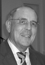 Gert Baumann合作论文数Klinik für Kardiologie, Angiologie und Intensivmedizin, Charité Universitätsmedizin Berlin10Hilmar W. Prange合作论文数10Wolfram Teske合作论文数10H. J. Klippe合作论文数10Hans Anton Adams合作论文数9A. Sarrafzadeh合作论文数9L. Lampl合作论文数9
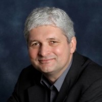 Ingolf Cascorbi合作论文数Faculty of Medicine, Christian-Albrechts-Universität zu Kiel;Department of Experimental and Clinical Pharmacology, University Hospital Schleswig-Holstein9S. Geiger合作论文数8U. Rolle合作论文数7D. Fischer合作论文数7D. Fries合作论文数7
 Uwe Trieschmann合作论文数7R Zander合作论文数7
 Axel R Heller合作论文数7
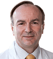 Gerald S. Werner合作论文数Medizinische Klinik I, Klinikum Darmstadt GmbH;Darmstadt Clinic, University of Frankfurt7
 Frank Hildebrand合作论文数7l ben mime合作论文数6E. Klar合作论文数6Khalil Markus合作论文数6A. Gaensslen合作论文数5
 Sascha Flohe合作论文数Dept Trauma Hand & Reconstruct Surg, Univ Hosp Essen5
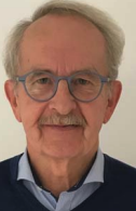 Rüdiger Eberhard Scharf合作论文数Boston Children's Hospital, Harvard Medical School5Bernhard Roth合作论文数5F Hitchcock合作论文数4
 Sabine Schickendantz合作论文数4K. Mehler合作论文数4Oliver Kretschmar合作论文数4W. T. Knoefel合作论文数4R. Gitter合作论文数3C. Ebener合作论文数3Hr Zerkowski合作论文数3E. de Haan合作论文数3Uwe Janssens合作论文数3
 Peter M. Vogt合作论文数Department of Plastic, Aesthetic, Hand and Reconstructive Surgery, Hannover Medical School, Medizinische Hochschule Hannover;Burn Center, Hannover Medical School, Medizinische Hochschule Hannover3Andreas Eicken合作论文数3J. Piek合作论文数3
 Felix Berger合作论文数3
 Thomas Paul合作论文数3
 Thorsten Wahlers合作论文数Department of Cardiac and Thoracic Surgery, University Hospital Cologne3
 Hans-Christoph Pape合作论文数Department of Traumatology, University Hospital Zürich;Medical Faculty, University of Zurich3
 Joachim Windolf合作论文数Heinrich-Heine-Universität Düsseldorf;Klinik für Unfall- und Handchirurgie am Universitätsklinikum Düsseldorf3Uwe Klima合作论文数3Roman Antonin Gebauer合作论文数3
 Jan-Hendrik Nurnberg合作论文数3




