Isolated skeletal muscle ryanodine receptors (RyRs) complexed with the modulatory ligands, calmodulin (CaM) or 12-kDa FK506-binding protein (FKBP12), have been characterized by electron cryomicroscopy and three-dimensional reconstruction. RyRs are composed of 4 large subunits (molecular mass 565 kDa) that assemble to form a 4-fold symmetric complex that, architecturally, comprises two major substructures, a large (≈80% of the total mass) cytoplasmic assembly and a smaller transmembrane assembly. Both CaM and FKBP12 bind to the cytoplasmic assembly at sites that are 10 and 12 nm, respectively, from the putative entrance to the transmembrane ion channel. FKBP12 binds along the edge of the square-shaped cytoplasmic assembly near the face that interacts in vivo with the sarcolemma/transverse tubule membrane system, whereas CaM binds within a cleft that faces the junctional face of the sarcoplasmic reticulum membrane at the triad junction. Both ligands interact with a domain that connects directly to a cytoplasmic extension of the transmembrane assembly of the receptor, and thus might cause structural changes in the domain which in turn modulate channel gating.
Ryanodine receptors (RyRs), a class of intracellular calcium release channels, are the largest ion channels known. Recently, cryoelectron microscopy and image reconstructions of isolated receptors have shown that most of the protein mass forms a porous, multidomain cytoplasmic assembly. Evidence is mounting that suggests that the cytoplasmic assembly communicates with the transmembrane regions over distances of 100 Å or greater. RyRs are centrally important in excitation-contraction coupling, which occurs at specialized regions where the sarcoplasmic reticulum, containing the RyRs, and the plasma membrane/transverse-tubule system form junctions. Numerous proteins are present at these junctions, some of which interact directly with the RyR.
The SPIDER system has evolved into a comprehensive tool set for image processing, making use of modern graphics interfacing in the VMS and UNIX environment. SPIDER and WEB handle the complementary tasks of batch processing and visualization of the results. The emphasis of the SPIDER system remains in the area of single particle averaging and reconstruction, although a variety of other application areas have been added. Novel features are a suite of operations relating to the determination, modeling, and correction of the contrast transfer function and the availability of the entire documentation in hypertext format.
A platform-like structure, which appears equivalent to the platform or lobe structure of the 30 S subunit of the eubacterial ribosome, is observed in the reconstruction of the small 40 S ribosomal subunit from images of ice-embedded particles. This cup-shaped structure, 15.0 nm in side length and 13.5 nm wide at its rim, extends obliquely upward on the back of the subunit. Other previously characterized features of the 40 S subunit can readily be identified: the head with its prominent beak structure, the body with its two back lobes expressed as relatively small-scale features, and the two widely separated feet that comprise the base of the subunit.
THE ribosome is formed by assembly of proteins and nucleic acids, and synthesizes proteins according to genetic instructions in all organisms. Many of the biochemical steps of this fundamental process are known, but a detailed understanding requires a well-defined structural model of the ribosome. Electron microscopy combined with image reconstruction of two-dimensional crystals or single ribosomes(4) has been the most promising technique, but the resolution of the resulting models has been insufficient. Here we report a 25-Angstrom reconstruction of the ribosome from Escherichia coli, obtained by combining 4,300 projections of ice-embedded single particles. Our new reconstruction reveals a channel in the small ribosomal subunit and a bifurcating tunnel in the large subunit which may constitute pathways for the incoming message and the nascent polypeptide chain, respectively. Based on these new findings, a three-dimensional model of the basic framework of protein synthesis is presented.
The morphology of the Escherichia coli ribosome, i.e., its shape at moderate to low (20-40 A (1 A = 0.1 nm)) resolution, provides important constraints in modeling both the folding of ribosomal RNA and the translational process. A new reconstruction, obtained by low-dose cryoelectron microscopy and image processing of single ribosomes, contains clues to the way in which the ribosome interacts with the key functional ligands: the mRNA and the A- and P-site tRNAs. It also suggests possible pathways of the nascent polypeptide chain. From an interpretation of these clues in the light of existing knowledge, a plausible model for the locations and interactions of key components of protein synthesis is suggested.
A method for alignment of projections with unknown projecting directions towards a three-dimensional reference has been developed. The technique has been applied to the three-dimensional reconstruction from images of a frozen-hydrated preparation of 50S ribosomal subunits from Escherichia coli recorded in the electron microscope. The algorithm as used here combines the Single Exposure Conical Reconstruction Technique (SECReT) with a three-dimensional orientation search. The algorithm allows for the refinement of a reconstruction obtained with SECReT by refinement of the true projection angles, and by the inclusion of projections with a priori unknown random orientation. With model data it is demonstrated that the algorithm works reliably even for signal-to-noise ratios lower than 1.
The nuclear pore complex spans the nuclear envelope and functions as a macromolecular transporter in the ATP-dependent process of nucleocytoplasmic transport. In this report, we present three dimensional (3D) structures for both membrane-associated and detergent-extracted Xenopus NPCs, imaged in frozen buffers by cryo-electron microscopy. A comparison of the differing configurations present in the 3D maps suggests that the spokes may possess an intrinsic conformational flexibility. When combined with recent data from a 3D map of negatively stained NPCs (Hinshaw, J. E., B. O. Carragher, and R. A. Milligan. 1992. Cell. 69:1133-1141), these observations suggest a minimal domain model for the spoke-ring complex which may account for the observed plasticity of this assembly. Moreover, lumenal domains in adjacent spokes are interconnected by radial arm dimers, forming a lumenal ring that may be responsible for anchoring the NPC within the nuclear envelope pore. Importantly, the NPC transporter is visualized as a centrally tapered cylinder that spans the entire width of the NPC, in a direction normal to the nuclear envelope. The central positioning, tripartite structure, and hollow nature of the transporter suggests that it may form a macromolecular transport channel, with a globular gating domain at each end. Finally, the packing of the transporter within the spokes creates a set of eight internal channels that may be responsible, in part, for the diffusion of ions and small molecules across the nuclear envelope.
An immunocomplex of the 4 x 6-meric hemocyanin of the scorpion Androctonus australis with the monoclonal Fab fragment L104 was reconstructed from electron micrographs of a negatively stained specimen, using the double-carbon-layer technique. The resulting structure enables a clear visualization of the Fab fragments bound to the four copies of the Aa6 subunit and directly confirms a previous localization of the L104 epitope deduced from two-dimensional image processing. Despite a strong flattening effect produced by the negative-staining technique the orientations of the Fab fragments are well characterized. Moreover, the observation of a central hole within the elbow bends of the Fab fragments provides information about the disposition of the Fabs around their main axis.
The random-conical reconstruction method has been highly successful in three-dimensional imaging of macromolecules under low-dose conditions. This article summarizes the different steps of this technique as applied to molecules prepared with negative staining or vitreous ice, and sketches out the current directions of development. We anticipate that by using new instrumental developments, transfer function correction and computational refinement techniques, a resolution in the range of 7-10 A could ultimately be achieved.
The native structure of the calcium release channel (ryanodine receptor) from rabbit skeletal muscle has been analyzed in two dimensions from electron micrographs of frozen hydrated specimens. Within a resolution of 3.0 nm there is excellent agreement between the structure as seen in vitreous water and in negative stained specimens. Features seen in the three-dimensional reconstruction of the negatively stained channel can be identified in the projection of the unstained receptor.
Single particles embedded in ice pose new challenges for image processing because of the intrinsically low signal-to-noise ratio of such particles in electron micrographs. We have developed new techniques that address some of these problems and have applied these techniques to electron micrographs of the Escherichia coli ribosome. Data collection and reconstruction follow the protocol of the random-conical technique of Radermacher et al. [J. Microscopy 146 (1987) 113]. A reference-free alignment algorithm has been developed to overcome the propensity of reference-based algorithms to reinforce the reference motif in very noisy situations. In addition, an iterative 3D reconstruction method based on a chi-square minimization constraint has been developed and tested. This algorithm tends to reduce the effects of the missing angular range on the reconstruction, thereby facilitating the merging of random-conical data sets obtained from differently oriented particles.
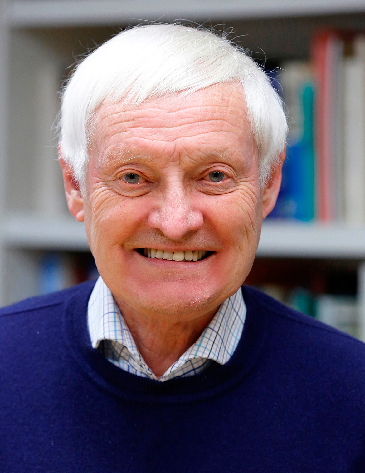 Joachim Frank (阿希姆·弗兰克)合作论文数Howard Hughes Medical Institute, Department of Biochemistry and Molecular Biophysics, College of Physicians and Surgeons, Columbia University;Department of Biological Sciences, Columbia University38
 Terence Wagenknecht合作论文数24Adriana Verschoor合作论文数17Robert A. Grassucci合作论文数Wadsworth Center, Empire State Plaza, Albany, NY 12201 USA13
 Sidney Fleischer合作论文数Department of Molecular Biology, Vanderbilt University8J M Carazo合作论文数6
 Pawel Penczek合作论文数Department of Biochemistry and Molecular Biology, McGovern Medical School, University of Texas Health Science Center at Houston5KR Lata合作论文数4M Inui合作论文数4Bf Mcewen合作论文数3M Boublik合作论文数3J Berkowitz合作论文数3A Saito合作论文数3
 Montserrat Samso合作论文数3
 Satish Kumar Srivastava合作论文数University of Texas Medical Branch3
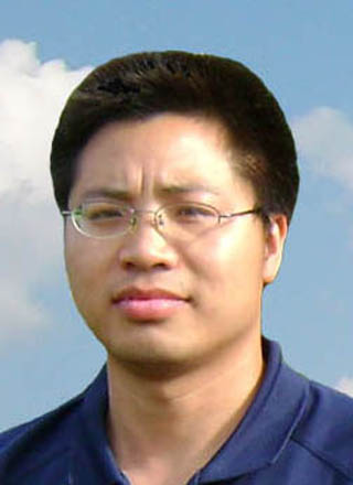 Yingbing Li (李英冰)合作论文数School of Geodesy and Geomatics, Wuhan University3V Rao合作论文数2Hb Xin合作论文数2Ap Timerman合作论文数2C CHADWICK合作论文数2Nicolas Boisset合作论文数2Ardean Leith合作论文数2
 Rajendra K. Agrawal合作论文数Department of Biomedical Sciences, State University of New York at Albany2Michael Marko合作论文数2Jean N. Lamy合作论文数2JN TURNER合作论文数1D. Glitz合作论文数1V Mandiyan合作论文数1Cs Miles合作论文数1Jc Taveau合作论文数1Df Parsons合作论文数1Martin Kessel合作论文数1B Nadalginard合作论文数1H Schindler合作论文数1Cc Chadwick合作论文数1Weiping Liu合作论文数1
 Christopher W. Akey合作论文数Chobanian & Avedisian School of Medicine, Boston University1
 Juan Ausio合作论文数University of Victoria1
 Andrew R. Marks合作论文数Department of Physiology & Cellular Biophysics, Vagelos College of Physicians and Surgeons, Columbia University;Department of Biomedical Engineering, The Fu Foundation School of Engineering and Applied Science, Columbia University1Conly L. Rieder合作论文数1
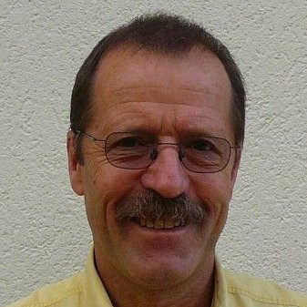 Andreas Engel合作论文数The Center for Molecular Life Sciences, University of Basel;cryoWrite1M. Rosenthal合作论文数1
 Nicholas Dixon合作论文数School of Chemistry and Molecular Bioscience, Faculty of Science, Medicine and Health, University of Wollongong1
 James N. Turner合作论文数1Lin Hymel合作论文数1C. San Martín合作论文数1T. Obrig合作论文数1Bettina Fedtke合作论文数1Nanyan Zhang合作论文数1




