Cell-cell communication underlies all emergent properties of the brain, including cognition, learning and memory. The physical basis for these communications is the synapse, a multi-component structure requiring coordinated interactions between diverse cell types. However, many aspects of three-dimensional (3D) synaptic organization remain poorly understood. Here, we developed an approach, seamless in situ trimming and milling (SISTM), to reliably fabricate sufficiently thin lamellae for mapping of the 3D nanoarchitecture of synapses in mouse, monkey and human brain tissue under near-native conditions via cryo-electron tomography (cryo-ET). We validated SISTM in a mouse model of Huntington's disease, demonstrating distinct 3D alterations to synaptic vesicles and mitochondria. By successfully applying SISTM to macaque brain, we described the 3D architecture of a tripartite synapse within the cortex. Subtomogram averaging (STA) enabled spatial mapping of astrocyte-neuron contacts within the tripartite synapse, revealing neurexin-neuroligin complexes as potential constituents that tether the two cell types. Finally, we showed that the defining features of synaptic nanoarchitecture were conserved across species and evident in human brain tissue obtained postmortem. Combining SISTM with cryo-ET and STA is a starting point for a new understanding of brain organization, disease-induced structural alterations and the development of rational, structure-guided therapeutics.
Mitochondria in mammalian cardiomyocytes display considerable structural heterogeneity, the significance of which is not currently understood. We use electron microscopic tomography to analyze a dataset of 68 mitochondrial subvolumes to look for correlations among mitochondrial size and shape, crista morphology and membrane density, and organelle location within rat cardiac myocytes. A tomographic analysis guided the definition of four classes of crista morphology: lamellar, tubular, mixed and transitional, the last associated with remodeling between lamellar and tubular cristae. Correlations include an apparent bias for mitochondria with lamellar cristae to be located in the regions between myofibrils and a two-fold larger crista membrane density in mitochondria with lamellar cristae relative to mitochondria with tubular cristae. The examination of individual cristae inside mitochondria reveals local variations in crista topology, such as extent of branching, alignment of fenestrations and progressive changes in membrane morphology and packing density. The findings suggest both a rationale for the interfibrillar location of lamellar mitochondria and a pathway for crista remodeling from lamellar to tubular morphology.
Cryo-electron microscopy (cryo-EM) enables the study of protein complexes, cytoskeletal elements, and organelles in three dimensions without the use of chemical fixation. Most cryo-EM studies focus on vitreously frozen individual cells separated from their native tissue contexts. This reliance on imaging of single cells is primarily due to technical challenges associated with preparing fresh tissue sections at a thinness sufficient for visualization via cryo-EM. Highly heterogenous and specialized tissues, such as brain, are especially affected by this limitation as the cellular, subcellular, and synaptic milieus can significantly vary across neuroanatomical locations. To address this limitation, we established new instrumentation and a workflow that consists of: 1) high-pressure freezing of fresh brain tissue; 2) tissue trimming followed by cryo-focused ion beam milling via the H-bar approach to generate ultrathin lamellae; and 3) cryo-EM imaging. Here, we apply this workflow to visualize the fine ultrastructural details of organelles, as well as cytoskeletal and synaptic elements that comprise the cortical neuropil within fresh, unfixed mouse brain tissue. Moreover, we present initial studies that apply principles of the above workflow to the analysis of postmortem human brain tissue. Overall, our work integrates the strengths of cryo-electron microscopy and tissue-based approaches to produce a generalizable workflow capable of visualizing subcellular structures within complex tissue environments.
Phase plates (PPs) are beneficial devices to improve the phase contrast of life-science objects in cryo-transmission electron microscopy (TEM). The development of the hole-free (HF) PP, which consists of a thin carbon film, has led to impressive results due to its ease in fabrication, implementation and application. However, the phase shift of the HFPP can be controlled only indirectly. The electrostatic Zach PP uses a strongly localized and adjustable electrostatic potential to generate well-defined and variable phase shifts between scattered and unscattered electrons. However, artifacts in phase-contrast TEM images are induced by the presence of the PP rod in the diffraction plane. We present a detailed analysis and comparison of the contrast-enhancing capabilities of both PP types and their emerging artifacts. For this purpose, cryo-TEM images of a standard T4-bacteriophage test sample were acquired with both PP types. Simulated images reproduce the experimental images well and substantially contribute to the understanding of contrast formation. An electrostatic Zach PP was used in this work to acquire cryo-electron tomograms with enhanced contrast, which are of similar quality as tomograms obtained by HFPP TEM.
An abstract is not available for this content so a preview has been provided. As you have access to this content, a full PDF is available via the ‘Save PDF’ action button.
Cryo-electron tomography (cryo-ET) is a well-established technique for studying 3D structural details of subcellular macromolecular complexes and organelles in their nearly native context in the cell. A primary limitation of the application of cryo-ET is the accessible specimen thickness, which is less than the diameters of almost all eukaryotic cells. It has been shown that focused ion beam (FIB) milling can be used to prepare thin, distortion-free lamellae of frozen biological material for high-resolution cryo-ET. Commercial cryosystems are available for cryo-FIB specimen preparation, however re-engineering and additional fixtures are often essential for reliable results with a particular cryo-FIB and cryo-transmission electron microscope (cryo-TEM). Here, we describe our optimized protocol and modified instrumentation for cryo-FIB milling to produce thin lamellae and subsequent damage-free cryotransfer of the lamellae into our cartridge-type cryo-TEM.
Phase plate (PP) imaging has proven to be valuable in transmission cryo electron microscopy of unstained, native-state biological specimens. Many PP types have been described, however until the recent implementation of the "hole-free" phase plate (HFPP), imaging has been challenging. We found the HFPP to be simple to construct and to set up in the transmission electron microscopy, but care in implementing automated data collection is needed. Performance may be variable, both initially and over time, thus it is important to monitor and evaluate image quality by observing the power spectrum. We found that while some HFPPs gave transfer to high resolution without CTF oscillation, most reached high resolution when operated with modest defocus.
Journal Article Analysis of Thin Phase-Shifter Films using Surface Analysis Techniques Get access Vincent S Smentkowski, Vincent S Smentkowski General Electric Global Research Center, 1 Research Circle, Niskayuna NY 12309 Search for other works by this author on: Oxford Academic Google Scholar Laurie LeTarte, Laurie LeTarte General Electric Global Research Center, 1 Research Circle, Niskayuna NY 12309 Search for other works by this author on: Oxford Academic Google Scholar Hong Piao, Hong Piao General Electric Global Research Center, 1 Research Circle, Niskayuna NY 12309 Search for other works by this author on: Oxford Academic Google Scholar Michael Marko Michael Marko Wadsworth Center, Empire State Plaza, P.O. Box 509, Albany NY 12201-0509 Search for other works by this author on: Oxford Academic Google Scholar Microscopy and Microanalysis, Volume 22, Issue S3, 1 July 2016, Pages 394–395, https://doi.org/10.1017/S1431927616002828 Published: 25 July 2016
Zernike phase-plate (ZPP) imaging greatly increases contrast in cryo-electron microscopy, however fringe artifacts appear in the images. A computational de-fringing method has been proposed, but it has not been widely employed, perhaps because the importance of de-fringing has not been clearly demonstrated. For testing purposes, we employed Zernike phase-plate imaging in a cryo-electron tomographic study of radial-spoke complexes attached to microtubule doublets. We found that the contrast enhancement by ZPP imaging made nonlinear denoising insensitive to the filtering parameters, such that simple low-frequency band-pass filtering made the same improvement in map quality. We employed sub-tomogram averaging, which compensates for the effect of the "missing wedge" and considerably improves map quality. We found that fringes (caused by the abrupt cut-on of the central hole in the phase plate) can lead to incorrect representation of a structure that is well-known from the literature. The expected structure was restored by amplitude scaling, as proposed in the literature. Our results show that de-fringing is an important part of image-processing for cryo-electron tomography of macromolecular complexes with ZPP imaging.
Cryo-electron tomography (cryo-ET) has emerged as perhaps the only practical technique for revealing nanometer-level three-dimensional structural details of subcellular macromolecular complexes in their native context, inside the cell. As currently practiced, the specimen should be 0.1- 0.2 microns in thickness to achieve optimal resolution. Thus, application of cryo-ET to intact frozen (vitreous) tissues, such as skeletal muscle, requires that they be sectioned. Cryo-ultramicrotomy is notoriously difficult and artifact-prone when applied to frozen cells and tissue, but a new technique, focused ion beam milling (cryo-FIB), shows great promise for "thinning" frozen biological specimens. Here we describe our initial results in applying cryo-FIB and cryo-ET to triad junctions of skeletal muscle.
Specimens prepared solely by vitreous freezing facilitate high-resolution imaging of macromolecules in-situ and in a near-native state. However, such samples are weakly scattering, suffering from low contrast and requiring phase-contrast imaging. In addition, high-resolution information is limited by electron-radiation damage. Cryo-TEM technology has steadily been improving so that improved image contrast can be obtained without increased electron dose.
Thin-film phase plates, which usually have a central hole for the unscattered beam, are made of various materials, using various methods [1,2]. For optimal use, they should be evaluated for (1) absence of charging, (2) correct phase shift, and (3) cut-on frequency. During imaging at the TEM, centering of the phase plate (PP) needs to be maintained, and post-processing of the images may often be desirable.
The use of focused-ion-beam milling (FIB) to thin vitreously frozen cells for application of cryo-electron tomography is an emerging technology. However, successful application of cryo-FIB milling and tomography to frozen biological tissue has, to our knowledge, not been reported. This is because bulk tissue cannot be effectively frozen by simple plunge freezing of EM grids, but rather must be prepared by high-pressure freezing, which requires extra steps. The cryo-transfers that are required at each step of the process can cause devitrification, excessive frost contamination and mechanical damage, consequently resulting in unacceptably high failure rates. To address these problems, we have devised a protocol and have designed new devices for manipulating the specimens as they are transferred from the high-pressure freezer, to a cryo-ultramicrotome (for rough trimming), to the FIB instrument, and finally to the transmission electron microscope. Using these improved procedures we have thinned toad fish swim bladder muscle to less than 150 nm, which is necessary to achieve resolutions of 2-3 nm, and we have determined the first cryo-tomograms of vitreously frozen triad junctions. The tomograms should reveal the three-dimensional architecture of the triad with an unprecedented fidelity and level of detail. (Supported by NIH).
Journal Article Artifact Correction for Zernike Phase-Plate Cryo-Electron Tomography Get access Haixin Sui, Haixin Sui Wadsworth Center, New York State Department of Health, Albany, NY 12201Dept. of Biomedical Sciences, School of Public Health, University at Albany, Albany, NY 12201 Search for other works by this author on: Oxford Academic Google Scholar Gregory Kishchenko, Gregory Kishchenko Wadsworth Center, New York State Department of Health, Albany, NY 12201 Search for other works by this author on: Oxford Academic Google Scholar Radostin Danev, Radostin Danev Okazaki Institute for Integrative Bioscience, National Institutes of Natural Sciences, Okazaki, JapanMax-Planck-Institute for Biochemistry, Martinsried 82152, Germany Search for other works by this author on: Oxford Academic Google Scholar Jie He, Jie He Wadsworth Center, New York State Department of Health, Albany, NY 12201 Search for other works by this author on: Oxford Academic Google Scholar Rebecca Fisher, Rebecca Fisher Wadsworth Center, New York State Department of Health, Albany, NY 12201 Search for other works by this author on: Oxford Academic Google Scholar Chyongere Hsieh, Chyongere Hsieh Wadsworth Center, New York State Department of Health, Albany, NY 12201 Search for other works by this author on: Oxford Academic Google Scholar Michael Marko Michael Marko Wadsworth Center, New York State Department of Health, Albany, NY 12201 Search for other works by this author on: Oxford Academic Google Scholar Microscopy and Microanalysis, Volume 20, Issue S3, 1 August 2014, Pages 234–235, https://doi.org/10.1017/S143192761400289X Published: 27 August 2014
In cryo-electron microscopy of vitreously frozen biological specimens, phase-plate imaging can greatly increase the signal-to-noise ratio of the micrographs. When routinely used, this imaging technique may be the method of choice for cryo-electron tomography, in which each image in the tilt-series data set suffers from a high noise background due to the low-electron-dose exposure. Utilizing the Zernike phase-contrast imaging technique, we have carried out an electron tomographic study of the axonemal microtubule doublets and their associated radial spokes. The reconstructed tomogram displays a strong contrast. The quality of the resultant density maps, obtained by sub-volume averaging of a single tomogram in this study, is comparable with that from the averaging of many cryo-tomograms without using the phase-plate.
Vitreous freezing offers a way to study cells and tissue in a near-native state by cryo-transmission electron microscopy (cryo-TEM), which is important when structural information at the macromolecular level is required. Many cells - especially those in tissue - are too thick to study intact in the cryo-TEM. Cryo focused-ion-beam (cryo-FIB) milling is being used in a few laboratories to thin vitreously frozen specimens, thus avoiding the artifacts and difficulties of cryo-ultramicrotomy. However, the technique is challenging because of the need to avoid devitrification and frost accumulation during the entire process, from the initial step of freezing to the final step of loading the specimen into the cryo-TEM. We present a robust workflow that makes use of custom fixtures and devices that can be used for high-pressure-frozen bulk tissue samples as well as for samples frozen on TEM grids.
Imaging with Zernike phase plates is increasingly being used in cryo-TEM tomography and cryo-EM single-particle applications. However, rapid ageing of the phase plates, together with the cost and effort in producing them, present serious obstacles to widespread adoption. We are experimenting with phase plates based on silicon chips that have thin windows; such phase plates could be mass-produced and made available at moderate cost. The windows are coated with conductive layers to reduce charging, and this considerably extends the useful life of the phase plates compared to traditional pure-carbon phase plates. However, a compromise must be reached between robustness and transmission through the phase-plate film. Details are given on testing phase-plate performance by means of imaging an amorphous thin film and evaluating the power spectra of the images.
Extended abstract of a paper presented at Microscopy and Microanalysis 2013 in Indianapolis, Indiana, USA, August 4 – August 8, 2013.
Extended abstract of a paper presented at Microscopy and Microanalysis 2012 in Phoenix, Arizona, USA, July 29 – August 2, 2012.
 Chyong-Ere Hsieh合作论文数61
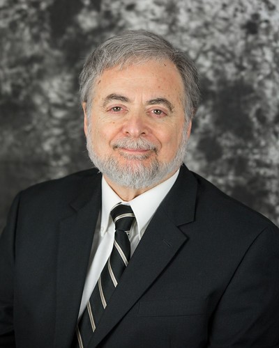 Carmen A. Mannella合作论文数Department of Physiology, School of Medicine, University of Maryland60
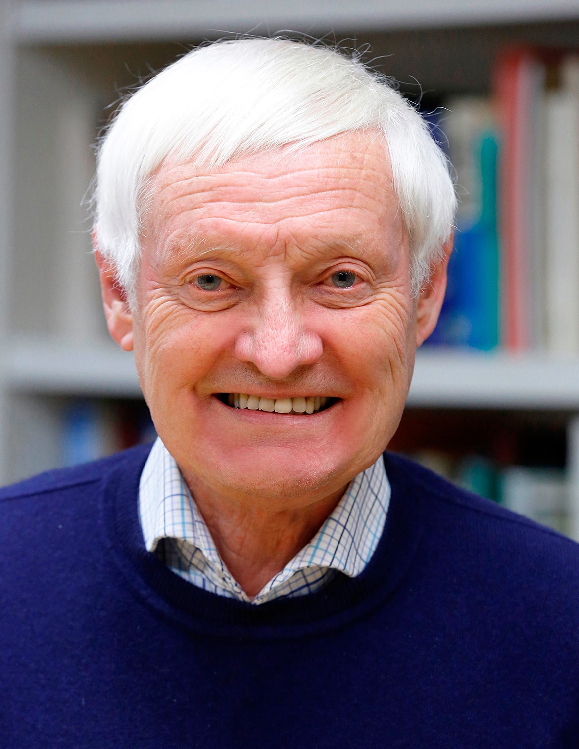 Joachim Frank (阿希姆·弗兰克)合作论文数Howard Hughes Medical Institute, Department of Biochemistry and Molecular Biophysics, College of Physicians and Surgeons, Columbia University;Department of Biological Sciences, Columbia University27K. Buttle合作论文数18Ardean Leith合作论文数18Df Parsons合作论文数16Bf Mcewen合作论文数8Terence Wagenknecht合作论文数8Ce Hsieh合作论文数7
 Radostin Danev合作论文数Graduate School of Medicine, University of Tokyo, S402, 7-3-1 Hongo, Bunkyo-ku, Japan 7Claire S. Ting合作论文数7C Renken合作论文数6
 Sesh A. Sundararaman合作论文数Departments of Medicine and;Perelman School of Medicine, University of Pennsylvania;Perelman School of Medicine, University of Pennsylvania6Rath B K合作论文数6K Tessitore合作论文数5
 Pawel Penczek合作论文数Department of Biochemistry and Molecular Biology, McGovern Medical School, University of Texas Health Science Center at Houston5Christian Renken合作论文数5KF Buttle合作论文数4Kishchenko Gregory合作论文数4Ronald J. Limberger合作论文数4Haixin Sui合作论文数4D'Arcangelis D合作论文数3Kj Wansor合作论文数3Charon Nyles W合作论文数3Xing Meng合作论文数3
 Rebecca Fisher合作论文数Australian Institute of Marine Science3
 Yuru Deng (邓瑜如)合作论文数Wenzhou Institute, University of Chinese Academy of Sciences3Jacques Izard合作论文数3D Darcangelis合作论文数2Karolyn Buttle合作论文数2M Rodriguez合作论文数2
 Jeffrey G Ault合作论文数2Thomas Schmelzer合作论文数2Jonathan Franks合作论文数2M Radermacher合作论文数2
 Jill Glausier合作论文数Department of Psychiatry, University of Pittsburgh School of Medicine2Martin Obermair合作论文数2
 Jiying Ning合作论文数Department of Structural Biology, University of Pittsburgh School of Medicine2Michael Radermacher合作论文数Department of Computer Science, College of Engineering and Mathematical Sciences, University of Vermont2
 Nicholas Mantis合作论文数Div Infect Dis, New York State Dept Hlth2
 Dagmar Gerthsen合作论文数Laboratory for Electron Microscopy, Karlsruhe Institute of Technology2Simon Hettler合作论文数Laboratory for Electron Microscopy, Karlsruhe Institute of Technology, Engesserstrasse 7, Karlsruhe 76131, Germany2He Jie合作论文数2Braun S J合作论文数2
 Charles W. Wolgemuth合作论文数Department of Physics, College of Science, The University of Arizona/Departments of Molecular and Cellular Biology, College of Science, The University of Arizona2Martinelli Daniel合作论文数2Mieczkowski M合作论文数2
 Peter G Sacks合作论文数2




