Magnesium and its alloys are promising biodegradable implant materials for developing orthopaedic implants. However, the drawback of pure Mg and Mg alloys is their high degradation rate in a biological environment. Thus, several strategies are implemented to enhance corrosion resistance and biocompatibility and achieve a controlled degradation rate for these implants by using surface modification. This systematic review addressed the importance of corrosion rate, mechanical properties, toxicity, and bone healing of the biodegradable metallic implant coated with other materials. From the review, the study indicates that chemical and physical coatings are among the most extensively studied strategies for enhancing the performance of orthopaedic implants. For example, a composite coating made of polycaprolactone (PCL), and amorphous calcium carbonate (ACC) particles has shown improved corrosion resistance when tested in simulated body fluid (SBF). Furthermore, bioactivity osteointegration could be enhanced by calcium phosphate (CaP) coating, such as brushite, β-tricalcium phosphate, and hydroxyapatite. Several studies have shown that a compact HA coating has excellent corrosion resistance and good biocompatibility, which are suitable for biodegradable metal orthopaedic implants. These findings highlight the various successful surface modification methods that could enhance the biodegradable metal implants.
Socket is one of the components that can determine the comfort of the user. Poor socket fit can induce pain and discomfort to the residual leg. Besides, poor selection of amputation morphological parameters such as length of bone and tibial bevelling angle can hinder the control of prosthesis. Thus, the surgeon needs to make a wise decision, or else it can cause problems later in prescribing prostheses for the amputee. The aim of the study is to investigate the effect of morphological variability of residual limbs such as tibial length, distal tibia bevelling angle, and stiffness of soft tissue. The residual leg of the volunteer was scanned using a 3D scanner, and a socket was designed based on the contour of the residual leg. The bone 3D model was derived from CT-scanned data and modelled as amputated bone, with the length of the tibia cut to 12.5 cm, 15 cm, and 17.5 cm and the distal is bevelled to 30°, 45°, and 60°. Stiffness of the soft tissue is defined as the D and C hyper-elastic values. From the result, tibial length of 12.5 cm, distal tibial bevelling angle of 45°, and stiffness of 55 kPa are found as the optimal parameters for the volunteer by selecting the lowest maximum von Mises stress of soft tissue for each category. No pain is predicted on the soft tissue during standing since the normal stress is not exceeding the pain threshold at fibular head region (0.89 MPa). The result indicates that the morphology of the residual limb affected the stress distribution on the soft tissue, where for this study specific patient, 15 cm tibial length, 45° tibial beveling angle, and contracted soft tissue are the optimum parameters.
Background:External fixation stabilizes fractured bones externally, ensuring proper bone union through optimal frame stability. Since different materials provide varying mechanical properties, this study investigated an external fixation system under two conditions: (1) fixator materials (stainless steel vs. titanium alloy) and (2) bone properties (healthy vs. osteoporotic). Method:A 3D fractured femoral bone model was reconstructed with a unilateral fixator and analyzed using the Finite Element Method (FEM) under stance phase loading. Findings:The findings revealed that both bone and fixator materials influenced fixation stability. A healthy bone combined with a titanium fixator showed lower stress at the pin-bone interface, potentially reducing the risk of pin tract infection. Interfragmentary movement was 104% higher in osteoporotic bone than in healthy bone, while the titanium fixator exhibited only 1.67% more movement compared to stainless steel, indicating comparable performance. Despite its lighter weight and biocompatibility, titanium's advantage in fixation stability was minimal. However, its lower interface stress may improve clinical outcomes. In contrast, external fixation in osteoporotic patients should be planned cautiously due to higher risk of secondary fractures arising from reduced bone strength and increased micromotion at the fracture site. Conclusion:This study demonstrates that fixator material significantly affects stability. It provides orthopaedic surgeons, clinicians, and researchers with insights that may support improved treatment strategies.
This study investigates the anisotropic properties of three different poly(lactic-co-glycolic acid) (PLGA)-based materials: PLGA with nano-calcium sulphate (nCS), PLGA with fucoidan (fu) and PLGA with both nCS and fu. Using finite element analysis (FEA), the study explores their potential applications in bone tissue engineering. Anisotropy, or the directional dependency of mechanical properties, is critical in designing biomaterials for bone regeneration due to the complex, hierarchical structure of natural bone. The objective was to evaluate the mechanical behaviour of each composite material under simulated physiological conditions, focusing on their anisotropic responses to loading. The findings indicate that PLGA-nCS exhibited the highest degree of anisotropy, with enhanced stiffness and strength along preferred load-bearing directions, making it suitable for applications requiring higher mechanical stability. In contrast, PLGA-nCS-fu demonstrated moderate mechanical strength but displayed isotropic behaviour, ensuring consistent compressive performance across all directions. The study highlights the synergistic effects of incorporating nCS and fu into PLGA-based materials. fu, a natural sulphated polysaccharide derived from brown seaweed, significantly enhances the biological performance of these composites.
This study investigates the relationship between the thickness of articular cartilage, subchondral, and cancellous bone with the biomechanical behaviour of the osteochondral unit, aiming to identify the dominant roles of these structures. The findings could assist in designing structures with compatible morphology, materials, and cartilage repair procedures for surgeons. Using bovine hindlegs, measurements of articular cartilage thickness were taken, and indentation and three-point bending tests were performed. The study found that the cartilage thickness was greater in the medial tibial plateau, particularly in the middle region, while the lateral plateau exhibited lower thickness but higher instantaneous modulus and stiffness, especially in the anterior region. Strong correlations were observed between osteochondral bone properties and the thickness of subchondral and cancellous bone in the medial tibial plateau. The biomechanical behaviour of the osteochondral unit showed regional variation, with bone thickness influencing the medial plateau cartilage thickness impacting the lateral plateau. This study provides a possible reference for surgeons to design effective strategies for treating cartilage damage.
The surface inertness of polyethylene terephthalate (PET) gives disadvantages including noncompliance, thrombogenicity, intimal hyperplasia, aneurysms, calcium deposition and infection also limited growth for small diameter vascular graft application thus limits their efficacy. To overcome this problem, electrospun PET fiber was treated with sodium hydroxide (NaOH) at 65oC for 1-hour. Fibrin gel was then coated onto NaOH treated samples and bare PET for comparison before seeding human umbilical vein endothelial cells (HUVECs) for 1, 7 and 14 days. The NaOH treatment resulted in reduced fibre diameter, enhanced hydrophilicity due to the existence of carboxyl and hydroxyl groups confirmed by FTIR results and contact angle measurements showed that the hydrophilicity of NaOH treated samples improved from 123° to 86°. After fibrin gel was coated on PET treated NaOH, in vitro tests showed increased cell adhesion and proliferation after 14 days of culture also formation of collagen after 21 days of culture. The PET treated with NaOH fibers coated with fibrin was proven attract HUVEC cells and promote endothelialization.
External fixators have been used effectively in damage control orthopaedic and open fractures management of various bones. It is well known that stability of external fixators is greatly influenced by its construct. Various rules have been documented to influence the stiffness and stability of external fixators. In this study, two clinical cases treated with a novel concept of cross self-locking rods external fixation construct were being described, coupled with biomechanical analysis of its stability in comparison with other constructs by using finite element study. These novel self-locking rods configuration proven improve strength by applying the same numbers of rod and pin with the delta frame construct in clinical practice. A validated three-dimensional (3D) model of the bone from a previous study was used and external fixator were designed via computer-aided design (CAD) modelling software, Solidworks. A 1500 N load representing the axial load compression during weight bearing was applied to the tibia with the distal segment of the fracture site secured without any movement. The clinical results showed bone healing process with both cases achieving bone union within the acceptable time. The results of the finite element study shows that the double cross self-locking rods construct had better stability since it showed optimum magnitude in relative micromotion (0.18 mm), lowest stress at the fracture site (189 MPa), displacement of fixator (13.4 mm), and stress at the fixator (687 MPa). In conclusion, double cross self-locking design could provide optimum stability of the external fixator construct by providing better stress distribution at the bone and external fixator, minimize displacement and micromotion at fracture fragments.
The clinical condition of joint pain and dysfunction induced by joint degeneration, osteoarthritis, affects more people than any other joint illness. Mechanical stress is a major contributor to the onset of osteoarthritis (OA). However, there is a difficulty of achieving direct quantitative measures of tissue behaviours during different grades of osteoarthritis and currently there is a lack of studies that explore the changes seen in cartilage effected by OA during swing and stance phases. Therefore, the purpose of this research is to look at the role of articular cartilage in the development of OA, as well as to evaluate and simulate the biomechanical behaviour of the knee joint under various boundary conditions by segmented knee joints from computed tomography datasets. Mimics software has been used to obtain the 3D model of the knee bones. In addition, the soft tissues were modelled using 3-matic software. Marc.Mentat software was used to correctly simulate the knee OA behaviour during the stance and swing phases for the nonlinear finite element analysis. During the stance and swing phases, the maximum von Mises stress and displacement on the femur, femoral cartilage, tibia, and tibial cartilage were collected for healthy, grade 1 and grade 2 osteoarthritis. The results reveal that when body weight load increased, so did stresses and displacements in articular cartilage and bones. This suggests that being overweight or obese may increase the risk of joint articular cartilage degeneration and osteoarthritis of the knee. In conclusion, the articular cartilage could be in a trouble if excessive forces are exerted towards it.
Polyethylene terephthalate (PET) hydrophobic nature may lead to problems, particularly protein absorption, when used for biomedical application. Thus, NaOH surface treatment was introduced towards electrospun PET fiber, to improve the hydrophilicity of the fiber. This investigation involved treating electrospun PET fiber with sodium hydroxide (NaOH) at 65°C for one hour. Fibrin gel was subsequently applied to NaOH-treated samples and uncoated PET for comparative analysis prior to the seeding of human umbilical vein endothelial cells (HUVECs) for durations of 2, 4, and 6 hours. The NaOH treatment led to increased hydrophilicity attributed to the presence of carboxyl and hydroxyl groups, as verified by EDS mapping data. Following the application of fibrin gel to NaOH-treated PET, in vitro culture with HUVEC cells demonstrated enhanced cellular growing up to 6 hours of culture. The PET fibers treated with NaOH and coated with fibrin were demonstrated to attract HUVEC cells and enhance endothelialization.
Bone scaffolds are widely used in orthopedics for tissue repair and regeneration, yet achieving optimal bone growth through porous scaffolds remains a significant challenge. In this study, the Moving Morphable Components (MMC) method was employed to design novel non-periodic biomimetic bone scaffolds. Four types of scaffolds were created to mimic different human bone tissues. Their average elastic moduli were evaluated, and found to closely match with those of the corresponding bone tissues. Compared to triply periodic minimal surface (TPMS) structures, the novel scaffolds exhibited significantly higher permeability _ up to 3.70 x 10_8 m2 at a porosity of 62 %. These scaffolds demonstrated not only suitable mechanical properties but also enhanced permeability. Furthermore, they showed a good manufacturability, making them practical for fabrication. Overall, the MMC-designed scaffolds present a promising solution with matched mechanical properties and superior permeability, potentially reducing stress shielding and promoting bone cell growth and regeneration in tissue engineering applications.
Previous research has primarily focused on pre-processing parameters such as design, material selection, and printing techniques to improve the strength of 3D-printed prosthetic leg sockets. However, these methods fail to address the major challenges that arise post-printing, namely failures at the distal end of the socket and susceptibility to shear failure. Addressing this gap, the study aims to enhance the mechanical properties of 3D-printed prosthetic leg sockets through post-processing techniques. Fifteen PLA + prosthetic leg sockets are fabricated and reinforced with four materials: carbon fiber, carbon-Kevlar fiber, fiberglass, and cement. Mechanical and microstructural properties of the sockets are evaluated through axial compression testing and scanning electron microscopy (SEM). Results highlight superior attributes of cement-reinforced sockets, exhibiting significantly higher yield strength (up to 89.57% more than counterparts) and higher Young’s modulus (up to 76.15% greater). SEM reveals correlations between microstructural properties and socket strength. These findings deepen the comprehension of 3D-printed prosthetic leg socket post-processing, presenting optimization prospects. Future research can focus on refining fabrication techniques, exploring alternative reinforcement materials, and investigating the long-term durability and functionality of post-processed 3D-printed prosthetic leg sockets.
Surface texturing is one of the proven approaches in the surface engineering field that can improve the tribological properties of mechanical components. It is important to achieve superior performance and durability when dealing with the surface of solid matter, as well as an implant device. However, during fabrication of the dimples, an unwanted crack could be produced on the dimple edge. Therefore, this study on crack formation is significant as cracks on the dimple edges can be the main source of the formation of wear particles and can cause fatigue and implant failure. This research conducted a study to evaluate the formation of cracks on EDMed-textured surface on sliding contact, especially in metal-on-metal (MoM) hip implant application for loads of 250, 500 and 1000 N. This research provides significant findings on crack formation for future work, especially in modeling and simulation, to estimate the lifespan of a hip with a textured surface. The best method for machining dimples on the hip implant surface could then be suggested, hence helping in improving the quality of life for the patients.
In clinical practice, the current medical device for total hip arthroplasty is made of a solid structural material. Unfortunately, stress shielding is developed as a result of the materials used in solid hip implants being stiffer than the surrounding bone, which can lead to hip implant failure. Therefore, many researchers devised a complex lattice structure embedded in the implant in order to address the aforementioned issues. However, the developed implant must undergo mechanical testing to ensure the model's reliability, as this approach can simulate the physiological conditions of human daily activities such as standing, walking, running, and leaping. In addition, the manufacturing method of the implant plays a significant role in ensuring that the procedure does not inhibit the mechanical properties and bone ingrowth of the model, as the porous lattice structure is extremely difficult to manufacture. This article reviewed the most recent mechanical tests conducted on porous hip implants constructed using additive manufacturing (AM), highlighting the boundary conditions, loading conditions, advantages, and disadvantages of the testing and AM methods. In conclusion, the use of lattice structure as well as the implementation of additive manufacturing processes for porous hip implant development are promising methods in terms of mechanical properties (31%-62% better than conventional). Nevertheless, further in-vivo study should be conducted to check the reliability of porous implant when treating femoral bone.
Previous works had successfully demonstrated the clinical effectiveness of unilateral external fixator in treating various types of fracture, ranging from the simple type, such as oblique and transverse fractures, to complex fractures. However, literature that investigated its biomechanical analyses to further justify its efficacy is limited. Therefore, this paper aimed to analyse the stability of unilateral external fixator for treating different types of fracture, including the simple oblique, AO32C3 comminuted, and 20 mm gap transverse fracture. These fractures were reconstructed at the distal diaphysis of the femoral bone and computationally analysed through the finite element method under the stance phase condition. Findings showed a decrease in the fixation stiffness in large gap fracture (645.2 Nmm-1 for oblique and comminuted, while 23.4 Nmm-1 for the gap fracture), which resulted in higher displacement, IFM and stress distribution at the pin bone interface. These unfavourable conditions could consequently increase the risk of delayed union, pin loosening and infection, as well as implant failure. Nevertheless, the stress observed on the fracture surfaces was relatively low and in controlled amount, indicating that bone unity is still allowable in all models. Briefly, the unilateral fixation may provide desirable results in smaller fracture gap, but its usage in larger gap fracture might be alarming. These findings could serve as a guide and insight for surgeons and researchers, especially on the biomechanical stability of fixation in different fracture types and how will it affect bone unity.
Polyvinyl alcohol (PVA) hydrogels have garnered significant attention in biomedical applications due to their tunable mechanical properties, biocompatibility, and non-toxic nature. This study focuses on the development of PVA hydrogels through the freeze-thaw (FT) method, investigating the impact of varying freezing and thawing durations on their compressive modulus. The hydrogels, prepared with 10 wt% and 15 wt% PVA concentrations, underwent FT cycles with two different thawing times: equal freeze-thaw durations (24F24T) and reduced thawing time (21F3T). The results reveal a direct correlation between PVA concentration, FT cycle count, and compressive modulus, with higher concentrations and increased cycles leading to enhanced mechanical properties and the percentage of increment varying between 17% to 58%. Conversely, shorter thawing times resulted in reduced compressive modulus, which up to 38%, due to the formation of larger, non-uniform pores and less dense network structures. The study underscores the importance of optimizing the freeze-thaw process to tailor the mechanical properties of PVA hydrogels for specific biomedical applications, such as artificial cartilage, where precise control over compressive modulus is crucial.
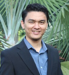 Muhammad Hanif Ramlee合作论文数Med. Devices & Technol. Group, Univ. Teknol. Malaysia;c;Med. Devices & Technol. Group, Univ. Teknol. Malaysia44
 Ardiyansyah Syahrom合作论文数Universiti Teknologi Malaysia Medical Implant Technology Group (MEDITEG), Faculty of Biomedical Engineering and Health Sciences Johor Malaysia40
 S. Izman合作论文数Faculty of Mechanical Engineering, Universiti Teknologi Malaysia34
 Tunku Kamarul Zaman合作论文数30
 Saiful Izwan Abd Razak合作论文数27
 Hong seng Gan合作论文数Dept. of Biotechnol. & Med. Eng., Univ. Teknol. Malaysia;c;Dept. of Biotechnol. & Med. Eng., Univ. Teknol. Malaysia24
 Kahar Osman合作论文数Faculty of Mechanical Engineering;Universiti Teknologi Malaysia;Faculty of Mechanical Engineering, Universiti Teknologi Malaysia24
 Muhamad Noor Harun合作论文数Faculty of Mechanical Engineering, Universiti Teknologi Malaysia22
 Nida Iqbal合作论文数Medical Devices &Faculty of Bioscience and Medical Engineering, Universiti Teknologi Malaysia;Faculty of Bioscience and Medical Engineering, Universiti Teknologi Malaysia18
 Mat Uzir Wahit合作论文数Faculty of Chemical and Natural Resources Engineering;Universiti Teknologi Malaysia;Faculty of Chemical and Natural Resources Engineering, Universiti Teknologi Malaysia18
 Mohd Hasbullah Idris合作论文数Faculty of Mechanical Engineering, Universiti Teknologi Malaysia18
 Andreas Oechsner合作论文数Esslingen University of Applied Sciences/Griffith University17
 Habibollah Haron合作论文数Fac Comp, Univ Teknol Malaysia17Jamal Kashani合作论文数Medical Implant Technology Group, Faculty of Biosciences &Medical Engineering, Universiti Teknologi Malaysia;Faculty of Biosciences & Medical Engineering, Universiti Teknologi Malaysia16
 Azli Yahya合作论文数14
 Mas Ayu Hassan合作论文数Faculty of Manufacturing and Mechatronic Engineering Technology, Universiti Malaysia Pahang13
 Nadirul Hasraf合作论文数13
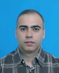 Hamid Reza Bakhsheshi-Rad合作论文数Najafabad Branch, Islamic Azad Univ13
 Abdul Hadi Abdul Wahab合作论文数13
 Nazri Kamsah合作论文数Faculty of Mechanical Engineering, University Technology Malaysia13
 Tan Tian Swee合作论文数12
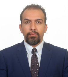 Shahrouz Zamani Khalajabadi合作论文数School of Chemical Engineering, Rowan University12
 Hendra Hermawan合作论文数Laboratory for Biomaterials and Bioengineering, Laval University12
 Muhammad Umar Aslam Khan合作论文数11
 Mohd Nazri Bajuri合作论文数Faculty of Health Science and Biomedical Engineering, Universiti Teknologi Malaysia11
 Rafaqat Hussain合作论文数Department of Chemistry, Universiti Teknologi Malaysia11
 Mohd. Saleh Jaafar合作论文数Faculty of Engineering, University Putra Malaysia11
 Irza Sukmana合作论文数10
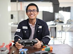 Fatihhi S.J.合作论文数Advanced Facilities Engineering Technology Research Cluster (AFET),, Universiti Kuala Lumpur Malaysian Institute of Industrial Technology10
 Mahmood Anwar合作论文数10
 Mina Alizadeh合作论文数10
 Amir Putra合作论文数10
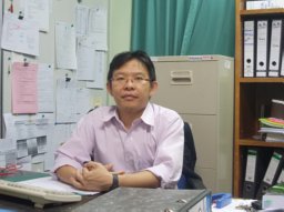 Samion Syahrullail合作论文数Department of Thermo-fluid, Universiti Teknologi Malaysia10
 Malliga Raman Murali合作论文数Department of Biochemistry, University of Madras10
 Nasrul Humaimi Mahmood合作论文数Cordless Charger Research Group;Faculty of Electrical Engineering;Cordless Charger Research Group|Faculty of Electrical Engineering10
 Sornambikai Sundaram合作论文数Department of Chemistry, Vellore Institute of Technology University10H. R. Bakhsheshi-Rad合作论文数9
 A. Shah合作论文数TAB, Universiti Pendidikan Sultan Idris9
 Asnida Abdul Wahab合作论文数University of Technology Malaysia9Noor Hayaty Abu Kasim合作论文数9
 Nasution Ahmad K合作论文数9
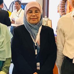 Esah Hamzah合作论文数Department of Materials Engineering9zohreh arabshahi合作论文数School of Chemistry, Physics and Mechanical Engineering, Queensland University of Technology8mohammad mahdi taheri合作论文数IBTN8
 Mohammadreza Daroonparvar合作论文数Department of Mechanical Engineering, University of Nevada Reno8
 Eshamsul Sulaiman合作论文数8
 Muhammad Ikman Ishak合作论文数Med. Implant Technol. Group, Univ. Teknol. Malaysia;c;Med. Implant Technol. Group, Univ. Teknol. Malaysia8
 Mohd Shafry Mohd Rahim合作论文数8
 Davood Almasi合作论文数Faculty of Mechanical Engineering, Universiti Teknologi Malaysia;Medical Devices &Faculty of Bioscience and Medical Engineering, Universiti Teknologi Malaysia;Universiti Teknologi Malaysia, Universiti Teknologi Malaysia8




