Abstract X-ray crystallographic fragment screening is a powerful strategy in modern drug discovery, enabling the identification of small-molecule starting points for rational hit-to-lead optimization. While highly effective for soluble proteins, its application to membrane proteins remains challenging due to low expression yields, high hydrophobicity, and the complexities of crystallization—particularly when using lipid cubic phase (LCP), which is often essential for high-resolution structural studies of targets like G-protein-coupled receptors (GPCRs). In this study, we present a methodology that integrates high-throughput X-ray crystallography with computational modeling and complementary biophysical validation to overcome these barriers. Using a thermostabilized human adenosine A 2A receptor crystallized in LCP as a test system, we screened 568 fragments and identified 23 initial hits. The work represents the first large-scale fragment screening effort targeting crystals of a membrane protein grown in LCP. Structure-guided virtual screening of these hits led to the design of 109 follow-up compounds, of which 56 yielded crystal structures. Of these, 19 were additionally confirmed to bind by grating-coupled interferometry (GCI), providing complementary biophysical validation. Our results demonstrated the feasibility and effectiveness of this integrated approach for fragment-based drug discovery on membrane proteins crystallized in LCP. Moreover, the detection of ligands at a previously uncharacterized intracellular pocket in a GPCR highlights the potential of this strategy to accelerate the discovery of therapeutically relevant compounds for challenging drug targets.
The macromolecular crystallography (MX) beamline X06DA-PXIII at the Swiss Light Source (SLS) has undergone a significant upgrade in preparation for SLS 2.0. After 15 years of operation and >2,500 PDB depositions, this highly productive superbend magnet beamline has been completely rebuilt with new optics and experimental hutches. The new design, which aims to exploit the ×37 reduced emittance of the SLS 2.0 storage ring, consists of a toroidal mirror close to the source to harvest a large solid angle of the bending magnet beam, a horizontal 1:2 focusing concept to minimize toroidal aberration and Kirkpatrick-Baez mirrors to focus the beam down to 10 × 10 microns2 with a flux of 2 × 10¹² ph/s at the sample position. The refurbished double channel-cut monochromator provides an energy range from 3 to 15 keV. While this makes the beamline highly suitable for identifying low-Z elements in macromolecular structures with data acquisition at low energy, we plan to center our main activity around high-throughput industrial applications for fragment-based drug discovery. We will perform fully autonomous and unattended experiments using the TELL sample changer, the multi-axis goniometer SmarGon, and the new Dectris PILATUS4 X 2 M detector. We also intend to automate experiments at room temperature, which will benefit from the unprecedented speed of data collection provided by the detector. In this presentation, I will present the upgrade of the X06DA-PXIII beamline and the planned experiments with the new SLS 2.0 storage ring. In addition, I will give an overview of the upgrade of the other 2 MX undulator beamlines.
Small Angle-X-ray Scattering Tensor Tomography (SAS-TT) is a relatively new but powerful technique for studying the multiscale architecture of hierarchical structures particularly relevant to life science applications. Currently, the technique is very demanding on synchrotron beamtime, which limits its applications, especially for cases requiring a statistically relevant number of samples. This study reports the first SAS-TT measurement at a macromolecular X-ray crystallography beamline, PX-I at the Swiss Light Source (SLS), with an improvement in acquisition time from 96 h/Mvoxel in the pilot experiments to 6 h/Mvoxel with comparable sampling, defining a new standard for fast SAS-TT with a micrometer beam size and allowing to record a full tomogram in 1.2 h. Measurements are performed on the long and lenticular process of the incus bone, one of the three human auditory ossicles. The main orientation and degree of alignment of the mineralised collagen fibrils are characterised, as well as the size and shape of the mineral particles which show relevant variations in different tissue locations. The study reveals three distinct regions of high fibril alignment, most likely important pathways of sound throughout the ossicular chain, and highlights the technique's potential to aid in future developments in middle ear reconstructive surgery.
In recent years, time-resolved serial crystallography has emerged as a transformative technique for unraveling the intricate dynamics of macromolecules at atomic resolution. By leveraging the high-intensity and ultra-short pulses of X-ray free electron lasers (XFELs) alongside the high brilliance of synchrotron light sources, this technique has enabled the observation of transient states in biomolecules as they catalyze chemical reactions.This presentation will highlight the advancements and applications of time-resolved serial crystallography in the study of macromolecular dynamic. We will discuss the light-sensitive membrane protein Nonlabens marinus halorhodopsin (NmHR) as an example of how this method enables us to capture the structural dynamics from femtoseconds to milliseconds after light activation. Through combining time-resolved studies at the X-ray free electron laser and synchrotron with spectroscopy and chemical simulation, we obtained a comprehensive understanding of the molecular mechanism that allows NmHR to catalyze ion transport across biological membranes. In addition to discussing the rich chemical information that can be obtained in time-resolved crystallographic studies, this talk will highlight how steady-state experiments can provide exciting structural insights while requiring only a limited amount of beamtime and a minimal setup.
The newly upgraded SLS 2.0 as a 4th generation synchrotron delivers unprecedented brilliance, enabling exploration of biological structures at vastly improved timescales and throughput. Our upgraded Macromolecular crystallography (MX) beamlines aim to support cutting-edge experiments including high-throughput fragment screening (FFCS),1 automated data collection,2 room temperature3, time-resolved serial crystallography,4 and X-ray scattering tensor tomography (SAS-TT)5—all generating massive data volumes requiring efficient management solutions.We are experimenting with different implementations including a multi-tiered approach utilizing HW-accelerated edge servers,6 PSI’s high- performance computing infrastructure, and DECTRIS CLOUD capabilities. The data processing pipelines operate both locally and remotely to support ongoing pilot experiments that will provide valuable insights into the performance and usability of these pipelines during user operation. This presentation will deliver firsthand experience from our active beamline commissioning in the beginning of SLS 2.0, sharing emerging strategies for handling high data rates. We will present our data acquisition, data processing and data reduction strategies. In addition, we will discuss the benefits and the challenges of our new data management systems. By sharing our current journey implementing these systems, we aim to contribute valuable perspectives to the MX community navigating similar challenges with increasing data volumes.
The SLS 2.0 upgrade, beamline improvements, and developments in charge-integrating X-ray detector technology [1] open new opportunities for scientists to investigate the dynamics of biological structures at shorter timescales and with higher throughput than ever before. However, handling many GBs of X-ray images per second is a big data challenge [2] that must be overcome in order to make collecting and processing diffraction images at rates up to 10 kHz feasible in the future (see Fig. 1). I will first present Jungfraujoch [3,4], a detector read-out system with FPGAs and GPUs capable of handling 40 GB/s data rates within a single server. The system also facilitates real-time image analysis to provide fast experimental feedback, including spot finding, indexing, and radial integration. We are currently working on applying modern machine-learning frameworks and optimizing them to run efficiently on GPUs and FPGAs. We have successfully integrated the Jungfraujoch into the PXI beamline at the SLS and achieved 2 kHz real-time data acquisition and analysis with a JUNGFRAU 9M detector. Through the utilization of JUNGFRAU's storage cell mode, we've achieved burst readout speeds of up to 100 kHz across 15 images, thus enabling a remarkable 10-microsecond time resolution in serial synchrotron crystallography. As we gain more experience with integrating Jungfraujoch, it could be a broadly applicable solution for more types of detectors, more beamlines, and more techniques at both Synchrotron and XFEL facilities.
This review highlights the development and evolution of three macromolecular crystallography (MX) beamlines at the Swiss Light Source (SLS) over the past two decades. We discuss key advancements in X-ray optics, detectors, goniometers, sample changers and MX methodology, emphasizing their impact on high-throughput and high-resolution structural biology. Our contributions are presented within the broader context of global efforts in synchrotron-based MX. Looking ahead, we explore the future experiments enabled by SLS 2.0 and new opportunities at SwissFEL to enhance experimental capabilities and drive scientific discoveries.
Advances in structural biology have relied heavily on synchrotron cryo-crystallography and cryogenic electron microscopy to elucidate biological processes and for drug discovery. However, disparities between cryogenic and room-temperature (RT) crystal structures pose challenges. Here, Cryo2RT, a high-throughput RT data-collection method from cryo-cooled crystals that leverages the cryo-crystallography workflow, is introduced. Tested on endothiapepsin crystals with four soaked fragments, thaumatin and SARS-CoV-2 3CLpro, Cryo2RT reveals unique ligand-binding poses, offers a comparable throughput to cryo-crystallography and eases the exploration of structural dynamics at various temperatures.
Light–oxygen–voltage (LOV) domains are small photosensory flavoprotein modules that allow the conversion of external stimuli (sunlight) into intracellular signals responsible for various cell behaviors (e.g. phototropism and chloroplast relocation). This ability relies on the light-induced formation of a covalent thioether adduct between a flavin chromophore and a reactive cysteine from the protein environment, which triggers a cascade of structural changes that result in the activation of a serine/threonine (Ser/Thr) kinase. Recent developments in time-resolved crystallography may allow the activation cascade of the LOV domain to be observed in real time, which has been elusive. In this study, we report a robust protocol for the production and stable delivery of microcrystals of the LOV domain of phototropin Phot-1 from Chlamydomonas reinhardtii (CrPhotLOV1) with a high-viscosity injector for time-resolved serial synchrotron crystallography (TR-SSX). The detailed process covers all aspects, from sample optimization to data collection, which may serve as a guide for soluble protein preparation for TR-SSX. In addition, we show that the crystals obtained preserve the photoreactivity using infrared spectroscopy. Furthermore, the results of the TR-SSX experiment provide high-resolution insights into structural alterations of CrPhotLOV1 from Δt = 2.5 ms up to Δt = 95 ms post-photoactivation, including resolving the geometry of the thioether adduct and the C-terminal region implicated in the signal transduction process.
Recent advances in automation have fostered the development of unattended data collection services at a handful of synchrotron facilities worldwide. At the Swiss Light Source, the installation of new high-throughput sample changers at all three macromolecular crystallography beamlines and the commissioning of the Fast Fragment and Compound Screening pipeline created a unique opportunity to automate data acquisition. Here, the DA+ microservice software stack upgrades, implementation of an automatic loop-centering service and deployment of the Smart Digital User (SDU) software for unattended data collection are reported. The SDU software is the decision-making software responsible for communications between services, sample and device safety, sample centering, sample alignment with grid based X-ray diffraction and, finally, data collection.
Both intensity and phase information are needed for structure determination by macromolecular X-ray crystallography. The diffraction experiment provides intensities. Phases must be accessed indirectly by molecular replacement, or by experimental phasing. A popular method for crystallising membrane proteins employs a lipid cubic mesophase (the in meso method). Monoolein is the most popular lipid for in meso crystallisation. Invariably, the lipid co-crystallises with the protein recapitulating the biomembrane from whence it came. We reasoned that such a lipid bearing a heavy atom could be used for experimental phasing. In this study, we replaced half the monoolein in the mesophase with a seleno-labelled analogue (Se-MAG), which has a selenium atom in the fatty acyl chain of the lipid. The lipid mixture formed the cubic mesophase and grew crystals by the in meso method of the alginate transporter, AlgE, and the lipoprotein N-acyltransferase, Lnt. Se-MAGs co-crystallised with both proteins and were used to obtain phases for high-resolution structure determination by the selenium single-wavelength anomalous diffraction method. The use of such a mixed lipid system may prove to be a general strategy for the experimental phasing part of crystallographic structure determination of membrane proteins that crystallise via the in meso method.
The first demonstration of 2 kHz time-resolved serial crystallography data acquisition at a fourth-generation synchrotron, using the JUNGFRAU 4M pixel detector.
Over the last two decades, fragment-based drug discovery has emerged as an effective and efficient method to identify chemical scaffolds for the development of novel lead compounds.The inherent "start small -elaborate efficiently" approach allows issues like compound-selectivity, toxicity and efficiency to be addressed from a very early development state on, while saving time and resources compared to classical high throughput screening of larger compounds.X-ray crystallography has been used for a long time as an important orthogonal method for validating binders discovered by higher throughput screening methods and to elucidate corresponding ligandtarget interactions.Advances in beamline-and crystal-harvestinginstrumentation have tremendously increased the throughput of X-ray crystallography in the last few years.This has facilitated the establishment of macromolecular crystallography as a powerful primary screening method for the identification of ligand binding.Combining fragment screening with the high information content derived from crystal-based fragment screening results in a powerful platform for structure-based drug discovery.Here, we proudly present the Fast Fragment and Compound Screening pipeline (FFCS) at the Swiss Light Source (PSI, Switzerland) and its application in the development of novel drugs 1 .
The JUNGFRAU 4-megapixel (4M) charge-integrating pixel-array detector, when operated at a full 2 kHz frame rate, streams data at a rate of 17 GB s(-1). To operate this detector for macromolecular crystallography beamlines, a data-acquisition system called Jungfraujoch was developed. The system, running on a single server with field-programmable gate arrays and general-purpose graphics processing units, is capable of handling data produced by the JUNGFRAU 4M detector, including conversion of raw pixel readout to photon counts, compression and on-the-fly spot finding. It was also demonstrated that 30 GB s(-1) can be handled in performance tests, indicating that the operation of even larger and faster detectors will be achievable in the future. The source code is available from a public repository.
The binding and release of ligands from their protein targets is central to fundamental biological processes as well as to drug discovery. Photopharmacology introduces chemical triggers that allow the changing of ligand affinities and thus biological activity by light. Insight into the molecular mechanisms of photopharmacology is largely missing because the relevant transitions during the light-triggered reaction cannot be resolved by conventional structural biology. Using time-resolved serial crystallography at a synchrotron and X-ray free-electron laser, we captured the release of the anti-cancer compound azo-combretastatin A4 and the resulting conformational changes in tubulin. Nine structural snapshots from 1 ns to 100 ms complemented by simulations show how cis-to-trans isomerization of the azobenzene bond leads to a switch in ligand affinity, opening of an exit channel, and collapse of the binding pocket upon ligand release. The resulting global backbone rearrangements are related to the action mechanism of microtubule-destabilizing drugs.
SARS-CoV-2 nsp3 is essential for viral replication and host responses. The SARS-unique domain (SUD) of nsp3 exerts its function through binding to viral and host proteins and RNAs. Herein, we show that SARS-CoV-2 SUD is highly flexible in solution. The intramolecular disulfide bond of SARS-CoV SUD is absent in SARS-CoV-2 SUD. Incorporating this bond in SARS-CoV-2 SUD allowed crystal structure determination to 1.35 Å resolution. However, introducing this bond in SARS-CoV-2 genome was lethal for the virus. Using biolayer interferometry, we screened compounds directly binding to SARS-CoV-2 SUD and identified theaflavin 3,3’-digallate (TF3) as a potent binder, K d 2.8 µM. TF3 disrupted the SUD-guanine quadruplex interactions and exhibited anti-SARS-CoV-2 activity in Vero E6-TMPRSS2 cells with an EC 50 of 5.9 µM and CC 50 of 98.5 µM. In this work, we provide evidence that SARS-CoV-2 SUD harbors druggable sites for antiviral development.
Bacterial lipoproteins (BLPs) decorate the surface of membranes in the cell envelope. They function in membrane assembly and stability, as enzymes, and in transport. The final enzyme in the BLP synthesis pathway is the apolipoprotein N -acyltransferase, Lnt, which is proposed to act by a ping-pong mechanism. Here, we use x-ray crystallography and cryo–electron microscopy to chart the structural changes undergone during the progress of the enzyme through the reaction. We identify a single active site that has evolved to bind, individually and sequentially, substrates that satisfy structural and chemical criteria to position reactive parts next to the catalytic triad for reaction. This study validates the ping-pong mechanism, explains the molecular bases for Lnt’s substrate promiscuity, and should facilitate the design of antibiotics with minimal off-target effects.
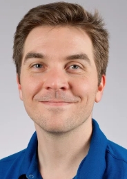 Vincent Olieric合作论文数Laboratory for Macromolecules and Bioimaging, Center for Photon Science, Paul Scherrer Institut43
 Ezequiel Panepucci合作论文数Neurology and Neurological Sciences, and Stanford Synchrotron Radiation Laboratory, Stanford University35
 Chia-Ying Huang合作论文数29
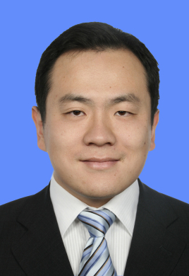 Sheng Cui (崔胜)合作论文数Institute of Pathogen Biology, Chinese Academy of Medical Sciences & Peking Union Medical College24
 Justyna A. Wojdyla合作论文数Department of Biology, University of York;Department of Biology, University of York24
 Martin Caffrey合作论文数School of Biochemistry and Immunology, The University of Dublin21Arthur Mar合作论文数21Bo Qin合作论文数18
 Tobias Weinert合作论文数Lab Biomol Res, Paul Scherrer Inst17Filip Leonarski合作论文数Photon Science Division, Paul Scherrer Institute16
 Dworkowski Florian S N合作论文数14Clemens Schulze-Briese合作论文数14
 Isabelle Martiel合作论文数Food and Soft Materials Science, Institute of Food, Nutrition &Health, ETH Zurich;Institute of Food, Nutrition & Health, ETH Zurich13
 Shibom Basu合作论文数Department of Chemistry and Biochemistry, Arizona State University13Takashi Tomizaki合作论文数12
 Kay Diederichs合作论文数Department of Biology, Universitat Konstanz12Daniel James合作论文数Department of Physics, Arizona State University11Sandro Waltersperger合作论文数11
 A. Mozzanica合作论文数Paul Scherrer Inst, CH-5232 Villigen, Switzerland10Robert McDonald合作论文数10
 Laura Vera合作论文数10Dmitry Ozerov合作论文数Paul Scherrer Institute10Claude Pradervand合作论文数9
 Przemyslaw Nogly合作论文数Laboratory of Biomolecular Research, Paul Scherrer Institute9
 Karol Nass合作论文数Center for Free-Electron Laser Science (CFEL), DESY, Notkestrasse 85, 22607 Hamburg, Germany.;Department of Physics, University of Hamburg;Center for Free-Electron Laser Science (CFEL),, University of Hamburg9
 Xiaopan Gao (高小攀)合作论文数9
 Oliver Bunk合作论文数9Shihua Wang合作论文数8
 Christopher Milne合作论文数European XFEL8Howe Nicole合作论文数8
 Xue Yao合作论文数College of Life Sciences, Nankai University8
 Roman Schneider合作论文数University of Trier8Wayne Glettig合作论文数7
 Lei Zheng合作论文数7Maia M. Cherney合作论文数7Antonia Furrer合作论文数Division of Biology and Chemistry, Paul Scherrer Institut7Dardan Gashi合作论文数Paul Scherrer Inst, SwissFEL, CH-5232 Villigen, Switzerland7b schmitt合作论文数Paul Scherrer Inst, CH-5232 Villigen, Switzerland7Rajendran Chitra合作论文数7
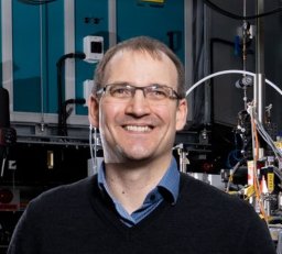 Standfuss Joerg合作论文数Laboratory of Biomolecular Research, Paul Scherrer Institut7
 Michel O. Steinmetz合作论文数Laboratory of Biomolecular Research, Division of Chemistry and Biology, Paul Scherrer Institut7
 Petr Skopintsev合作论文数Deutsches Elektronen-Synchrotron DESY7
 Rangana Warshamanage合作论文数Department of Chemistry, University of Zürich7Natacha Olieric合作论文数Biomolecular Research, Paul Scherrer Institut7
 Santosh Panjikar合作论文数Australian Synchrotron7
 Sophie Redford合作论文数Sensirion7Marcus Müller合作论文数6May E Sharpe合作论文数6L Chan合作论文数6




