216 Background: Colorectal cancer (CRC) is the second leading cause of cancer-related deaths. We previously found that delayed diagnosis due to lack of radiological identification results in significantly worse outcome for patients. We had developed a rudimentary, AI second observer which demonstrated potential for detecting CRC on routine CT abdomen/pelvis (CTAP). However, the AI algorithm detected many false positives. In this study, we analyzed the data using TCIA as test cases and evaluated whether patient peritoneal fat content influenced the false positive rate. This could serve as a guide for future training of AI second observer to minimize false positive detection. Methods: 2D U-Net convolutional neural network (CNN) containing 31 million trainable parameters was trained with 58 CRC CT images from Banner MD Anderson (AZ) and MD Anderson Cancer Center (TX) (51 used for training and 7 for validation) and 59 normal CT scans from Banner MD Anderson Cancer Center. 18 of the 25 CRC cases from public domain data (The Cancer Genome Atlas) were used to evaluate the performance of the models (5 had no identifiable cancer and 2 were rejected for having no contrast). The CRC was segmented using ITK-SNAP open-source software (v. 3.8). To apply the deep ensemble approach, five CNN models were trained independently with random initialization using the same U-Net architect and the same training data. Given a testing CT scan, each of the five trained CNN models was applied to produce tumor segmentation for the testing CT scan. The tumor segmentation results produced by the trained CNN models were then fused using a simple majority voting rule (up to 2 voters) to produce consensus tumor segmentation results. The segmentation was analyzed for the number and location of false positives per case. The peritoneal fat content was classified at the level of aortic bifurcation by the distance of fat between adjacent small bowel loops (≤ or > 1 cm). Chi-square test was performed testing fat volume and number of voters as the intervention. Results: Our results showed that the higher volume of peritoneal fat (> 1 cm, N=6) decreases the rate of false positive compared with low volume (≤ 1 cm, N=12, p=0.013). When comparing between having one voter and two voter ensemble using low fat volume data, two voter ensemble also decreased the number of false positives but not statistically significant (p=0.286). Conclusions: Our results show that AI-based second observer generates more false positives when patients have lower peritoneal fat volume; this implies that future training may require higher percentage of cases with low peritoneal fat to improve second observer precision. Our analysis also showed that increasing the number of voter in the ensemble also decrease the number of false positives per case.
643 Background: Although modern cancer treatment has significantly improved overall survival, this comes with a high financial cost. Based on an NIH report on Financial Burden of Cancer Care, treating pancreatic adenocarcinoma (PDAC) costs $108K, $18K, and $125K for initial treatment, during follow-up, and in last year of life, respectively. During the course of treatment, patients will visit the hospital for acute symptoms for which cross-sectional imaging is obtained within 45 days of subsequent scheduled follow-up imaging. This presents an opportunity for cost saving if data could be collected to identify an algorithm to avoid unnecessary duplication of imaging in a short interval. The purpose of this pilot study is to evaluate tumor growth trajectory within this short time span of 45 days to guide future trials to clarify the circumstances for which a subsequent follow up study is necessary. Methods: We retrospectively identified patients from our tumor registry from 2016 to 2019 who were diagnosed with metastatic PDAC (mPDAC). Subjects were included for analysis if they met the following criteria: 1) diagnosed with mPDAC; 2) had at least 2 CT abdomen/pelvis (CTAP) within 45 days of each other with one of the studies being obtained in the emergency department; and 3) had at least 1-3 measurable lesions as defined by RECIST 1.1 criteria. Tumor dimension was measured on axial images with the largest cross-section. For our primary outcome we used paired t-tests to evaluate the difference in mean size of all lesions (primary and metastatic). We also performed sub-group analyses comparing mean size of primary and metastatic lesions separately. An alpha level of 0.05 was used to determine statistical significance. Results: We screened 383 subjects from the tumor registry and identified 28 subjects suitable for our study. 18 subjects were female and 10 were male. We identified 69 target lesions, 23 of which were primary and 46 were metastatic. The mean sum of the largest dimension of target lesions was 2.82 cm on the initial study and 3.23 on the latter study (p<0.001). The mean sum of the largest dimension of the primary lesion was 3.62 cm on the initial study and 3.89 on the latter study (p=0.01). For metastatic lesions, the initial sum was 2.38 cm while the latter was 2.85 (p<0.001). Conclusions: Our finding of increase in tumor size in 45 days for mPDAC suggests that mPDAC is aggressive and can grow even in short time span. This suggests that if progression is identified on a study obtained within 45 days of scheduled follow-up, the follow-up study could be cancelled to avoid added cost to the patient. However, if the earlier study is stable or shows response, the scheduled follow-up may still be necessary to demonstrate continued stability/response.
BACKGROUNDMissing occult cancer lesions accounts for the most diagnostic errors in retrospective radiology reviews as early cancer can be small or subtle, making the lesions difficult to detect. Second-observer is the most effective technique for reducing these events and can be economically implemented with the advent of artificial intelligence (AI). AIMTo achieve appropriate AI model training, a large annotated dataset is necessary to train the AI models. Our goal in this research is to compare two methods for decreasing the annotation time to establish ground truth: Skip-slice annotation and AI-initiated annotation. METHODSWe developed a 2D U-Net as an AI second observer for detecting colorectal cancer (CRC) and an ensemble of 5 differently initiated 2D U-Net for ensemble technique. Each model was trained with 51 cases of annotated CRC computed tomography of the abdomen and pelvis, tested with 7 cases, and validated with 20 cases from The Cancer Imaging Archive cases. The sensitivity, false positives per case, and estimated Dice coefficient were obtained for each method of training. We compared the two methods of annotations and the time reduction associated with the technique. The time differences were tested using Friedman's two-way analysis of variance. RESULTSSparse annotation significantly reduces the time for annotation particularly skipping 2 slices at a time (P < 0.001). Reduction of up to 2/3 of the annotation does not reduce AI model sensitivity or false positives per case. Although initializing human annotation with AI reduces the annotation time, the reduction is minimal, even when using an ensemble AI to decrease false positives. CONCLUSIONOur data support the sparse annotation technique as an efficient technique for reducing the time needed to establish the ground truth.
Supplementary Tables 1-2 from Detection of Tumor DNA at the Margins of Colorectal Cancer Liver Metastasis
Supplementary Figure 2 from Detection of Tumor DNA at the Margins of Colorectal Cancer Liver Metastasis
Supplementary Figure 1 from Detection of Tumor DNA at the Margins of Colorectal Cancer Liver Metastasis
693 Background: Although modern cancer treatment has significantly prolonged patient’s life, it comes with a high financial cost. Based on an NIH report on Financial Burden of Cancer Care, treating PDAC costs $108K, $18K, and $125K for initial treatment, during follow up, and in last year of life, respectively. Of these, CA19-9 and CT studies are obtained concurrently to evaluate tumor burden whose cost can be reduced by decreasing imaging frequency when CA19-9 remains stable. As a pilot study, we evaluated whether changes in CA19-9, an inexpensive laboratory study commonly used for monitoring treatment response, correlates with tumor size change on imaging studies. A positive correlation may lead us to design future studies to evaluate whether stable CA19-9 can serve as an indicator for stable disease on CT scan such that imaging frequency (and cost) may be reduced. Methods: We retrospectively identified PDAC patients from our tumor registry from 2016 to 2019. Subjects were included if they met the following criteria: 1) diagnosed with PDAC 2) had at least 2+ CA19-9 and CT/MR follow up assessments with the laboratory and imaging studies within one month of each other, and 3) had measurable lesions as defined by RECIST 1.1 criteria. Due to limited resources, we limited the maximum number of lesions to 4 instead of the usual 5 for RECIST 1.1. Two-dimensional orthogonal tumor sizes were measured on axial images with the largest cross-section. Changes in the values were obtained serially (from the immediately prior study). Nonparametric correlations (Spearman’s rho) were used to evaluate monotonicity. The significance level was set at p < 0.05. Results: We screened 300 subjects from the tumor registry and identified 23 subjects suitable for our study. 13 subjects were female and 10 were male with ages ranging from 35 to 84. We identified 55 target lesions and 128 assessments (with both CA19-9 and imaging) from the 23 subjects. We analyzed the log absolute and log relative change in CA19-9 with change in tumor area. When absolute CA19-9 value, absolute change in CA19-9, and relative change in CA19-9 were tested against the absolute change in tumor size, the correlation was significant for the absolute value and absolute change of CA19-9 (p < 0.01), but not for relative change of CA19-9 (p < 0.11). When tested against relative change in tumor size, all three values were highly significant (p < 0.001). Conclusions: Our finding of direct correlations between changes of CA19-9 and relative change of tumor size on imaging suggests that CA19-9 can serve as a surrogate measure of relative change of tumor burden for patients undergoing treatment. This provides motivation for future studies evaluating the possibility that stable CA19-9 can represent stable tumor burden such that expensive imaging studies may be obtained at longer time intervals to reduce financial cost to patients.
Carcinoid crisis is a potentially fatal condition characterized by various symptoms, including hemodynamic instability, flushing, and diarrhea. The incidence of carcinoid crisis is unknown, in part due to inconsistency in definitions across studies. Triggers of carcinoid crisis include general anesthesia and surgical procedures, but drug-induced and spontaneous cases have also been reported. Patients with neuroendocrine tumors (NETs) and carcinoid syndrome are at risk for carcinoid crisis. The pathophysiology of carcinoid crisis has been attributed to secretion of bioactive substances, such as serotonin, histamine, bradykinin, and kallikrein by NETs. The somatostatin analog octreotide has been considered the standard of care for carcinoid crisis due to its inhibitory effect on hormone release and relatively fast resolution of carcinoid crisis symptoms in several case studies. However, octreotide's efficacy in the treatment of carcinoid crisis has been questioned. This is due to a lack of a common definition for carcinoid crisis, the heterogeneity in clinical presentation, the paucity of prospective studies assessing octreotide efficacy in carcinoid crisis, and the lack of understanding of the pathophysiology of carcinoid crisis. These issues challenge the classical physiologic model of carcinoid crisis and its common etiology with carcinoid syndrome and raise questions regarding the utility of somatostatin analogs in its treatment. As surgical procedures and invasive liver-directed therapies remain important treatment modalities in patients with NETs, the pathophysiology of carcinoid crisis, potential benefits of octreotide, and efficacy of alternative treatment modalities must be studied prospectively to develop an effective evidence-based treatment strategy for carcinoid crisis.
141 Background: Colorectal cancer (CRC) is the second leading cause of cancer-related deaths, and survival can be improved if early, suspect imaging features on CT of the abdomen and pelvis (CTAP) can be routinely identified. At present, up to 40% of these features are undiagnosed on routine CTAP, but this can be improved with a second observer. In this study, we developed a deep ensemble learning method for detecting CRC on CTAP to determine if increasing agreement between ensemble models can decrease the false positives detected by artificial intelligence (AI) second-observer. Methods: 2D U-Net convolutional neural network (CNN) containing 31 million trainable parameters was trained with 58 CRC CT images from Banner MD Anderson (AZ) and MD Anderson Cancer Center (TX) (51 used for training and 7 for validation) and 59 normal CT scans from Banner MD Anderson Cancer Center. 20 of the 25 CRC cases from public domain data (The Cancer Genome Atlas) were used to evaluate the performance of the models. The CRC was segmented using ITK-SNAP open-source software (v. 3.8). To apply the deep ensemble approach, five CNN models were trained independently with random initialization using the same U-Net architect and the same training data. Given a testing CT scan, each of the five trained CNN models was applied to produce tumor segmentation for the testing CT scan. The tumor segmentation results produced by the trained CNN models were then fused using a simple majority voting rule to produce consensus tumor segmentation results. The segmentation was analyzed by the percentage of correct detection, the number of false positives per case, and the Dice similarity coefficient (DSC). If parts of the CRC were flagged by AI, then it was considered correct. A detection was considered false positive if the marked lesion did not overlap with any CRC; contiguous false positives across different slices of CT image were considered a single false positive. DSC measures the quality of the segmentation by measuring the overlap between the ground-truth and AI detected lesion. Results: Our results showed that increasing the agreement between the 5 models dramatically decreases the number of false positives per CT at the expense of slight decrease in accuracy and DSC. This is described in the table. Conclusions: Our results show that AI-based second observer can potentially detect CRC on routine CTAP. Although the initial result yields high false positives per case, ensemble voting is an effective method for decreasing the false positives with a slight decrease in accuracy. This technique can be further improved for eventual clinical application.[Table: see text]
142 Background: Colorectal cancer (CRC) is the second leading cause of cancer-related deaths, and its outcome can be improved with better detection of incidental early CRC on routine CT of the abdomen and pelvis (CTAP). AI-second observer (AI) has the potential as shown in our companion abstract. The bottleneck in training AI is the time required for radiologists to segment the CRC. We compared two techniques for accelerating the segmentation process: 1) Sparse annotation (annotating some of the CT slice containing CRC instead of every slice); 2) Allowing AI to perform initial segmentation followed by human adjustment. Methods: 2D U-Net convolutional neural network (CNN) containing 31 million trainable parameters was trained with 58 CRC CT images from Banner MD Anderson (AZ) and MD Anderson Cancer Center (TX) (51 used for training and 7 for validation) and 59 normal CT scans from Banner MD Anderson Cancer Center. Twenty of the 25 CRC cases from public domain data (The Cancer Genome Atlas) were used to evaluate the performance of the models. The CRC was segmented using ITK-SNAP open-source software (v. 3.8). For the first objective, 3 separate models were trained (fully annotated CRC, every other slice, and every third slice). The AI-annotation on the TCGA dataset was analyzed by the percentage of correct detection of CRC, the number of false positives, and the Dice similarity coefficient (DSC). If parts of the CRC were flagged by AI, then it was considered correct. A detection was considered false positive if the marked lesion did not overlap with CRC; contiguous false positives across different slices of CT image were considered a single false positive. DSC measures the quality of the segmentation by measuring the overlap between the ground-truth and AI detected lesion. For the second objective, the time required to adjust the AI-produced annotation was compared to the time required for annotating the entire CRC without AI assistance. The AI-models were trained using ensemble learning (see our companion abstract for details of the techniques). Results: Our results showed that skipping slices of tumor in training did not alter the accuracy, false positives, or DSC classification of the model. When adjusting the AI-observer segmentation, there was a trend toward decreasing the time required to adjust the annotation compared to full manual segmentation, but the difference was not statistically significant (Table; p=0.121). Conclusions: Our results show that both skipping slices of tumor as well as starting with AI-produced annotation can potentially decrease the effort required to produce high-quality ground truth without compromising the performance of AI. These techniques can help improve the throughput to obtain a large volume of cases to train AI for detecting CRC.[Table: see text]
Background: Hepatic angiosarcoma (AS) and hepatic epithelioid hemangioendothelioma (HEHE) are rare primary hepatic vascular malignancies (PHVM) that remain poorly understood. To guide management, we sought to identify factors and trends predicting survival after surgical intervention using a national database. Materials and methods: In a retrospective analysis of the National Cancer Database patients with a diagnosis of PHVM were identified. Clinicopathologic factors were extracted and compared. Overall survival (OS) was estimated and predictors of survival were identified. Results: Three hundred ninty patients with AS and 216 with HEHE were identified. Only 16% of AS and 36% of HEHE patients underwent surgery. The median OS for patients who underwent surgical intervention was 97 months, with 5-year OS of 30% for AS versus 69% for HEHE patients (P < 0.001). Tumor biology strongly impacted OS, with AS histology (Hazard Ratio [HR] of 3.61 [1.55-8.42]), moderate/poor tumor differentiation (HR = 3.86 [1.03-14.46]) and tumor size (HR = 1.01 [1.00-1.01]) conferring worse prognosis. The presence of metastatic disease in the surgically managed cohort (HR = 5.22 [2.01-13.57]) and involved surgical margins (HR = 3.87 [1.59-9.42]), were independently associated with worse survival. Conclusions: In this national cohort of PHVM, tumor biology, in the form of angiosarcoma histology, tumor differentiation and tumor size, was strongly associated with worse survival after surgery. Additionally, residual tumor burden after resection, in the form of positive surgical margins or the presence of metastasis, was also negatively associated with survival. Long-term clinical outcomes remain poor for patients with the above high-risk features, emphasizing the need to develop effective forms of adjuvant systemic therapies for this group of malignancies. Published by Elsevier Inc.
Objective: Pancreatic ductal adenocarcinoma (PDAC) has the best survival when detected early with 5-year survival near 40% for small, resectable PDAC. We evaluate the undiagnosed PDAC imaging features on routine CT and their impact on resectability. Methods: 76 of the screened 134 CTs from 1/1/2012 to 12/31/2018 using our tumor registry were obtained prior to PDAC diagnosis for other indications at least one month before presentation. Each cross-sectional study was reviewed for features of early PDAC: pancreatic mass, pancreatic ductal dilatation, perivascular/peripancreatic soft-tissue infiltration, omental lesions/ascites, and lymphadenopathy. When such features were detectible by the reviewing radiologists, the original CT readings were classified as concordant/discrepant. Descriptive statistics are reported for discrepant reads, tumor resectability, and tumor size. Results: Of the 76 cases from 46 unique subjects (30 male/16 female), 25 CTs (33%) had undetected PDAC imaging features: masses (15/19 unreported), ductal dilatation (16/20 unreported), and peripancreatic/perivascular soft-tissue infiltration (20/36 unreported). 63% of early PDAC features were not identified initially. One year before clinical diagnosis, 75-80% of the PDAC cases were resectable; at < 6 months before clinical diagnosis, only 29% were resectable. Conclusion: Improving early detection of key PDAC features on routine CT examinations can potentially improve patient outcomes.
Objective Current National Comprehensive Cancer Network guidelines for gastroenteropancreatic neuroendocrine tumors (GEPNETs) recommend complete (R0) surgical resection of the primary tumor and metastases, if feasible. However, large multicenter studies of recurrence patterns of GEPNETs after resection have not been performed. Methods Patients 18 years or older who presented to 7 participating National Comprehensive Cancer Network institutions between 2004 and 2008 with a new diagnosis of a small bowel, pancreas, or colon/rectum neuroendocrine tumor (NET) and underwent R0 resection of the primary tumor, and synchronous metastases, if present, were included in this analysis. Descriptive statistics and Kaplan-Meier estimates were used to calculate recurrence rates and time-associated end points, respectively. Results Of 294 patients with GEPNETs, 50% were male, 88% were White, and 99% had Eastern Cooperative Oncology Group performance status 0 to 1. The median age was 55 years (range, 20–90). The median follow-up time from R0 resection was 62.1 months. Recurrence rates were 18% in small bowel NETs (n = 110), 26% in pancreatic NETs (n = 141), and 10% in colon/rectum NETs (n = 50). The frequency of surveillance imaging was highly variable. Conclusions R0 resection was associated with variable risk of recurrence across subtypes. Further research to inform refinement of guidelines for the appropriate duration of surveillance after R0 resection is needed.
380 Background: Pancreatic ductal adenocarcinoma (PDAC) is the third leading cause of cancer-related deaths. At present, the best 5-year survival is 25% for resectable PDAC. For small (1 cm) stage 1 PDAC, resection has resulted in much better survival. The goal of this study was to evaluate the appearance and location of early undiagnosed PDAC on computed tomography scans (CT) prior to diagnosis with the goal of minimizing missing early PDAC. We also categorize the errors as either perceptive or cognitive. Methods: PDAC cases were retrospectively reviewed from 1/1/2012 through 12/31/2018 from our tumor registry, identifying 81 cases with paired CT scans both at the time of and prior to diagnosis. Among these, 31 contained imaging features considered diagnostic or suspicious for early PDAC(38%). These “errors” were classified by radiologic features and as well as by location. In addition, errors were classified into “perceptive errors" when the first study was read as normal, and as “cognitive errors” when the report noted an abnormality but failed to note suspicion for malignancy. Results: Among the 31 undiagnosed PDAC, 18 had features of an identifiable mass (58%), 9 had pancreatic ductal dilatation (29%), and 4 had evidence of perivascular soft tissue (13%). 44% of undiagnosed tumors were located in the head-neck, 39% in the body, and 17% in the tail. Perceptive errors were found in 58% and 42% were cognitive. No significant differences were seen between perceptive and cognitive errors based on suspicious features. Conclusions: Radiologic findings of early PDAC was retrospectively evident in more than one third of cases in which prior imaging was performed. These findings are most often masses or ductal dilatation. Location of these undiagnosed tumors were distributed throughout the gland. This study identifies the radiologic features of undiagnosed PDAC which may provide an opportunity for future prospective studies and improved technology which may improve early detection of pancreatic cancer.
Preoperative chemotherapy for potentially resectable colorectal liver metastases can lead to the disappearance of up to 25% of lesions on cross-sectional imaging, a phenomenon termed “disappearing liver metastasis” (DLM). While radiologic response from preoperative therapy can be seen as a desirable outcome, DLM can be a management challenge for the hepatic surgeon. Specifically, even when a metastasis “disappears,” viable malignancy remains present in up to 80% of cases. Therefore, all initially identified macroscopic disease should be resected or ablated if a curative intent strategy is being considered. Patients with smaller lesions and those deep within the hepatic parenchyma, as well as those who receive a longer duration of preoperative therapy, are at higher risk for DLM development. Early multidisciplinary case discussion of involving the hepatic surgeon is crucial to limit this conundrum. High-quality contrast-enhanced imaging prior to initiation of chemotherapy is important in order to identify all disease. Limiting the duration of chemotherapy can be useful particularly when lesions are small and when sub-lobar resections or ablations are anticipated. In some cases with small tumors which are initially resectable, proceeding directly to surgery rather than using neoadjuvant chemotherapy should be considered. While many DLM can be identified intraoperatively with a thorough examination and intraoperative ultrasonography, an attempt to treat all original sites of disease should be made, when possible. When not achievable and occult sites are left behind, these can often be followed and managed upon recurrence with a staged approach.
Background: Patients with cholangiocarcinoma often have indwelling biliary stents or catheters which are prone to obstructions and/or infections; studies show that 20–40% present with fever and/or jaundice requiring urgent treatment in the outpatient setting for which there are no uniform guidelines. The goal was to develop an expert panel consensus on this topic using the modified RAND/UCLA Delphi process to rate treatment appropriateness. Methods: Thirteen expert physicians from relevant specialties, geography, and practice settings were recruited for the panel. Patient scenarios were developed and panelists rated the therapies before and after a face-to-face discussion. The appropriateness of various therapies was rated on a scale from 1–9 and classified as appropriate, inappropriate, or uncertain. Scenarios with greater than 2 (>2) ratings of 1–3 (inappropriate) and greater than 2 (>2) ratings of 7–9 (appropriate) were considered to have disagreement and were not assigned an appropriateness rating. Results: Panelists were from all US regions and the UK (8%) and had practiced for a mean 16.5 years (4–33 years). Panelists rated 480 scenarios before the meeting and re-rated 288 of the clinical scenarios after the meeting. The panelists agreed that ongoing treatment with chemotherapy did not influence decision-making and, therefore, 192 scenarios were excluded from the final list. Disagreement decreased from 37.5% before to 10.4% after the meeting. Consensus on stent/tube manipulation and inpatient antibiotic therapy was obtained and summarized in patients as “appropriate” or “maybe appropriate” based on a patient’s bilirubin level at presentation. Conclusions: The Delphi process produced consensus guidelines to fill an unmet need in the urgent management of ascending cholangitis in patients with cholangiocarcinoma.
BACKGROUND:Despite an increasing number of women in the field of surgery, bias regarding cognitive or technical ability may continue to affect the experience of female trainees differently than their male counterparts. This study examines the differences in the degree of operative autonomy given to female compared with male general surgery trainees.METHODS:A smartphone app was used to collect evaluations of operative autonomy measured using the 4-point Zwisch scale, which describes defined steps in the progression from novice ("show and tell") to autonomous surgeon ("supervision only"). Differences in autonomy between male and female residents were compared using hierarchical logistic regression analysis.RESULTS:A total of 412 residents and 524 faculty from 14 general surgery training programs evaluated 8,900 cases over a 9-month period. Female residents received less autonomy from faculty than did male residents overall (P < .001). Resident level of training and case complexity were the strongest predictors of autonomy. Even after controlling for potential confounding factors, including level of training, intrinsic procedural difficulty, patient-related case complexity, faculty sex, and training program environment, female residents still received less operative autonomy than their male counterparts. The greatest discrepancy was in the fourth year of training.CONCLUSION:There is a sex-based difference in the autonomy granted to general surgery trainees. This gender gap may affect female residents' experience in training and possibly their preparation for practice. Strategies need to be developed to help faculty and residents work together to overcome this gender gap.
In robotic needle steering, flexible asymmetric-tip needles can steer around obstacles to reach targets deep within tissue. Due to tissue inhomogeneity and needle flexibility, needle buckling can occur, preventing accurate placement. This paper focuses on detecting needle buckling using axial force and needle-tip position readings from sensors. Our algorithm uses errors between the force readings and a predictive force model generated from those readings to track rapid changes in the measured forces. Using this prediction error and needle-tip position, the algorithm detects unexpected force increase, strict needle buckling, and buckling with sliding events at the needle-tip. The metrics for the detections are derived using a standard three-sigma rule and a sigmoid function to ensure generalizability of this method to a variety of tissue types. Our algorithm was tested using insertions into a gelatin tissue with an embedded rectangular obstacle designed to elicit buckling events. Needle buckling was detected at a maximum of 2[Formula: see text]mm after collision with the obstacle. Our algorithm was tested for robustness with insertions in an ex vivo tissue under different boundary conditions. Our algorithm was also able to detect buckling events 1–2[Formula: see text]s sooner than human detection times, showing significance for future autonomous control.
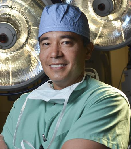 Richard Schulick合作论文数Division of Surgical Oncology, Department of Surgery, School of Medicine, University of Colorado128
 Timothy M Pawlik合作论文数Department of Surgery, College of Medicine, The Ohio State University Wexner Medical Center116
 Christopher Wolfgang合作论文数Department of Surgery, School of Medicine, New York University;Johns Hopkins School of Medicine85
 John L. Cameron合作论文数Johns Hopkins School of Medicine;Sidney Kimmel Comprehensive Cancer Center, Johns Hopkins University;Skip Viragh Center for Pancreatic Cancer, Johns Hopkins University77
 Barish Edil合作论文数45
 Timothy M. Pawlik合作论文数Department of Surgery, The Ohio State University Wexner Medical Center37Mansour John C合作论文数Div Surg Oncol, Univ Texas Southwestern Med Ctr Dallas34
 Ralph Hruban合作论文数Department of Oncology, Johns Hopkins University School of Medicine;The Sol Goldman Pancreatic Cancer Research Center, Johns Hopkins University34
 Hari Nathan合作论文数General Surgery, Department of Surgery, Medical School, University of Michigan-Ann Arbor33
 Emad Boctor合作论文数Division of Medical Imaging Physics, Department of Radiology and Radiological Science, Johns Hopkins University;Department of Computer Science, Whiting School of Engineering, Johns Hopkins University;Laboratory for Computational Sensing and Robotics, The Malone Center for Engineering in Healthcare, Johns Hopkins University31
 Joseph M Herman合作论文数30Polanco Patricio M合作论文数Division of Surgical Oncology, University of Pittsburgh Medical Center30Yopp Adam C合作论文数Univ Texas Southwestern Med Ctr Dallas28
 Al B Benson合作论文数Division of Hematology and Oncology, Department of Medicine, Feinberg School of Medicine, Northwestern University;Robert H. Lurie Comprehensive Cancer Center, Feinberg School of Medicine, Northwestern University26Porembka Matthew R合作论文数Department of Surgery, Washington University25Mechteld C. De Jong合作论文数Johns Hopkins University School of Medicine25
 Michael S. Torbenson合作论文数Mayo Clinic23
 Rebecca M. Minter合作论文数Division of Surgical Oncology, Department of Surgery, University of Wisconsin-Madison21Sam C. Wang合作论文数Center for Cardiovascular Development;Baylor College of Medicine;Department of Medicine;Department of Molecular and Cellular;Biology;Department of Molecular and Human Genetics;Winters Center for Heart Failure Research;Baylor College of;Medicine;Methodist Hospital, DeBakey Heart;Center;Department of Surgery;Baylor College of Medicine, Baylor College of Medicine21
 Alan P. Venook合作论文数Department of Medicine, School of Medicine, University of California San Francisco;Helen Diller Family Comprehensive Cancer Center, University of California San Francisco21
 Charles J. Yeo合作论文数Department of Surgery, Sidney Kimmel Medical College, Thomas Jefferson University20Mathew M Augustine合作论文数20
 Leonard Saltz合作论文数Memorial Sloan Kettering Cancer Center;Weill Medical College, Cornell University19
 David A Bluemke合作论文数Department of Radiology, School of Medicine and Public Health, University of Wisconsin- Madison;Department of Medical Physics, School of Medicine and Public Health, University of Wisconsin- Madison19
 Nassour Ibrahim合作论文数19
 Paul F. Engstrom合作论文数Fox Chase Cancer Center19
 Carlo Pulitano合作论文数School of Medicine, Faculty of Medicine and Health, The University of Sydney18
 Charles S. Fuchs合作论文数Genentech and Roche;EvolveImmune Therapeutics18John M. Skibber合作论文数18Lucille A. Leong合作论文数18
 Bert Vogelstein合作论文数Department of Molecular Biology and Genetics, Johns Hopkins School of Medicine17
 Kenneth W. Kinzler合作论文数Department of Oncology, School of Medicine, Johns Hopkins University;The Sidney Kimmel Comprehensive Cancer Center, Johns Hopkins University School of Medicine17
 Ana Gleisner合作论文数Division of Surgical Oncology, Department of Surgery, School of Medicine, University of Colorado17
 Luca Antonio Aldrighetti合作论文数16
 Keith D Lillemoe合作论文数Brigham and Women's Hospital;Massachusetts General Hospital;Harvard Medical School16
 Marwan G. Fakih合作论文数Department of Medical Oncology&Therapeutics Research, City of Hope;Comprehensive Cancer Center, City of Hope;Briskin Center for Clinical Research, City of Hope16David Shibata合作论文数16Harry S. Cooper合作论文数16
 Mary F. Mulcahy合作论文数Division of Hematology and Oncology, Department of Medicine, Feinberg School of Medicine, Northwestern University15
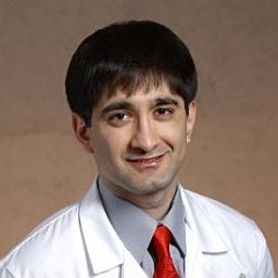 Hyder Omar合作论文数15Jean L. Grem合作论文数15
 Russell Taylor合作论文数Department of Computer Science, Whiting School of Engineering, Johns Hopkins University;Department of Mechanical Engineering, Whiting School of Engineering, Johns Hopkins University;Laboratory for Computational Sensing and Technology, Johns Hopkins University;NSF Engineering Research Center for Computer-Integrated Surgical Systems and Technology, Johns Hopkins University15
 Christopher G. Willett合作论文数Department of Radiation Oncology, Duke University School of Medicine;Duke Cancer Institute, Duke University15
 Peter C. Enzinger合作论文数Center for Esophageal and Gastric Cancer, Dana-Farber Cancer Institute;Harvard Medical School15Constantinos T. Sofocleous合作论文数15
 Ryan David P合作论文数14
 Yi-Jen Chen合作论文数Department of Radiation Oncology, City of Hope14Lia Assumpcao合作论文数Johns Hopkins School of Medicine, University of Puerto Rico14Ihab R. Kamel合作论文数14




