Methotrexate (MTX) has long been a first-line anti-rheumatic agent due to its efficacy and cost-effectiveness. While generally safe at the recommended weekly dosage of 5-25 mg for rheumatoid arthritis, medication errors can lead to acute toxicity, severe adverse effects, and potentially fatal outcomes. We report a case of a 51-year-old male who experienced fatal pancytopenia following an MTX medication error during treatment for rheumatoid arthritis. His blood MTX concentration measured 0.04 μmol/L 11 days after discontinuation. Postmortem examination revealed MTX-related toxicity in multiple organs, including the liver and intestines. The spleen showed a prominent increase in plasma cells, including numerous Mott cells. Forensic analysis established the cause of death as acute MTX intoxication, directly stemming from a chain of medical negligence involving prescribing, verification, and administration failures. This case underscores the necessity of rigorous therapeutic drug monitoring, individualized dosing, and robust systemic safeguards to prevent iatrogenic fatalities, highlighting its critical medicolegal and educational implications for forensic and clinical practice.
Insulin is an important protein hormone that participates in multiple metabolic pathways. Biosynthetic insulin has been widely used in the treatment of type 1 and type 2 diabetes. Currently, the number of reported cases of insulin overdose both at home and abroad is gradually increasing, and insulin homicide is no longer a means of “committing murder without leaving a trace”. At present, there are no systematic protocols for the identification of insulin overdose in the field of forensic medicine in China. This article introduces the causes, toxicological characteristics, forensic examination, laboratory testing methods and indicator reference of insulin overdose. Based on the identification practice and research results and referring to relevant studies on insulin overdose at home and abroad, this paper aims to provide recommendations and references for the formulation of forensic identification guidelines for insulin overdose cases.
Severe fever with thrombocytopenia syndrome virus (SFTSV) causes severe disease in humans, yet the pathogenesis remains poorly understood. A hallmark of fatal SFTS cases is the marked depletion of T cells. Previous studies on T cell depletion have predominantly focused on alterations in blood and peripheral lymphoid organs, while the thymus, a critical site for T cell development, has remained largely overlooked. In this study, we employed a lethal murine infection model to investigate the impact of SFTSV on thymic function. Our results revealed that SFTSV infected the thymus and induced severe cortical atrophy in type I interferon receptor-deficient (IFNAR-/-) mice, characterized by a dramatic depletion of CD4+CD8+ double-positive (DP) thymocytes. Transcriptomic analysis indicated that thymic damage is likely attributable to impaired thymocyte proliferation and increased apoptosis, which may be a consequence of SFTSV-induced alterations in the thymic microenvironment. We found SFTSV-infected macrophages and dendritic cells in the thymus, which accumulated in the cortex and exhibited elevated secretion of IFN-γ, a cytokine commonly associated with acute thymic atrophy. These results demonstrate thymic atrophy caused by SFTSV infection and suggest potential therapeutic strategies for restoring thymic function and promoting T cell reconstitution.
Infective endocarditis (IE) and myocardial abscesses caused by Rhodococcus erythropolis are rare clinical manifestations in healthy individuals. We report the autopsy of a 43-year-old male who suffered from IE caused by R. erythropolis infection and died of an atrioventricular node abscess, arrhythmia, and heart failure. This case indicates that the possibility of infection affecting the cardiac conduction system should be considered in arrhythmia-related IE cases. Our findings suggest that clinicians should consider the possibility of R. erythropolis infection in arrhythmia cases associated with IE.
The high risk of SARS-CoV-2 infection and reinfection and the occurrence of post-acute pulmonary sequelae have highlighted the importance of understanding the mechanism underlying lung repair after injury. To address this concern, comparative and systematic analyses of SARS-CoV-2 infection in COVID-19 patients and animals were conducted. In the lungs of nine patients who died of COVID-19 and one recovered from COVID-19 but died of unrelated disease in early 2020, damage-related transient progenitor (DATP) cells expressing CK8 marker proliferated significantly. These CK8+ DATP cells were derived from bronchial CK5+ basal cells. However, they showed different cell fate toward differentiation into type I alveolar cells in the deceased and convalescent patients, respectively. By using a self-limiting hamster infection model mimicking the dynamic process of lung injury remodeling in mild COVID-19 patients, the accumulation and regression of CK8+ cell marker were found to be closely associated with the disease course. Finally, we examined the autopsied lungs of two patients who died of infection by the recent Omicron variant and found that they only exhibited mild pathological injury with no CK8+ cell proliferation. These results indicate a clear pulmonary cell remodeling route and suggest that CK8+ DATP cells play a primary role in mediating alveolar remodeling, highlighting their potential applications as diagnostic markers and therapeutic targets.
As a primary target of severe acute respiratory syndrome coronavirus 2, lung exhibits heterogeneous histopathological changes following infection. However, comprehensive insight into their protein basis with spatial resolution remains deficient, which hinders further understanding of coronavirus disease 2019 (COVID-19)-related pulmonary injury. Here, we generate a region -resolved proteomic atlas of hallmark pathological pulmonary structures by integrating histological examination, laser microdissection, and ultrasensitive proteomics. Over 10,000 proteins are quantified across 71 post-mortem specimens. We identify a spectrum of pathway dysregulations in alveolar epithelium, bronchial epithelium, and blood vessels compared with non-COVID-19 controls, providing evidence for transitional -state pneumocyte hyperplasia. Additionally, our data reveal the region -specific enrichment of functional markers in bronchiole mucus plugs, pulmonary fibrosis, airspace inflammation, and alveolar type 2 cells, uncovering their distinctive features. Furthermore, we detect increased protein expression associated with viral entry and inflammatory response across multiple regions, suggesting potential therapeutic targets. Collectively, this study provides a distinct perspective for deciphering COVID-19-caused pulmonary dysfunction by spatial proteomics.
Since 2019, Coronavirus Disease 2019(COVID-19) has affected millions of people worldwide. Except for acute respiratory distress syndrome, dysgeusis is also a common symptom of COVID-19 that burdens patients for weeks or permanently. However, the mechanisms underlying taste dysfunctions remain unclear. Here, we performed complete autopsies of five patients who died of COVID-19. Integrated tongue samples, including numerous taste buds, salivary glands, vessels, and nerves were collected to map the pathology, distribution, cell tropism, and receptor distribution of severe acute respiratory syndrome coronavirus 2 (SARS-CoV-2) in the tongue. Our results revealed that all patients had moderate lymphocyte infiltration around the salivary glands and in the lamina propria adjacent to the mucosa, and pyknosis in the epithelia of taste buds and salivary glands. This may be because the serous acini, salivary gland ducts, and taste buds are the primary sites of SARS-CoV-2 infection. Multicolor immunofluorescence showed that SARS-CoV-2 readily infects Keratin (KRT)7+ taste receptor cells in taste buds, secretory cells in serous acini, and inner epithelial cells in the ducts. The major receptors, angiotensin-converting enzyme 2 (ACE2) and transmembrane protease serine subtype 2 (TMPRSS2), were both abundantly expressed in these cells. Viral antigens and receptor were both rarely detected in vessels and nerves. This indicates that SARS-CoV-2 infection triggers pathological injury in the tongue, and that dysgeusis may be directly related to viral infection and cellular damage.
Liquid-phase microextraction (LPME) possesses a high potential to isolate organic substances from different sample matrices. In this work, LPME was applied for the first time to investigate the biodistribution of diphenidol in different biofluids, organs, and brain regions using a fatal poisoning case. Since the LPME of diphenidol hasn't been reported, the effect of supported liquid membrane (SLM), acceptor and donor phases, and extraction time on LPME performance was investigated first. The solvents of 2-nonanone and 2-nitrophenyl octyl ether (NPOE) were found to be stable and efficient SLMs for LPME of diphenidol from biofluids and tissue samples, respectively. At steady state, the LPME recoveries for different sample matrices were in the range of 87 %-91 %. Due to the clean-up capability of LPME and the relatively high concentration of diphenidol in the fatal poisoning case, the proposed LPME systems were validated with related sample matrices using HPLC-UV for the determination. The methods displayed good linearity (R2 >= 0.9943), and the limits of detection were 0.30 mg L-1, 0.28 mg L- 1, and 2.7 mu g g- 1 for blood, urine, and liver samples, respectively. Meanwhile, the precision (<= 13%), accuracy (90-110%), and matrices effect (+/- 15%) were satisfactory at low, medium, and high concentrations. In addition, the stability, carryover, and dilution integrity met the requirements of ASB Standard 036. Finally, the proposed method was successfully applied to evaluate the biodistribution of diphenidol in five different biofluids, five organs, and six brain regions from a fatal poisoning case. Generally, the distribution of diphenidol in biofluids was lower than that in the organs and brain regions, and the highest concentration of diphenidol was observed in the liver, which is very important for the selection of inspection samples in forensic toxicological analysis. Therefore, LPME was proved to be a powerful tool for the investigation of biodistribution and postmortem redistribution in the fields of forensics.
ABSTRACT The lung, as a primary target of SARS-CoV-2, exhibits heterogeneous microenvironment accompanied by various histopathological changes following virus infection. However, comprehensive insight into the protein basis of COVID-19-related pulmonary injury with spatial resolution is currently deficient. Here, we generated a region-resolved quantitative proteomic atlas of seven major pathological structures within the lungs of COVID-19 victims by integrating histological examination, laser microdissection, and ultrasensitive proteomic technologies. Over 10,000 proteins were quantified across 71 dissected FFPE post-mortem specimens. By comparison with control samples, we identified a spectrum of COVID-19-induced protein and pathway dysregulations in alveolar epithelium, bronchial epithelium, and pulmonary blood vessels, providing evidence for the proliferation of transitional-state pneumocytes. Additionally, we profiled the region-specific proteomes of hallmark COVID-19 pulmonary injuries, including bronchiole mucus plug, pulmonary fibrosis, airspace inflammation, and hyperplastic alveolar type 2 cells. Bioinformatic analysis revealed the enrichment of cell-type and functional markers in these regions (e.g. enriched TGFBI in fibrotic region). Furthermore, we identified the up-regulation of proteins associated with viral entry, host restriction, and inflammatory response in COVID-19 lungs, such as FURIN and HGF. Collectively, this study provides spatial proteomic insights for understanding COVID-19-caused pulmonary injury, and may serve as a valuable reference for improving therapeutic intervention for severe pneumonia.
虚拟解剖在我国法医病理学检案方面尚处于初步探索与应用阶段,其中虚拟解剖联合传统尸检鉴定新生儿死亡的案例报道数量较少.本单位受理一例尸表检验未见明显损伤的新生儿死亡案例,在传统尸检的基础上,尝试采取虚拟解剖,最终鉴定为高坠他杀,本文对传统尸检与虚拟解剖结果进行比较分析,为今后虚拟解剖对新生儿死亡的法医鉴定提供借鉴.
Historically, hydrogen sulfide (H2S) poisoning has extremely high and irreparable mortality. Currently, the identification of H2S poisoning needs to combine with the case scene analysis in forensic medicine. The anatomy of the deceased seldom had obvious features. There are also a few reports about H2S poisoning in detail. As a result, we give a comprehensive analysis of the related knowledge on the forensic aspect of H2S poisoning. Furthermore, we provide the analytical methods of H2S and its metabolite-which may assist in H2S poisoning identification.
Arsenic is a valuable component in tumor treatment and traditional Chinese medicine and has seen widespread use in processing, manufacturing, and agriculture. Although rare, arsenic poisoning can occur in forensic practice. Elusive pathological changes, as well as obscure clinical signs, may cause arsenic poisoning to go unrecognized. Here, we report four cases of fatal acute arsenic poisoning, with careful observation of pathological changes and collection of postmortem specimens for arsenic concentration analysis. Additionally, we reviewed six cases of fatal arsenic poisoning in the past 20 years. In the present study, microvesicular steatosis in the peripheral areas of the hepatic lobules and acute splenitis were observed, which are rare findings in acute arsenic poisoning. This study summarizes the histopathological features of arsenic poisoning and presents data on arsenic distribution. Arsenic concentrations in the liver and kidneys can increase the reliability of identifying arsenic poisoning. Furthermore, in traditional Chinese medicine-related deaths, arsenic poisoning needs more attention.
结节病(Sarcoidosis)在我国法医学实践非常少见,容易导致漏诊及误诊.本单位曾尸检一例"肺中叶综合征"肺切除术后大出血死亡的医疗纠纷案件,法医病理学检查证实死者患有肺、心、脾等多器官结节病,参考国内外文献对结节病的病理诊断标准及法医鉴定注意事项报道如下.
Severe fever with thrombocytopenia syndrome (SFTS) caused by a novel bunyavirus (SFTSV) is an emerging infectious disease with up to 30% case fatality. Currently, there are no specific antiviral drugs or vaccines for SFTS. Here, we constructed a reporter SFTSV in which the virulent factor nonstructural protein (NSs) was replaced by eGFP for drug screening. First, we developed a reverse genetics system based on the SFTSV HBMC5 strain. Then, the reporter virus SFTSV-delNSs-eGFP was constructed, rescued, and characterized in vitro. SFTSV-delNSs-eGFP showed similar growth kinetics with the wild-type virus in Vero cells. We further detected the antiviral efficacy of favipiravir and chloroquine against wild-type and recombinant SFTSV by the quantification of viral RNA, and compared the results with that of fluorescent assay using high-content screening. The results showed that SFTSV-delNSs-eGFP could be used as a reporter virus for antiviral drug screening in vitro. In addition, we analyzed the pathogenesis of SFTSV-delNSs-eGFP in interferon receptor-deficient (IFNAR−/−) C57BL/6J mice and found that unlike the fatal infection of the wild-type virus, no obvious pathological change or viral replication were observed in SFTSV-delNSs-eGFP-infected mice. Taken together, the green fluorescence and attenuated pathogenicity make SFTSV-delNSs-eGFP a potent tool for the future high-throughput screening of antiviral drugs.
Traumatic axonal injury (TAI) is one of the most common pathological features of severe traumatic brain injury (TBI). Our previous study using proteomics suggested that peripherin (PRPH) should be a potential candidate as a biomarker for TAI diagnosis. This study is to further elucidate the role and association of PRPH with TAI. In the animal study, we performed immunohistochemistry, ELISA and morphological analysis to evaluate PRPH level and distribution following a severe impact. PRPH-positive regions were widely distributed in the axonal tract throughout the whole brain. Axonal injuries with PRPH inclusion were observed post-TBI. Besides, PRPH was significantly increased in both cerebral spinal fluid and plasma at the early phase post-TBI. Colocalization analysis based on microscopy revealed that PRPH represents an immunohistological biomarker in the neuropathological diagnosis of TAI. Brain samples from patients with TBI were included to further test whether PRPH is feasible in the real practice of neuropathology. Immunohistochemistry of PRPH, NFH, APP and NFL on human brain tissues further confirmed PRPH as an immunohistological biomarker that could be applied in practice. Collectively, we conclude that PRPH mirrors the cytoskeleton injury of axons and could represent a neuropathological biomarker for TAI.
Bungarus multicinctus (B. multicinctus) is one of the top ten venomous snakes in China, ranking first in lethality at 26.9-33.3%. However, to our knowledge, no forensic autopsy-related cases of death from B. multicinctus bite poisoning have been reported. There are surprisingly few reported cases of death from poisoning by other species of neurotoxic snakes. Neurotoxic snake venom is often highly toxic, and death can quickly occur when bitten in the wild if victims are not taken to a doctor in time. We presented a case of an adult female in Fujian Province of China who was bitten by a poisonous snake while digging for bamboo shoots in the mountains and died from the bite of B. multicinctus confirmed by enzyme-linked immunosorbent assays (ELISA) results. The autopsy's results, histopathological findings, and ELISA results reported here can be helpful for future forensic practice in B. multicinctus venom poisoning; we also briefly review the pathological changes of neurotoxin poisoning, which may be useful in other types of neurotoxin snake venom poisoning.
Pathophysiological Model of SARS-CoV-2 Infection Human-induced pluripotent stem cell (hiPSC)-derived skin organoids can be used to investigate the susceptibility of skin cells to SARS-CoV-2 infection, providing the possibility to identify various pathological mechanisms and drug screening strategies. In article number 2104192, Shuyang Zhang, Yiwu Zhou, Manli Wang, Ling Leng, and co-workers, established an experimental skin organoid model from hiPSC with hair follicles and nervous system and proved KRT17+ hair follicles and different types of nervous cells can be infected by SARS-CoV-2, providing the evidence for association between COVID-19 and hair loss.
涉及人员死亡的骗保案件在法医病理学鉴定中较为罕见,本文根据受理的3例涉及死亡的骗保案件及文献报道2例法医病理学死亡鉴定结果,综合分析涉及死亡骗保案件基本规律、法医学鉴定注意事项及对策,同时讨论了法医学科建设在保险方面存在的缺陷,希望能够给涉及死亡骗保案件中的法医鉴定提供帮助.
Allan-Herndon-Dudley syndrome (AHDS) is a very rare, X-linked psychomotor disability syndrome with delayed myelination, almost exclusively affecting boys. We present a case of a 4-year-old boy with AHDS who was found cyanotic, with intermittent vomiting and paroxysmal convulsions about 4 h after his parents went out, and was then taken to the hospital, where he eventually died the next day. The autopsy revealed foreign bodies in the tiny bronchi and alveoli of the deceased, congestion, and punctate hemorrhage in multiple organs, consistent with the diagnosis of asphyxia. Compared with a normally developing 4-year-old boy, the deceased showed cerebral atrophy and cerebral edema, and Luxol Fast Blue (LFB) stain indicated delayed cerebellar, hippocampal, and basal ganglia development and myelination. A novel frameshift mutation c.584delG in the SLC16A2 gene was detected. Family lineage investigation showed that the mutation was also detected in the deceased’s 8-year-old brother and biological mother. The present work enriches the profile mutations in SLC16A2 related to AHDS and emphasizes the importance of autopsy and postmortem genetic analysis in such cases.
The incidence of paraquat poisoning has significantly decreased with the addition of odorizer and emetics to the liquid concentrate. Paraquat poisonings are usually attributed to suicidal and accidental or occupational exposure. Here, we report an unusual fatal case of homicidal paraquat poisoning. An intoxicated, a 37-year-old man consumed a mixture of white wine and paraquat prepared by his wife. This resulted in intermittent vomiting, which he attributed to being intoxicated. The man was admitted to the hospital for treatment 3 days later. Due to the lack of knowledge of paraquat exposure, the man did not receive effective treatment and died of respiratory failure 22 days later. High-performance liquid chromatography-tandem mass spectrometry (HPLC-MS/MS) was applied to detect paraquat in 16 postmortem specimens: kidney (1.31 ug/g), urine (0.91 ug/ml), liver (0.62 ug/g), lung (0.39 ug/g), muscle (0.35 ug/g), bile (0.32 ug/ml), heart (0.28 ug/g), brain (0.22 ug/g), pancreas (0.22 ug/g), spleen (0.18 ug/g), cardiac blood (0.15 ug/ml), cerebrospinal fluid (0.14 ug/ml), pericardial effusion (0.12 ug/ml), pleural effusion (0.09 ug/ml), peripheral blood (0.08 ug/ml), and vitreous humor (0.06 ug/ml). The highest concentration of paraquat was detected in the kidney followed by the urine in all tissues and body fluids. At present, although the cases of paraquat poisoning have decreased, the high mortality rate resulting from its irreversible lung damage and respiratory failure makes paraquat poisoning, especially occult paraquat poisoning, still needs to be carefully identified in forensic practice and clinical diagnosis.
 Yiwu Zhou (周亦武)合作论文数Tongji Medical College, Huazhong University of Science and Technology28
 Shuquan Zhao (赵枢泉)合作论文数Zhongshan School of Medicine, Sun Yat-sen University12
 Qing SHI (石青)合作论文数11
 Manli Wang (王曼丽)合作论文数University of Chinese Academy of Sciences;Wuhan Institute of Virology, Chinese Academy of Sciences6
 Zhihong Hu (胡志红)合作论文数Wuhan Institute of Virology, Chinese Academy of Sciences6Zihao Liu合作论文数4Jia Liu合作论文数4
 Huang Fang (方煌)合作论文数Tongji Hospital, Tongji Medical College, Huazhong University of Science and Technology4
 Liang Ren (任亮)合作论文数Department of Forensic Medicine, Tongji Medical College, Huazhong University of Science and Technology4
 Liang Liu (刘良)合作论文数Department of Forensic Medicine, Tongji Medical College, Huazhong University of Science and Technology4Jiaxin Zhang合作论文数3Hengrui Hu合作论文数3Min Yin (尹敏)合作论文数3Longlong Zhu合作论文数3
 Fang Tong (童昉)合作论文数Tongji Medical College, Huazhong University of Science and Technology3YuLuo Liu (刘育洛)合作论文数3WeiNian Deng (邓伟年)合作论文数3
 Qian Liu (刘谦)合作论文数School of Biomedical Engineering, Hainan University3Yunping Zhu (朱云平)合作论文数2Xiao Li合作论文数2Luye Lv合作论文数2Dunqin Gao合作论文数2Qi Wang合作论文数2Tian-Yi Dong合作论文数2Qing Shi合作论文数2
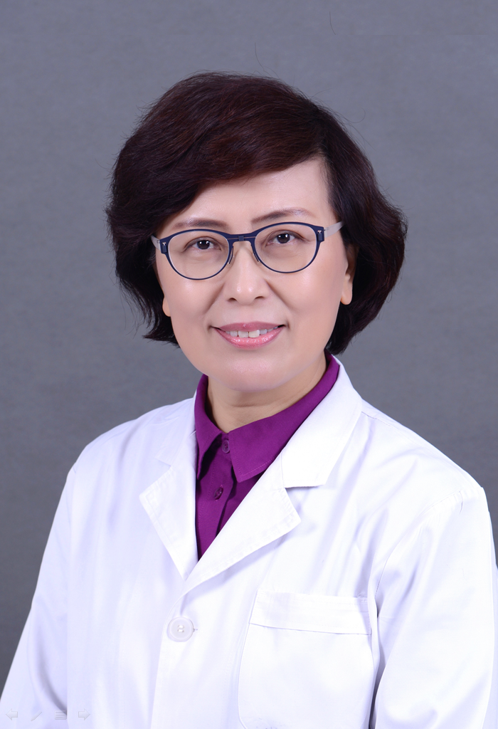 Shuyang Zhang (张抒扬)合作论文数Peking Union Medical College Hospital;Chinese Academy of Medical Sciences & Peking Union Medical College2Xin Tong合作论文数2Jie Ma合作论文数2Tian Yu (田禹)合作论文数2Fang Huang (黄芳)合作论文数2Qiyu Zhang合作论文数2Yiheng Mao合作论文数2Weina Gao合作论文数Southern Univ Sci & Technol, Dept Chem, Shenzhen 518055, Peoples R China2Yujie Wang合作论文数2An He合作论文数Southern Univ Sci & Technol, Dept Chem, Shenzhen 518055, Peoples R China2Rong LIANG (梁蓉)合作论文数2Xi Liu (喜六)合作论文数2Yue Liang (梁悦)合作论文数2Qian Wang (王茜)合作论文数2
 Zhengli Shi (石正丽)合作论文数Guangzhou Laboratory2
 Fei Deng (邓菲)合作论文数Wuhan Institute of Virology, Chinese Academy of Sciences2
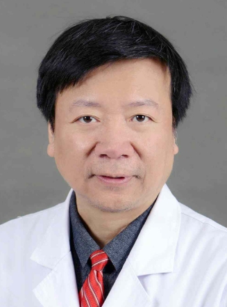 Zhihong Wu (吴志宏)合作论文数Peking Union Medical College Hospital2
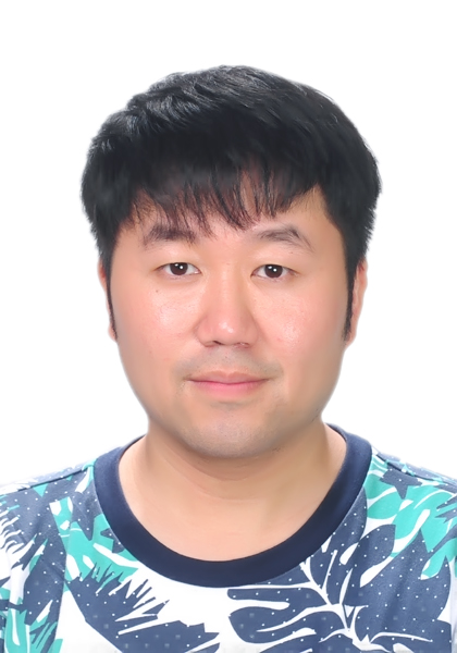 Ben Hu (胡犇)合作论文数Wuhan Institute of Virology, Chinese Academy of Sciences2
 Peng Zhou (周鹏)合作论文数Guangzhou Laboratory2
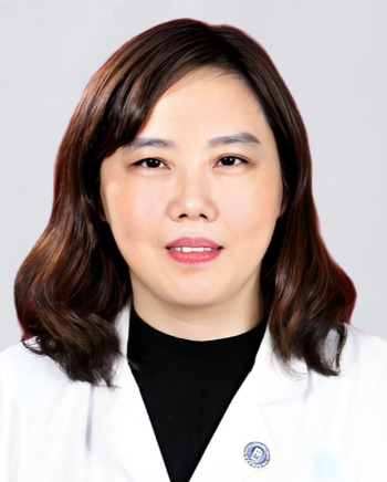 Lian Yang (杨炼)合作论文数Union Hospital, Tongji Medical College, Huazhong University of Science and Technology2
 Ling Leng (冷泠)合作论文数Peking Union Medical College Hospital2
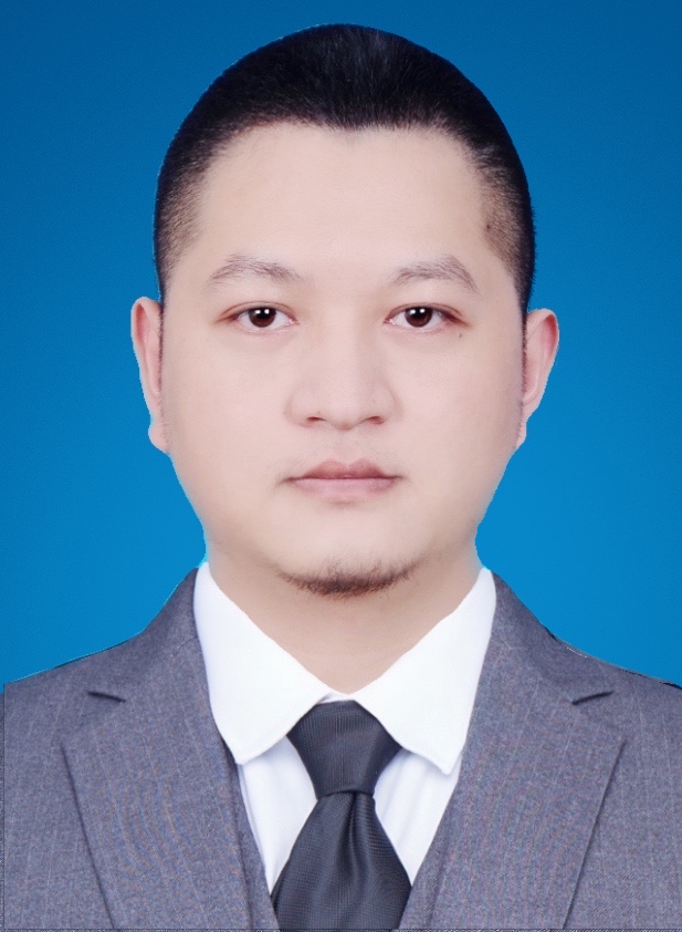 Yuan Li (李源)合作论文数School of Basic Medical Sciences, Southern Medical University2
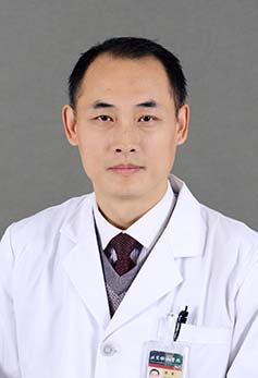 Jun Li (李军)合作论文数Beijing Union Medical College Hospital2




