Juvenile mantises can jump towards targets by rapidly extending their middle and hind legs. Here, we investigate how mantises can perform jumps from smooth surfaces such as those found on many plants. Stagmomantis theophila mantises possess two distinct types of attachment pads on each foot: three small proximal euplantulae (“heel pads”) with microscopic cuticular ridges and one smooth large distal pair of euplantulae (“toe pad”). Microscopy showed that the surface contact of heel pads is strongly load-dependent; at low normal forces, they make only partial surface contact due to the ridges, but at higher loads they switch to larger areas in full contact. By analysing the kinematics of 64 jumps of 23 third-instar nymphs from glass surfaces and the foot contact areas of their accelerating legs, we show that heel and toe pads fulfil distinct roles. During the acceleration phase of jumps, the contact area of the hind legs’ heel pads tripled, while that of the toe pad decreased strongly, and the toe pad sometimes detached completely before take-off. Although the middle legs also contribute to the jump, they showed a less consistent pattern; the contact areas of their heel and toe pads remained largely unchanged during acceleration. Our findings show that jumping mantises accelerate mainly by pushing with their hind legs and produce grip on smooth surfaces primarily with the heel pads on their proximal tarsus.
Resilin, an elastomeric protein with remarkable physical properties that outperforms synthetic rubbers, is a near-ubiquitous feature of the power amplification mechanisms used by jumping insects. Catapult-like mechanisms, which incorporate elastic energy stores formed from a composite of stiff cuticle and resilin, are frequently used by insects to translate slow muscle contractions into rapid-release recoil movements. The precise role of resilin in these jumping mechanisms remains unclear, however. We used RNAi to reduce resilin deposition in the principal energy-storing springs of the desert locust ( Schistocerca gregaria ) before measuring jumping performance. Knockdown reduced the amount of resilin-associated fluorescence in the semilunar processes (SLPs) by 44% and reduced the cross-sectional area of the tendons of the hind leg extensor-tibiae muscle by 31%. This affected jumping in three ways: First, take-off velocity was reduced by 15% in knockdown animals, which could be explained by a change in the extrinsic stiffness of the extensor-tibiae tendon caused by the decrease in its cross-sectional area. Second, knockdown resulted in permanent breakages in the hind legs of 29% of knockdown locusts as tested by electrical stimulation of the extensor muscle, but none in controls. Third, knockdown locusts exhibited a greater decline in distance jumped when made to jump in rapid succession than did controls. We conclude that stiff cuticle acts as the principal elastic energy store for insect jumping, while resilin protects these more brittle structures against breakage from repeated use.
To understand brain function it is necessary to characterize both the underlying structural connectivity between neurons and the physiological integrity of these connections. Previous research exploring insect brain connectivity has typically used electron microscopy techniques, but this methodology cannot be applied to living animals and so cannot be used to understand dynamic physiological processes. The relatively large brain of the desert locust, Schistercera gregaria (Forksȧl) is ideal for exploring a novel methodology; micro diffusion magnetic resonance imaging (micro-dMRI) for the characterization of neuronal connectivity in an insect brain. The diffusion-weighted imaging (DWI) data were acquired on a preclinical system using a customised multi-shell diffusion MRI scheme optimized to image the locust brain. Endogenous imaging contrasts from the averaged DWIs and Diffusion Kurtosis Imaging (DKI) scheme were applied to classify various anatomical features and diffusion patterns in neuropils, respectively. The application of micro-dMRI modelling to the locust brain provides a novel means of identifying anatomical regions and inferring connectivity of large tracts in an insect brain. Furthermore, quantitative imaging indices derived from the kurtosis model that include fractional anisotropy (FA), mean diffusivity (MD) and kurtosis anisotropy (KA) can be extracted. These metrics could, in future, be used to quantify longitudinal structural changes in the nervous system of the locust brain that occur due to environmental stressors or ageing.
ABSTRACT Lantern bugs are amongst the largest of the jumping hemipteran bugs, with body lengths reaching 44 mm and masses reaching 0.7 g. They are up to 600 times heavier than smaller hemipterans that jump powerfully using catapult mechanisms to store energy. Does a similar mechanism also propel jumping in these much larger insects? The jumping performance of two species of lantern bugs (Hemiptera, Auchenorrhyncha, family Fulgoridae) from India and Malaysia was therefore analysed from high-speed videos. The kinematics showed that jumps were propelled by rapid and synchronous movements of both hind legs, with their trochantera moving first. The hind legs were 20–40% longer than the front legs, which was attributable to longer tibiae. It took 5–6 ms to accelerate to take-off velocities reaching 4.65 m s−1 in the best jumps by female Kalidasa lanata. During these jumps, adults experienced an acceleration of 77 g, required an energy expenditure of 4800 μJ and a power output of 900 mW, and exerted a force of 400 mN. The required power output of the thoracic jumping muscles was 21,000 W kg−1, 40 times greater than the maximum active contractile limit of muscle. Such a jumping performance therefore required a power amplification mechanism with energy storage in advance of the movement, as in their smaller relatives. These large lantern bugs are near isometrically scaled-up versions of their smaller relatives, still achieve comparable, if not higher, take-off velocities, and outperform other large jumping insects such as grasshoppers.
The volume of the hind femora in the adult male flower beetle Oedemera nobilis is 38 times greater than in adult females. To determine what advantage limbs with swollen femora might provide, the behaviour of these insects was analysed with high-speed videography. First, because large hind legs are often associated with jumping and take-off, the performance of this behaviour by the two sexes was determined. Take-off was generated by a series of small-amplitude wing beats followed by larger ones, with the hind legs contributing little or no propulsion. The mean acceleration time to take-off was not significantly different in males (46.2 ms) and females (45.5 ms), but the mean take-off velocity of males was 10% higher than in females. Second, to determine if enlarged hind legs were critical in specifically male behaviour, interactions between males and females, and between males were videoed. The male mounted a female and then encircled her abdomen between the enlarged femora and tibiae of both his hind legs. The joint between these leg parts acted like a mole wrench (vice grip) so that when the tibia was fully flexed, a triangular space of 0.3 mm2 remained, in which a female abdomen (cross-sectional area 0.9 mm2) could be compressed and restrained firmly without inflicting damage. The flexor tibiae muscle in a male hind femur was 5.9 times larger than the extensor. In interactions between males, attempts to achieve a similar entrapment were frequently thwarted by the pursued male extending his hind legs vertically.
Analysis of the kinematics of take-off in the planthopper Proutista moesta (Hemiptera, Fulgoroidea, family Derbidae) from high-speed videos showed that these insects used two distinct mechanisms involving different appendages. The first was a fast take-off (55.7% of 106 take-offs by 11 insects) propelled by a synchronised movement of the two hind legs and without participation of the wings. The body was accelerated in 1 ms or less to a mean take-off velocity of 1.7 m s(-1) while experiencing average forces of more than 150 times gravity. The power required from the leg muscles implicated a power-amplification mechanism. Such take-offs propelled the insect along its trajectory a mean distance of 7.9 mm in the first 5 ms after take-off. The second and slower take-off mechanism (44.3% of take-offs) was powered by beating movements of the wings alone, with no discernible contribution from the hind legs. The resulting mean acceleration time was 16 times slower at 17.3 ms, the mean final velocity was six times lower at 0.27 m s(-1), the g forces experienced were 80 times lower and the distance moved in 5 ms after take-off was 7 times shorter. The power requirements could be readily met by direct muscle contraction. The results suggest a testable hypothesis that the two mechanisms serve distinct behavioural actions: the fast take-offs could enable escape from predators and the slow take-offs that exert much lower ground reaction forces could enable take-off from more flexible substrates while also displacing the insect in a slower and more controllable trajectory.
This paper analyses with high speed videos if and how adults of a winged species of scorpion fly (Mecoptera, Panorpa communis) jump and determines whether they use the same mechanism as that of the only other mecopteran known to jump, the wingless snow flea, Boreus hyemalis. Adult females are longer and heavier than males and have longer legs but which are of the same relative proportions. The middle legs are 20% longer and the hind legs 60% longer than the front legs. A jump starts with the middle and hind legs in variable positions, but together by depressing their coxo-trochanteral and extending their femoro-tibial joints they accelerate the body in 16-19 ms to mean take-off velocities of 0.7 - 0.8 m s−1; performances in males and females were not significantly different. Depression of the wings accompanies these leg movements, but clipping them does not affect jump performance. Smooth transition to flapping flight occurs once airborne with little loss of energy to body rotation. 90 % of the jumps analysed occurred without an observable stimulus; the remaining 10 % were in response to a mechanical touch. The performance of these jumps was not significantly different. In its fastest jumps a scorpion fly experiences an acceleration of 10 g, expends 23 µJ of energy and requires a power output less than 250 W kg−1 of muscle that can be met by direct muscle contractions without invoking an indirect power amplification mechanism. The jumping mechanism is like that of snow fleas.
The realisation has been rather slow in coming that the mechanics of an animal’s body — its skeleton, muscles and tendons — play a huge role in shaping and defining locomotion. The nervous system has previously received major attention and subsequent accolades for its role in processing sensory information, assessing the surrounding world and then generating motor patterns appropriate to the prevailing context. Combining an understanding of the biomechanical and neural factors is more likely to result in better holistic explanations of how animals move and interact with each other and their environment. David Hu’s book attempts to do two admirable things without quite making up its mind about its target audience. First, it seeks to describe and explain a range of movements by many different kinds of animals — with very different body forms and modes of locomotion — that depend on their physical attributes for their performance. All of these movements operate at the boundaries of our understanding of the natural world. Second, it endeavours to reach out to and enthuse a wide audience by explaining these issues in a comprehensive way. The book also integrates robotics into its narrative. Attempts by robots to close a drawer smoothly or maintain their balance during and after a jump over an object point to the magnitude of the problems that animals have solved and what they potentially can teach us about our designs for machines. Our experimental approach always carries the danger of anthropogenic influences no matter how hard we try to suppress them; such expectations that animals would do things in the same way as us if faced with similar challenges are frequently wrong. We are then surprised when we carry out detailed analyses of movements to find that our suppositions about mechanisms prove so often to be wide of the mark. Retaining these assumptions also increases the likelihood of failing to reveal the true, underlying principles of the real mechanisms. Who would have suspected that some insects would use gears to synchronise their propulsive legs precisely when jumping? At the same time, however, we seem more ready to believe that, if a flea were scaled to the size of a human, it would be able to jump over the Eiffel Tower in Paris. This is because we do not readily intuit the consequences of physical scaling. We might as well enter the world of nursery rhymes and consider the notion of a cow (the resulting approximate mass that linearly extrapolating the size of a flea to a human would give) jumping over a tall building, let alone the moon. True understanding only comes from entering the world of careful observation and seeing the interactions of animals with their surroundings. This also requires knowledge of the physical world together with the consequences that arise from the size, shape and body design of animals. The movements that are described in this book prove just how wrong some assumptions are and how illuminating careful observations and clear experiments can be. An eclectic range of animal movements and their interactions with the natural world is covered. These include insects walking on water, snakes gliding from treetops, lizards and snakes squirming through sand, worms burrowing in mud, mosquitoes flying in the rain, cockroaches running in dark, compressed spaces and ants building scaffolds by linking themselves together. Other topics include the functional role of eyelashes (through which I learnt about Elizabeth Taylor’s two rows of eyelashes), the structure of shark skin and a confusing description of how the nervous system generates the motor patterns underlying rhythmic movements. Surprisingly, however, I could find nothing on climbing up walls despite the promises of the book’s title, which had first attracted me to the book. I suspect that a common expectation of general readers who pick up this book would be to learn how geckos and flies manage to walk on the walls and ceilings of their houses. Each chapter is enticingly titled but often morphs into something rather different. ‘Flying in the rain’ moves into squashing cockroaches into tight spaces and ‘the shape of a flying snake’ starts with the shapes of bladders and urethras and changes to pulsating jellyfish before arriving at snakes leaping from trees: only then is a unifying link made between them. The subjects are treated more as a narrative that follows the research path of the author and his associates. For example, the discussion of moving on water does not consider the different ways that insects solve this problem, and the remarkable example of Jesus lizards is brushed aside in just a few words in the introduction. Each new behaviour has a long introduction with too much information about the people involved, and this then puts constraints on the space that is available for the more interesting explanations of the movements. The challenge of writing for a wide audience has prompted a pervasive use of analogies: always problematic devices that can draw attention away from a focus on the direct issue at hand. Many of these analogies to events in our everyday lives are unhelpful, rely heavily on a particular, cultural background that not all readers will share or just complicate rather than simplify. Comparing the body shapes of lizards to a loaf of Italian bread (p. 49), a cockroach to a motorised water stress ball (p. 150), the tail fin of a shark to a “hockey stick” or the orientation of its scales as having “a grain similar to a cat’s fur” (p. 100) all fail to enhance our understanding of these features. Similarly, extrapolations to our own experiences can also be unhelpful. How many of us fit “someone who crashes their car into a tree or lamppost” or feels “a million snowflakes falling at once” and then still cares to think about animal locomotion? Why extrapolate and scale to cars when considering the speed of running in a cockroach — “it can run at 25 body lengths per second, or the equivalent of a car traveling at 280 mph” (p. 148 and repeated on pp. 162–163) — given all the difficulties and misinterpretations that inappropriate scaling comparisons can introduce? By my reckoning, the cockroach is simply travelling at about 1 m/s (2 mph). The author could have been better served by his publisher. The presentation is not attractive; the text could so easily have been made more readable by the use of subheadings. The figures are disappointingly reproduced: many have no calibrations and legends frequently fail to explain adequately what is being illustrated. Appropriate copy editing could have picked up many small mistakes — 9 m/s is 20 mph not 30 mph (p. 81) — and corrected the more ambiguous sentences, e.g. “when you go see a doctor with urinary problems…” (p. 205), or ugly sentences: “the fluid around the object can be discretized into a number of layers” (p. 98). Nevertheless, the author presents a thoroughly intriguing approach to understanding the movement of animals that promises to deliver much more through future expositions that are similar to this one. The final conclusions are encouraging, insightful and reassuring; I take some solace in knowing that I am not alone in fretting about how to collect, analyse and store massive data sets and then communicate them with others.
There was an error published in J. Exp. Biol. (2017) 220, 3812-3825 (doi: 10.1242/jeb.161463).Two of the wasp species analysed were misidentified: Amblyteles armatorius should be Ichneumon xanthorius and Netelia testacea should be Ophion sp.The authors apologise for any inconvenience this may have caused.
A quick guide to the springs used by insects to achieve remarkable feats of jumping.
The order Hemiptera includes jumping insects with the fastest takeoff velocities, all generated by catapult mechanisms. It also contains the large family Miridae or plant bugs. Here, we analysed the jumping strategies and mechanisms of six mirid species from high-speed videos and from the anatomy of their propulsive legs, and conclude that they use a different mechanism in which jumps are powered by the direct contractions of muscles. Three strategies were identified. First, jumping was propelled only by movements of the middle and hind legs, which were, respectively, 140% and 190% longer than the front legs. In three species with masses ranging from 3.4 to 12.2 mg, depression of the coxo-trochanteral and extension of femoro-tibial joints accelerated the body in 8-17 ms to take-off velocities of 0.5-0.8 m s(-1). The middle legs lost ground contact 5-6 ms before take-off so that the hind legs generated the final propulsion. The power requirements could be met by the direct muscle contractions so that catapult mechanisms were not implicated. Second, other species combined the same leg movements with wing beating to generate take-off during a wing downstroke. Third, up to four wingbeat cycles preceded take-off and were not assisted by leg movements. Take-off velocities were reduced and acceleration times lengthened. Other species from the same habitat did not jump. The lower take-off velocities achieved by powering jumping by direct muscle contractions may be offset by eliminating the time taken to load catapult mechanisms.
Many hemipteran bugs can jump explosively from plant substrates, which can be very smooth. We therefore analysed the jumping performance of froghoppers (Philaenus spumarius,Aphrophoridae) and leafhoppers (Aphrodes bicinctus/makarovi,Cicadellidae) taking off from smooth (glass) and rough (sandpaper, 30 µm asperity size) surfaces. On glass, the propulsive hind legs ofPhilaenusfroghoppers slipped, resulting in uncontrolled jumps with a fast forward spin, a steeper angle and only a quarter of the velocity compared with jumps from rough surfaces. By contrast,Aphrodesleafhoppers took off without their propulsive hind legs slipping, and reached low take-off angles and high velocities on both substrates. This difference in jumping ability from smooth surfaces can be explained not only by the lower acceleration of the long-legged leafhoppers, but also by the presence of 2–9 soft pad-like structures (platellae) on their hind tarsi, which are absent in froghoppers. High-speed videos of jumping showed that platellae contact the surface briefly (approx. 3 ms) during the acceleration phase. Friction force measurements on individual hind tarsi on glass revealed that at low sliding speeds, both pushing and pulling forces were small, and insufficient to explain the recorded jumps. Only when the tarsi were pushed with higher velocities did the contact area of the platellae increase markedly, and high friction forces were produced, consistent with the observed jumps. Our findings show that leafhoppers have special adhesive footpads for jumping from smooth surfaces, which achieve firm grip and rapid control of attachment/detachment by combining anisotropic friction with velocity dependence.
Jumping in planthopper and froghopper insects is propelled by a catapult-like mechanism requiring mechanical storage of energy and its quick release to accelerate the hind legs rapidly. To understand the functional biomechanics involved in these challenging movements, the internal skeleton, tendons and muscles involved were reconstructed in 3-D from confocal scans in unprecedented detail. Energy to power jumping was generated by slow contractions of hind leg depressor muscles and then stored by bending specialised elements of the thoracic skeleton that are composites of the rubbery protein resilin sandwiched between layers of harder cuticle with air-filled tunnels reducing mass. The images showed that the lever arm of the power-producing muscle changed in magnitude during jumping, but at all joint angles would cause depression, suggesting a mechanism by which the stored energy is released. This methodological approach illuminates how miniaturized components interact and function in complex and rapid movements of small animals.
High-speed video analyses of the natural behaviour of parasitoid wasps revealed three strategies used to launch the insects into the air. Which strategy is the most energy efficient? In Pteromalus puparum, 92% of take-offs by were propelled entirely by movements of the middle and hind legs, which were depressed at their coxotrochanteral and extended at their femoro-tibial joints. The front legs left the ground first, followed by the hind legs, so that the middle legs provided the final propulsion. Second, in other species of a similar mass, Cotesia glomerata and Leptopilina boulardi, all take-offs were propelled by a mean of 2.8 and 3.8 wingbeats, respectively, with little or no contribution from the legs. The first strategy resulted in take-off times that were four times shorter (5 versus 22.8 ms) and take-off velocities that were four times faster (0.8 versus 0.2 m s(-1)). Calculations from the kinematics indicate that propulsion by the legs was the most energy-efficient strategy, because more energy is put into propulsion of the body, whereas in take-off propelled by repetitive wing movements energy is lost to generating these movements and moving the air. In heavier species such as Netelia testacea and Amblyteles armatorius, take-off was propelled by the combined movements of the middle and hind legs and wingbeats. In A. armatorius, this resulted in the longest mean take-off time of 33.8 ms but an intermediate take-off velocity of 0.4 m s(-1). In all three strategies the performance could be explained without invoking energy storage and power amplification mechanisms.
Locusts jump by using a catapult mechanism in which energy produced by slow contractions of the extensor tibiae muscles of the hind legs is stored in distortions of the exoskeleton, most notably (1) the two semi-lunar processes at each knee joint and (2) the tendons of the extensor muscles themselves. The energy is then suddenly released from these stores to power the rapid, propulsive movements of the hind legs. The reliance on the mechanical storage of energy is likely to impact on jumping because growth occurs by a series of five moults, at each of which the exoskeleton is replaced by a new one. All developmental stages (instars) nevertheless jump as a means of forward locomotion, or as an escape movement. Here, I show that in each instar, resilin is added to the semi-lunar processes and to the core of the extensor tendons so that their thickness increases. As the next moult approaches, a new exoskeleton forms within the old one, with resilin already present in the new semi-lunar processes. The old exoskeleton, the tendons and their resilin are discarded at moulting. The resilin of the semi-lunar processes and tendons of the new instar is initially thin, but a similar pattern of deposition results in an increase of their thickness. In adults, resilin continues to be deposited so that at 4 weeks old the thickness in the semi-lunar processes has increased fourfold. These changes in the energy stores accompany changes in jumping ability and performance during each moulting cycle.
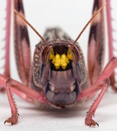
Many insects such as fleas, froghoppers and grasshoppers use a catapult mechanism to jump and a direct consequence of this is that their take-off velocities are independent of their mass. In contrast, insects such as mantises, caddis flies and bush crickets propel their jumps by direct muscle contractions. What constrains the jumping performance of insects that use this second mechanism? To answer this question, the jumping performance of the mantis, Stagmomantis theophila, was measured through all its developmental stages, from 5 mg first instar nymphs to 1200 mg adults. Older and heavier mantises have longer hind and middle legs and higher take-off velocities than younger and lighter ones. The length of the propulsive hind and middle legs scaled approximately isometrically with body mass (exponent, 0.29 and 0.32 respectively). The front legs, which do not contribute to propulsion, scaled with an exponent of 0.37. Take-off velocity increased with increasing body mass (exponent, 0.12). Time to accelerate increased and maximum acceleration decreased but the measured power that a given mass of jumping muscle produced remained constant throughout all stages. Mathematical models were used to distinguish between three possible limitations to the scaling relationships; first, an energy-limited model (which explains catapult jumpers); second, a power-limited model; third, an acceleration-limited model. Only the model limited by muscle power explained the experimental data. Therefore, the two biomechanical mechanisms impose different limitations on jumping; those involving direct muscle contractions (mantises) are constrained by muscle power, catapult mechanisms by muscle energy.
The desert locust, Schistocerca gregaria, shows a strong phenotypic plasticity. It can develop, depending upon population density, into either a solitarious or gregarious phase that differs in many aspects of behaviour, physiology and morphology. Prominent amongst these differences is that solitarious locusts have proportionately longer hind femora than gregarious locusts. The hind femora contain the muscles and energy-storing cuticular structures that propel powerful jumps using a catapult-like mechanism. We show that solitarious locusts jump on average 23% faster and 27% further than gregarious locusts, and attribute this improved performance to three sources: first, a 17.5% increase in the relative volume of their hind femur, and hence muscle volume; second, a 24.3% decrease in the stiffness of the energy-storing semi-lunar processes of the distal femur; and third, a 4.5% decrease in the stiffness of the tendon of the extensor tibiae muscle. These differences mean that solitarious locusts can generate more power and store more energy in preparation for a jump than can gregarious locusts. This improved performance comes at a cost: solitarious locusts expend nearly twice the energy of gregarious locusts during a single jump and the muscular co-contraction that energises the cuticular springs takes twice as long. There is thus a trade-off between achieving maximum jump velocity in the solitarious phase against the ability to engage jumping rapidly and repeatedly in the gregarious phase.
 Gregory Sutton合作论文数School of Biological Sciences,University of Bristol20
 Steve Rogers合作论文数Department of Zoology, University of Cambridge12
 M. V. S. Siegler合作论文数12
 Swidbert R Ott合作论文数Department of Zoology, University of Cambridge11
 Gilles Laurent合作论文数Department of Neural Systems and Coding, Max Planck Institute for Brain Research10AHD WATSON合作论文数9
 Hans-Joachim Pflüger合作论文数Institut für Biologie, Neurobiologie, Fachbereich Biologie, Chemie, Pharmazie, Freien Universität Berlin9Thomas Matheson合作论文数7Dorosenko Marina合作论文数7
 Adrian Horridge合作论文数7
 Cullen Darron A合作论文数School of Biological Sciences, The University of Sydney6G Hoyle合作论文数5Peter Bräunig合作论文数5Watkins B L合作论文数4Dorosenko M合作论文数4Graham Hoyle合作论文数4
 Philip L Newland合作论文数4
 Stephen Simpson合作论文数Charles Perkins Centre, The University of Sydney;School of Life and Environmental Sciences, The University of Sydney3Maurice R. Elphick合作论文数3
 Jeremy Niven合作论文数Sch Life Sci, Univ Sussex3
 Alan Hugh Watson合作论文数3
 Walter Federle合作论文数Department of Zoology, University of Cambridge;Jesus College, University of Cambridge3Michael L Anstey合作论文数2
 Tom Matheson合作论文数2De Brunn合作论文数2
 Abin Ghosh合作论文数2Lh Field合作论文数2Hanns Hagen Goetzke合作论文数Univ Cambridge, Dept Zool, Downing St, Cambridge CB2 3EJ, England2Harald Wolf合作论文数Inst Neurobiol, Ulm Univ2Yeshwanth H. M.合作论文数2Hale J P合作论文数2
 Ken Sasaki合作论文数2W. J. Heitler合作论文数2Stephen R. Shaw合作论文数2
 Ian A. Meinertzhagen合作论文数Department of Psychology and Neuroscience, Faculty of Science, Dalhousie University2
 Emma Despland合作论文数Biology Department, Concordia University2
 Holger G. Krapp合作论文数Department of Bioengineering, Imperial College London2Christopher M. Graham合作论文数2Patricia Mcneill合作论文数2S. P. Sane合作论文数1
 Tim Dodgson合作论文数1James M R Bullock合作论文数1Joanna Riley合作论文数1Laura Blackburn合作论文数Department of Zoology, University of Cambridge, U.K.1
 Fabrizio Gabbiani合作论文数Department of Neuroscience, Baylor College of Medicine1
 David Labonte合作论文数Department of Bioengineering, Faculty of Engineering, Imperial College London1
 Christofer Clemente合作论文数School of Science, Technology and Engineering, University of the Sunshine Coast1Caroline H. Brighton合作论文数1Bayley T G合作论文数1




