As a first step towards an acoustic localisation device for coronary stenosis to provide a non-invasive means of diagnosing arterial disease, measurements are reported for an agar-based tissue mimicking material (TMM) of the shear wave propagation velocity, attenuation and viscoelastic constants, together with one dimensional quasi-static elastic moduli and Poisson's ratio. Phase velocity and attenuation coefficients, determined by generating and detecting shear waves piezo-electrically in the range 300 Hz-2 kHz, were 3.2-7.5 ms(-1) and 320 dBm(-1). Quasi-static Young's modulus, shear modulus and Poisson's ratio, obtained by compressive or shear loading of cylindrical specimens were 150-160 kPa; 54-56 kPa and 0.37-0.44. The dynamic Young's and shear moduli, derived from fitting viscoelastic internal variables by an iterative statistical inverse solver to freely oscillating specimens were 230 and 33 kPa and the corresponding relaxation times, 0.046 and 0.036 s. The results were self-consistent, repeatable and provide baseline data required for the computational modelling of wave propagation in a phantom.
Heightened perception of gastrointestinal sensation is termed visceral hypersensitivity (VH) and is commonly observed in patients with gastrointestinal disorders. VH is thought to be a major contributory factor in oesophageal disease, particularly gastro-oesophageal reflux disease that does not respond to standard (proton pump inhibitor) treatment, and in functional heartburn. Clinical tools that can help phenotype according to the mechanism of chronic pain and thus allow targeted drug treatment (e. g. with pain modulator therapy) would be very desirable. A technique that produces repeatable and controllable thermal stimuli within the oesophagus could meet this need. The aims of this study were to develop a method for linear control of the heat stimulation in the oesophagus, to assess the reproducibility of this method, and obtain normal thermal sensitivity values in the distal and proximal oesophagus. The 7 mm diameter Peltier-based thermal device was investigated on 27 healthy subjects using a heating ramp of 0.2 degrees C s(-1). The pain detection threshold (PDT) temperature was recorded. To assess the reproducibility of the device, each subject underwent the procedure twice, with a minimum of two weeks between each procedure. The mean PDT temperature measured in the distal oesophagus, was 53.8 +/- 2.9 degrees C and 53.6 +/- 2.6 degrees C, for visits 1 and 2 respectively. The mean PDT temperature measured in the proximal oesophagus was 54.1 +/- 2.4 degrees C and 54.0 +/- 2.8 degrees C, for visits 1 and 2 respectively. The reproducibility of the PDT temperature in the distal and proximal oesophagus, was good (intraclass correlation >0.6). Future studies should be aimed to determine whether
•Composite speed was measured in carotid plaque embedded in a tissue mimicking material (TMM).•Individual speed values for five components were derived; TMM, elastin, fibrous/collagen, calcification and lipid.•The fibrous tissue showed a mean value of 1584ms−1 at 37°C.•The calcification had higher sound speed than the other plaque components at 1760–2000ms−1.
Introduction Investigation of oesophageal sensitivity might help better improve phenotyping of patients with GERD, functional heartburn and non-obstructive dysphagia. Studies on oesophageal thermal sensitivity have used balloon techniques with circulating water. This lacks linear control of heating, has relative heat loss, and provokes mechanical stimulation by the distended balloon. Oesophageal mucosal integrity is important in symptoms perception and can be studied using impedance “in vivo”. We developed a catheter device incorporating a “thermal stimulator” using Peltier heating technology and impedance electrodes to assess mucosal contact and integrity. We aimed to investigate oesophageal heat sensation/pain thresholds and their relationship with mucosal integrity in healthy human subjects. Methods 21 healthy volunteers underwent thermal stimulation using an oesophageal catheter with a 7 mm electronic Peltier heater, to deliver a ramp heating protocol up to 60°C at 5 and 15 cm above LOS. Symptoms were recorded by computerised Visual Analogue Score (VAS). The delivered temperature at Pain Detection Threshold (PDT), time to PDT was measured and the area under the heating curve (AUC) was calculated. Oesophageal impedance was assessed over 10 minutes pre (basal), during and post heating in the distal and proximal oesophagus. The protocol was repeated after 2 weeks to assess reproducibility. Results Temperature at PDT showed a very low SD in the distal and proximal oesophagus and Bland-Altman Tests showed good reproducibility (–0.1905 95% CI –1.4132 to 1.0327). There were no significant differences in thermal sensitivity parameters between distal and proximal oesophagus. Due to the low inter-individual variability in thermal sensitivity between healthy subjects, we could not find a significant correlation between impedance and perception parameters in the proximal or distal oesophagus. Conclusion A novel Peltier-based thermal stimulator device can accurately and reproducibly determine oesophageal thermal sensitivity. In healthy subjects we could not demonstrate a correlation between basal impedance and sensitivity to heating. Current experiments are assessing this correlation in NERD patients with hypersensitivity to acid. Disclosure of Interest None Declared
Flow in the wake of a coronary artery stenosis induces a bruit in the 300–1500 Hz range that can be heard at the chest wall. It has been hypothesised that this sound is caused by turbulence-induced shear waves which travel through the soft tissue of the thorax. This contribution describes a computational mathematical ‘forward solve’ method to simulate these shear waves in a virtual chest of tissue mimicking agarose gel. As the first stage in the development of a noninvasive diagnostic tool we also describe initial results towards the solution of the mathematical inverse problem. That is: to identify the source of the bruit given the surface measured signal. Objectives To demonstrate proof-of-concept of a novel biotechnology that will use mathematical simulations to provide a non-invasive screening tool for coronary artery disease. Methods Finite element based forward solvers for soft tissue response (given the source, generate the signal); optimisation-based inverse solver (given the signal, determine the source). Results For a simple, small scale, and axisymmetric cylindrical gel configuration, and for a source at 500 Hz, the forward solve generates signals that agree with experimental data (using Kelvin-Voigt viscoelasticity). Also, with surface signals generated by simulated sources in this virtual environment the inverse algorithm is able to identify this source given only chest surface measurements, and an adequate initial datum from which to start the computation. Conclusions While enormous challenges remain we have shown that this approach offers considerable promise in delivering a noninvasive diagnostic or screening tool.
Background: Turbulent flow downstream of atherosclerotic plaques produces low amplitude shear waves which travel through the chest and can be measured by skin sensors. This acoustic signature may provide a cheap non-invasive way to diagnose arterial disease. We report measurements of shearing oscillations and flow-induced turbulence in soft tissue-mimicking gels which provide input to a numerical model of soft tissue behaviour described in a companion presentation. Methods: Cylindrical specimens of 3% agarose gel were cast around an axial rod and bead connected to an electromechanical vibrator (figure 1), to generate shear-waves of known characteristics and location (frequency 250–750 Hz, amplitude 10–50 μm). Displacement was mapped optically by tracking the movement of carborundum particles on the surface. In the flow study (figure 2) a silicone rubber tube (i.d. 4.5mm) containing a stenosis was embedded in a cuboidal gel phantom and lateral displacement of the gel surface was mapped by a piezo-electric accelerometer. Results: Forced oscillations produced movement in the same direction at the gel surface, amplitude 10–50% of the bead’s movement. Amplitude modulation (≈5%) at around 40Hz, probably due to resonance in the gel, was also seen. Lateral movement (200–800Hz) of the gel surface caused by flow-induced turbulence increased monotonically with turbulence magnitude. Conclusions: The methods described above provide internally consistent and repeatable data, validating the numerical models. The next steps will compare computational results with measurements in progressively more realistic representations of the chest aiming ultimately to produce a device suitable for screening/diagnosis of coronary artery disease. Figure. 1. Forced vibration rig. Gels cast with bead in various positions. Laser measures bead movement; camera measures surface movement Figure 2. Steady flow rig. Measurements made at various flow rates, tube depths and stenosis severity. Accelerometer position varied.
Introduction Investigation of oesophageal sensitivity might help better improve phenotyping of patients with GERD, functional heartburn and non-obstructive dysphagia. Studies on oesophageal thermal sensitivity have used balloon techniques with circulating water. This lacks linear control of heating, has relative heat loss, and provokes mechanical stimulation by the distended balloon. Oesophageal mucosal integrity is important in symptoms perception and can be studied using impedance “in vivo”. We developed a catheter device incorporating a “thermal stimulator” using Peltier heating technology and impedance electrodes to assess mucosal contact and integrity. We aimed to investigate oesophageal heat sensation/pain thresholds and their relationship with mucosal integrity in healthy human subjects. Methods 21 healthy volunteers underwent thermal stimulation using an oesophageal catheter with a 7 mm electronic Peltier heater, to deliver a ramp heating protocol up to 60°C at 5 and 15 cm above LOS. Symptoms were recorded by computerised Visual Analogue Score (VAS). The delivered temperature at Pain Detection Threshold (PDT), time to PDT was measured and the area under the heating curve (AUC) was calculated. Oesophageal impedance was assessed over 10 minutes pre (basal), during and post heating in the distal and proximal oesophagus. The protocol was repeated after 2 weeks to assess reproducibility. Results Temperature at PDT showed a very low SD in the distal and proximal oesophagus and Bland-Altman Tests showed good reproducibility (–0.1905 95% CI –1.4132 to 1.0327). There were no significant differences in thermal sensitivity parameters between distal and proximal oesophagus. Due to the low inter-individual variability in thermal sensitivity between healthy subjects, we could not find a significant correlation between impedance and perception parameters in the proximal or distal oesophagus. Conclusion A novel Peltier-based thermal stimulator device can accurately and reproducibly determine oesophageal thermal sensitivity. In healthy subjects we could not demonstrate a correlation between basal impedance and sensitivity to heating. Current experiments are assessing this correlation in NERD patients with hypersensitivity to acid. Disclosure of Interest None Declared
Objective To use finite element analysis animated simulations to investigate factors affecting velopharyngeal closure. Design A coronal section multicomponent finite element analysis model of a human soft palate was created in Simulia Abaqus 6.5–1 from high resolution WIR I images of a single adult female subject, interpreted by reference to published anatomic dissections. Tissues were assigned hyperelastic property coefficients for neo-Hookean behavior, with gravity at 9.8 ms –2 in the y-axis. Vector forces based on estimations in previous publications were applied throughout levator veli palatini and palatopharyngeus muscles, using a nonlinear analysis algorithm, to produce animated simulations of velopharyngeal space closure. Variation of levator veli palatini angle from 60° to 49°, the contribution of palatopharyngeus muscle, and the effect of submucous cleft were investigated for their effects on velopharyngeal closure. Results The animated simulations showed anthropomorphic behavior and supported the previously suggested effects of the levator veli palatini angle, with reduced effectiveness of velopharyngeal closure as levator veli palatini angle decreases. Palatopharyngeus action reduced the efficiency of closure for a levator veli palatini angle of 60°, and a submucous cleft reduced this for both our normal subject and for a levator veli palatini angle of 60°, but both palatopharyngeus action and a submucous cleft enhanced closure for a levator veli palatini angle of 49°. Conclusions This study advances soft palate finite element analysis to a real-subject-based multicomponent hyperelastic model that demonstrates anthropomorphic behavior. Animated simulations using the model demonstrate the possible effects of levator veli palatini angle, a submucous cleft, and the contribution of the palatopharyngeus.
OBJECTIVE:The objective of this study was to demonstrate soft palate MRI at 1.5 and 3 T with high temporal resolution on clinical scanners.METHODS:Six volunteers were imaged while speaking, using both four real-time steady-state free-precession (SSFP) sequences at 3 T and four balanced SSFP (bSSFP) at 1.5 T. Temporal resolution was 9-20 frames s(-1) (fps), spatial resolution 1.6 × 1.6 × 10.0-2.7 × 2.7 × 10.0 mm(3). Simultaneous audio was recorded. Signal-to-noise ratio (SNR), palate thickness and image quality score (1-4, non-diagnostic-excellent) were evaluated.RESULTS:SNR was higher at 3 T than 1.5 T in the relaxed palate (nasal breathing position) and reduced in the elevated palate at 3 T, but not 1.5 T. Image quality was not significantly different between field strengths or sequences (p=NS). At 3 T, 40% acquisitions scored 2 and 56% scored 3. Most 1.5 T acquisitions scored 1 (19%) or 4 (46%). Image quality was more dependent on subject or field than sequence. SNR in static images was highest with 1.9 × 1.9 × 10.0 mm(3) resolution (10 fps) and measured palate thickness was similar (p=NS) to that at the highest resolution (1.6 × 1.6 × 10.0 mm(3)). SNR in intensity-time plots through the soft palate was highest with 2.7 × 2.7 × 10.0 mm(3) resolution (20 fps).CONCLUSIONS:At 3 T, SSFP images are of a reliable quality, but 1.5 T bSSFP images are often better. For geometric measurements, temporal should be traded for spatial resolution (1.9 × 1.9 × 10.0 mm(3), 10 fps). For assessment of motion, temporal should be prioritised over spatial resolution (2.7 × 2.7 × 10.0 mm(3), 20 fps). Advances in knowledge Diagnostic quality real-time soft palate MRI is possible using clinical scanners and optimised protocols have been developed. 3 T SSFP imaging is reliable, but 1.5 T bSSFP often produces better images.
Objective: To measure biomechanical properties of the human soft palate and the variation across anatomic regions. Design: Ex vivo analysis of human tissue.Patients/participants: Ten palates harvested from 10 normal adult human cadavers (age range, 37 to 90 years).Interventions: Computer-controlled uniaxial stress-relaxation mechanical properties tested in physiological saline at 37 degrees C.Main Outcome Measures: Measurement of Young modulus, Poisson ratio, and determination of viscoelastic constants C, tau(1), and tau(2) by curve-fitting of the reduced relaxation function to the data.Results: One hundred sections were tested from the 10 palates, representative of 10 anatomic zones. The mean Young modulus range was 585 Pa at the posterior free edge to 1409 Pa at regions of attachment. The mean Poisson ratio in the inferior-superior direction was 0.45 (SD 0.26) and in the lateral direction, was 0.30 (SD 0.21). The mean viscoelastic constants for 1-mm extensions were C = -0.1056 (+/- 0.1303), tau(1) = 11.0369 (+/- 9.1865) seconds, and tau(2) = 0.2128 (+/- 0.0792) seconds, and for 2-mm extensions were C = -0.1111 (+/- 0.1466), tau(1) = 14.3725 (+/- 5.2701) seconds, and tau(2) = 0.2094 (+/- 0.0544) seconds.Conclusions: The results show agreement with values of the Young modulus estimated by authors (Ettema and Kuehn, 1994; Berry et al., 1999) undertaking finite element modeling of the palate. However, other modulus measurements based on closing pressure are considerably different. The spatial distribution of viscoelastic parameters across the palate shows good consistency.
BACKGROUND:Stents are used abundantly to maintain ureteral patency. The majority are plastic tubes that adjust easily to upper urinary-tract motion. Recently, a coiled-wire lumenless stent was introduced (ZebraStent, Neo Medical, Munich, Germany) to facilitate expulsion of stone fragments after lithotripsy. Its metal core is composed of Nitinol, with the soft J ends being of titanium. The thin shape considerably increases the extraluminal space. The ZebraStent stretches the ureter and also provides a surface for the fragments to glide along. In our 18-month experience with the ZebraStent, two of them fractured along the shaft. We sought to learn whether this complication resulted from a defect in stent design or from material fatigue secondary to constant movement.MATERIALS AND METHODS:Our model is powered by an electric motor that produces a constant displacement similar to stent movements in vivo. The whole ZebraStent is embedded in a 37 degrees C waterbath to simulate physiological conditions within the ureter. We used an average displacement of 16 mm. The average frequency of ventilatory-cycle simulation was 20 times that in vivo, allowing us to collect data in a shorter time.RESULTS:All 10 stents broke within the proximal Nitinol shaft at the equivalent of 4 to 6 months (125-179 days).CONCLUSIONS:Our preliminary results show that all stents break after the equivalent of 4 or more months. The fact that this occurs in the homogenous proximal Nitinol shaft rather than at the welding point between the shaft and the titanium curl implies that breakage is secondary to material fatigue and not design error. Extensive testing is under way to confirm material fatigue as the cause of breakage. We hope to determine a safe dwelling time for these stents, which at the moment should not exceed 3 months.
The purpose of this study was to characterize the ultrasonic properties of agar-based tissue-mimicking materials (TMMs) at ultrasound frequencies centered around 20 MHz. The TMM acoustic properties measured are the amplitude attenuation coefficient a (dB cm(-1)MHz(-1)), the speed of sound (ms(-1)) and the backscattered power spectral density (distribution of power per unit frequency normalized to the total received power) characteristics of spectral slope (dB MHz(-1)), y-axis intercept (dB) and reflected power (dB). The acoustic properties are measured over a temperature range of 22 to 37 degrees C. An intercomparison of results between two independent ultrasound measurement laboratories is also presented. A longitudinal study of the acoustic properties over a period of two years is also detailed, and the effect of water immersion on the acoustic properties of TMM is measured. In addition, the physical parameters of mass density rho (kg m(-3)) and specific heat capacity C (J kg(-1) K-1) are included. The measurement techniques used were based on the substitution technique using both broadband and narrowband pulses centered on 20 MHz. Both the attenuation coefficient and speed of sound (both group and phase) showed good agreement with the expected values of 0.5 dB cm(-1) MHz(-1) and 1540 ms-1, respectively, with average values over the three-year period of 0.49 dBcm(-1) MHz(1) (SD +/- 0.05) and 1540.9 ms(-1) (SD +/- 8.7). These results also showed agreement between the two independent measurement laboratories. Speed of sound and attenuation coefficient were shown to change with temperature with rates of + 2.1 in s(-1) degrees C-1 and -0.005 dB cm(-1) MHz(-1) degrees C-1, respectively. Attenuation changed linearly with frequency at the high frequency range of 17 to 23 MHz, and speed of sound was found to be independent of frequency in this range. The spectral slope of relative backscattered power for the material increased with frequency at typically 1.5 dB MHz(-1). This compared favorably with theoretical spectral slope values, calculated for a variety of scatterer sizes, albeit at a lower frequency range. It is also noticed that, on extrapolation back to lower frequencies, the backscatter is comparable with that measured at 7 MHz. Overall, this non-commercial agar-based TMM is shown to perform as expected at the higher frequency range of 11 to 23 MHz and is seen to retain its acoustic properties of attenuation and speed of sound over a three-year period.
Purpose: Ureteral stenting plays a vital role in urology but still entails complications such as migration, encrustation, and blockage. We present our experience with a novel ureteral stent (Zebrastent) that had the complication of breakage which we explored by fatigue testing. Materials and Methods: We developed a device that can reproduce the movements of the Zebrastent in vitro (bending and cranio-caudal movements). We estimated the number of movements of the stent during 1 day and we thus calculated how much stress would result if it were in place for 9 months. Results: We estimate that the stent will not break when in place up to 9 months in a kidney with average curvature. However, the stent broke when it was used in a kidney with a sharper-than-normal curvature. Conclusions: The Zebrastent may be a valuable addition to the armamentarium of the endourologist, provided that the appropriate size is selected.
Miniature microwave helical antennas for use in thermal therapy applications are usually investigated using muscle-equivalent phantoms. In this paper, an alternative method using an electromagnetic solver based on the finite integration technique is used to simulate a range of 915 MHz helical antennas within a medium with the dielectric properties of muscle. By avoiding the stair-casing effect associated with many solvers, this method enables accurate simulations of non-orthogonal geometric objects such as helical antennas to be achieved. The effects of coil-spacing and insertion depth on the SAR distribution produced by the antennas were characterized and showed good agreement with previously published results obtained using a muscle phantom and a thermographic camera. The simulations confirm that the performance of helical antennas depends on insertion depth. Modification of the coil density demonstrated improvement of the return loss characteristics and changes to the resulting SAR profile.
PurposeTo built an experimental system that can create ultrasound velocity maps for human tissues.Methods and resultsWe describe a new experimental system and its use to examine ex vivo specimens of carotid atherosclerotic plaque.ConclusionThis new ultrasound system is promising but requires further testing and development in the laboratory.
Barrett's oesophagus is considered to increase the risk of cancer 30 fold. A set of helical microwave antennas was designed to investigate their potential use in the thermal therapy of Barrett's oesophagus. For treatment, a balloon filled with muscle-equivalent material encapsulates the antenna. The effects of insertion depth and coil-spacing on the thermal distribution produced by the antennas (20–35 mm) were characterized. The 35 mm helical antenna, with a coil-spacing of 3.6 mm resulted in uniform heating for an insertion depth of 40 mm. It was observed that the resultant temperature distribution produced, by the antennas, was dependent on the insertion depth within the phantom. For all antennas studied, deeper insertion resulted in two high intensity regions, approximately 1/4 and 3/4 along the antenna length. In contrast, shallow insertion resulted in predominant tip heating with undesirable heating at the phantom entry point. However, by manipulating the coil-spacing of the helix, uniform temperature profiles were achieved for a range of insertion depths.
 Mark Brewin合作论文数Salisbury District Hospital11
 Jonathan W Reeves合作论文数Department of Clinical Physics, Barts and The London NHS Trust9
 Stephen E Greenwald合作论文数Blizard Institute, Queen Mary University of London6
 Simon Shaw合作论文数Brunel University5
 Kruse Carola合作论文数Brunel University Uxbridge5
 John Whiteman合作论文数5P.D. Srodon合作论文数4Jonathan S Reeves合作论文数Max Planck Institute for Evolutionary Anthropology, Leipzig, Germany.3
 Qasim Aziz合作论文数Barts and The London School of Medicine and Dentistry, Queen Mary, University of London3
 Daniel Sifrim合作论文数Lab G-I Physiopathology, Katholieke Universiteit Leuven3
 Tom Banks合作论文数Department of Mathematics, North Carolina State University;Center for Research in Scientific Computation, North Carolina State University3Zackary R. Kenz合作论文数3
 Philip Woodland合作论文数Barts and the London School of Medicine and Dentistry, Queen Mary University of London3c m gregory合作论文数2Mohannad Al Zinaty合作论文数2Noor Buchholz合作论文数2Cp Swain合作论文数2Marc Miquel合作论文数2
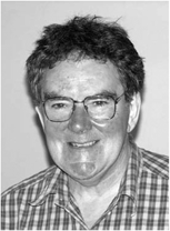 J.V. Hatfield合作论文数2
 Stephen J. Carey合作论文数University of Manchester2S Meeson合作论文数2L. C. Pike合作论文数1Stefanos Almpanis合作论文数1M Al-Zinaty合作论文数1Collier, R.合作论文数1Mayur Patel合作论文数1Keiichi Ikeda合作论文数1S Dindyal合作论文数1Munro, K.合作论文数1Nikos Bafaloukas合作论文数1Constantinos Kyriakides合作论文数1A. Thrush合作论文数1
 Andrew D Scott合作论文数Imperial College London1Dimitrios Staios合作论文数1
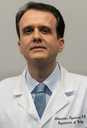 Athanasios Papatsoris合作论文数National & Kapodistrian University of Athens1Redha Boubertakh合作论文数London Chest Hospital1
 Jeff Hand合作论文数King's College London1
 Roger Feakins合作论文数Royal Free London NHS Foundation Trust1




