Double-strand breaks (DSBs) are toxic lesions that lead to genome instability. While canonical DSB repair pathways typically operate independently of RNA, growing evidence suggests that RNA:DNA hybrids and nearby transcripts can influence repair outcomes. However, whether transcript RNA can directly serve as a template for DSB repair in human cells remains unclear. In this study, we develop fluorescence and sequencing-based assays to show that RNA-containing oligonucleotides and messenger RNA can serve as templates during DSB repair. We conduct a CRISPR/Cas9-based genetic screen to identify factors that promote RNA-templated DSB repair (RT-DSBR). Of the candidate polymerases, we identify DNA polymerase zeta (Polζ) as a potential reverse transcriptase that facilitates RT-DSBR. Furthermore, analysis of cancer genome sequencing data reveals whole intron deletions - a distinct genomic signature of RT-DSBR that occurs when spliced mRNA guides repair. Altogether, our findings highlight RT-DSBR as an alternative pathway for repairing DSBs in transcribed genes, with potential mutagenic consequences.
Collisions of the transcription and replication machineries on the same DNA strand can pose a significant threat to genomic stability. These collisions occur in part due to the formation of RNA-DNA hybrids termed R-loops, in which a newly transcribed RNA molecule hybridizes with the DNA template strand. This study investigated the role of RAD52, a known DNA repair factor, in preventing collisions by directing R-loop formation and resolution. We show that RAD52 deficiency increases R-loop accumulation, exacerbating collisions and resulting in elevated DNA damage. Furthermore, RAD52's ability to interact with the transcription machinery, coupled with its capacity to facilitate R-loop dissolution, highlights its role in preventing collisions. Lastly, we provide evidence of an increased mutational burden from double-strand breaks at conserved R-loop sites in human tumor samples, which is increased in tumors with low RAD52 expression. In summary, this study underscores the importance of RAD52 in orchestrating the balance between replication and transcription processes to prevent collisions and maintain genome stability. Collisions of transcription and replication machineries on the same DNA strand threaten genomic stability. Here, the authors show that RAD52 prevents these collisions by regulating R-loop formation and resolution. RAD52 deficiency leads to increased R-loops, exacerbated collisions, DNA damage, and higher mutational burden in tumors.
Chromosome-containing micronuclei are a hallmark of aggressive cancers. Micronuclei frequently undergo irreversible collapse, exposing their enclosed chromatin to the cytosol. Micronuclear rupture catalyzes chromosomal rearrangements, epigenetic abnormalities, and inflammation, yet mechanisms safeguarding micronuclear integrity are poorly understood. In this study, we found that mitochondria-derived reactive oxygen species (ROS) disrupt micronuclei by promoting a noncanonical function of charged multivesicular body protein 7 (CHMP7), a scaffolding protein for the membrane repair complex known as endosomal sorting complex required for transport III (ESCRT-III). ROS retained CHMP7 in micronuclei while disrupting its interaction with other ESCRT-III components. ROS-induced cysteine oxidation stimulated CHMP7 oligomerization and binding to the nuclear membrane protein LEMD2, disrupting micronuclear envelopes. Furthermore, this ROS-CHMP7 pathological axis engendered chromosome shattering known to result from micronuclear rupture. It also mediated micronuclear disintegrity under hypoxic conditions, linking tumor hypoxia with downstream processes driving cancer progression.
Abstract Chromosomal instability, a hallmark of aggressive cancers, disrupts genome integrity through multiple hits by ongoing missegregation of chromosomes. These chromosomes, inherited only by one daughter cell, are subsequently encapsulated in micronuclei (MNi). Micronucleation is detrimental for replication fidelity not only because it sustains chromosomal missegregation, but also because MNi frequently undergo irreversible collapse during interphase exposing their enclosed chromatin to the cytosol. This exposure catalyzes chromosomal rearrangements and heritable epigenetic abnormalities that have been shown to further bolster cancer evolution and therapeutic resistance. Moreover, MNi collapse is known to promote distant metastasis and poor prognosis through eliciting a non-canonical response of otherwise inflammatory signaling pathways. Despite the fundamental role played by MNi catastrophe in compromising genome integrity and sustaining cancer progression, and the subsequent therapeutic potential of targeting this process, the mechanisms underlying MNi collapse are poorly understood. Here, we identify mitochondria-derived reactive oxygen species (ROS) as the main cause of MNi rupture. Notably, we observe that MNi that locate closer to mitochondria are more prone to rupture. Accordingly, increasing ROS chemically and by H2O2 addition increment rupture in a panel of 5 different tumor cell lines, while decreasing ROS using pan-cellular or mitochondrial specific scavengers reduce the frequency of ruptured MNi. By using a combination of advanced super-resolution microscopy, proteomics, transcriptomics, in vitro biochemistry assays, and extensive mutagenesis, we reveal the exact pathway leading to MNi collapse. We demonstrate that ROS promote a noncanonical function of the membrane repair ESCRT-III complex scaffolding protein, CHMP7. ROS reduce CHMP7 interaction with ESCRT-III promoting CHMP7 oligomerization and its binding to the inner nuclear membrane protein, LEMD2. CHMP7, while aggregating, physically pulls the micronuclear envelope together with the LEMD2-associated lamina, thereby disrupting MNi integrity. Finally, we show that hypoxic conditions promote ROS-dependent CHMP7-LEMD2 interaction, inducing MNi rupture and inflammatory signaling. Thus, we observe that human tumors characterized by hypoxia have a significantly increased predominance of ruptured MNi, providing a mechanistic link between tumor hypoxia and downstream processes that drive cancer progression. Citation Format: Melody Di Bona, Yanyang Chen, Albert Agustinus, Matthew Deyell, Mercedes A. Duran, Christy Hong, James Hickling, Daniel Bronder, Sara Martin, Nadeem Riaz, Bill Diplas, Manisha Jalan, Nancy Lee, Alban Ordureau, Benjamin Izar, Ashley Laughney, Simon Powell, Stefano Santaguida, John Maciejowski, Thomas Jeitner, Samuel Bakhoum. Collapse of cancer cell micronuclei from oxidative damage [abstract]. In: Proceedings of the American Association for Cancer Research Annual Meeting 2024; Part 1 (Regular Abstracts); 2024 Apr 5-10; San Diego, CA. Philadelphia (PA): AACR; Cancer Res 2024;84(6_Suppl):Abstract nr 1260.
It is thought that many of the simple and complex genomic rearrangements associated with congenital diseases and cancers stem from mistakes made during the restart of collapsed replication forks by recombination enzymes. It is hypothesised that this recombination-mediated restart process transitions from a relatively accurate initiation phase to a less accurate elongation phase characterised by extensive template switching between homologous, homeologous and microhomologous DNA sequences. Using an experimental system in fission yeast, where fork collapse is triggered by a site-specific replication barrier, we show that ectopic recombination, associated with the initiation of recombination-dependent replication (RDR), is driven mainly by the Rad51 recombinase, whereas template switching, during the elongation phase of RDR, relies more on DNA annealing by Rad52. This finding provides both evidence and a mechanistic basis for the transition hypothesis.
When replication forks encounter DNA lesions that cause polymerase stalling a checkpoint pathway is activated. The ATR-dependent intra-S checkpoint pathway mediates detection and processing of sites of replication fork stalling to maintain genomic integrity. Several factors involved in the global checkpoint pathway have been identified, but the response to a single replication fork barrier (RFB) is poorly understood. We utilized the E.coli -based Tus- Ter system in human MCF7 cells and showed that the Tus protein binding to TerB sequences creates an efficient site-specific RFB. The single fork RFB was sufficient to activate a local, but not global, ATR-dependent checkpoint response that leads to phosphorylation and accumulation of DNA damage sensor protein γH2AX, confined locally to within a kilobase of the site of stalling. These data support a model of local management of fork stalling, which allows global replication at sites other than the RFB to continue to progress without delay.
Genome instability has long been considered the primary driver of most cancer types. A double strand break (DSB) in DNA can have deleterious consequences for a cell, which if not repaired faithfully, can lead to mutations and chromosomal rearrangements, or even cell death. DSBs can be processed by several DNA repair pathways, of which homologous recombination (HR) is the preferred method due to its error-free nature. HR uses an intact homologous DNA sequence as a template for recovering the information lost at the break site. A significant proportion of all cancers, especially triple-negative breast, ovarian pancreatic and prostate cancers, have loss of function alterations affecting genes involved in HR-mediated DNA repair. Alternate repair pathways operate when HR is defective in tumors, but the pathways operative in this context remain a matter of contention. Previous work in vivo in yeast and in vitro systems has established a new role of RNA in DNA repair. Owing to its abundance in the cell and its sequence similarity to parental DNA, we sought to define whether RNA can act as a template for the repair of DSBs in human cells. We developed a novel high throughput assay to test if DNA breaks can be repaired using RNA as an alternative template in mammalian cells. Human cells were transfected with a guide RNA cloned in a Cas9 expression vector to generate a site-specific DSB at the AAVS1 locus, a safe harbour, in the human genome. Furthermore, a donor template in the form of DNA or RNA (homologous to the DSB locus) containing a unique mutational signature was provided at the time of transfection. The unique mutational signature enables us to determine if the donor has been utilized as a template for DNA repair. Using this assay, we demonstrate that cells can use a spliced RNA transcript as a functional template to repair a DSB. We have identified that Rev3L, a key component of the translesion synthesis polymerase Pol Zeta (ζ), has a novel reverse-transcriptase activity in human cells and can help repair the DSB using RNA as a template. Further characterization of this repair pathway and its associated mutational scar will provide new insights into the mutational signatures seen in HR-defective cancers, enabling a better understanding of the DNA repair pathways upregulated in these tumours. The proposed studies could help prioritize novel therapeutic approaches by exploiting synthetic lethality in HR-deficient cancers as well as HR-proficient cancers when used in combinatorial cancer therapy. Citation Format: Manisha Jalan, Juber Patel, Kyrie S Olsen, Sana Ahmed-Seghir, Daniel S Higginson, Jorge S Reis-Filho, Nadeem Riaz, Simon N Powell. RNA-mediated DNA repair: A novel repair pathway in homologous recombination-deficient cancers [abstract]. In: Proceedings of the American Association for Cancer Research Annual Meeting 2022; 2022 Apr 8-13. Philadelphia (PA): AACR; Cancer Res 2022;82(12_Suppl):Abstract nr 5688.
ABSTRACT A major constituent of the nuclear basket region of the nuclear pore complex (NPC), nucleoporin Tpr, plays roles in regulating multiple important processes. We have previously established that Tpr is phosphorylated in both a MAP-kinase-dependent and MAP-kinase-independent manner, and that Tpr acts as both a substrate and as a scaffold for ERK2 (also known as MAPK1). Here, we report the identification of S2059 and S2094 as the major novel ERK-independent phosphorylation sites and T1677, S2020, S2023 and S2034 as additional ERK-independent phosphorylation sites found in the Tpr protein in vivo. Our results suggest that protein kinase A phosphorylates the S2094 residue and that the site is hyperphosphorylated during mitosis. Furthermore, we find that Tpr is phosphorylated at the S2059 residue by CDK1 and the phosphorylated form distinctly localizes with chromatin during telophase. Abrogation of S2059 phosphorylation abolishes the interaction of Tpr with Mad1, thus compromising the localization of both Mad1 and Mad2 proteins, resulting in cell cycle defects. The identification of novel phosphorylation sites on Tpr and the observations presented in this study allow better understanding of Tpr functions.
Intranuclear position of several genes is dynamically altered during development concordant with their activation. To understand this dynamic, but non-random, nuclear organization, it is important to identify the relevant regulatory elements and trans acting factors. Murine TCRb locus gets activated during thymic development. Enhancer Eb is important for VDJ recombination at TCRb locus as it is critically required for establishment of recombination center. Our analysis revealed that TCRb locus gets located out of the chromosome territory specifically in developing thymocytes. Further, CRISPR/Cas9 based deletion mutagenesis established an unambiguous role of enhancer Eb in defining TCRb location relative to chromosome territory. The ability to reposition the target locus relative to chromosome territory highlights a novel aspect pertaining to activity of enhancers which may contribute to their ability to regulate gene expression. Additionally, our observations have implications for understanding the role of enhancers in three-dimensional genome organization and function.
Double strand break (DSB) repair primarily occurs through 3 pathways: non-homologous end-joining (NHEJ), alternative end-joining (Alt-EJ), and homologous recombination (HR). Typical methods to measure pathway usage include integrated cassette reporter assays or visualization of DNA damage induced nuclear foci. It is now well understood that repair of Cas9-induced breaks also involves NHEJ, Alt-EJ, and HR pathways, providing a new format to measure pathway usage. Here, we have developed a simple Cas9-based system with validated repair outcomes that accurately represent each pathway and then converted it to a droplet digital PCR (ddPCR) readout, thus obviating the need for Next Generation Sequencing and bioinformatic analysis with the goal to make Cas9-based system accessible to more laboratories. The assay system has reproduced several important insights. First, absence of the key Alt-EJ factor Pol θ only abrogates ∼50% of total Alt-EJ. Second, single-strand templated repair (SSTR) requires BRCA1 and MRE11 activity, but not BRCA2, establishing that SSTR commonly used in genome editing is not conventional HR. Third, BRCA1 promotes Alt-EJ usage at two-ended DSBs in contrast to BRCA2. This assay can be used in any system, which permits Cas9 delivery and, importantly, allows rapid genotype-to-phenotype correlation in isogenic cell line pairs.
Loss of RAD52 is synthetically lethal in BRCA-deficient cells, owing to its role in backup homologous recombination (HR) repair of DNA double-strand breaks (DSBs). In HR in mammalian cells, DSBs are processed to single-stranded DNA (ssDNA) overhangs, which are then bound by replication protein A (RPA). RPA is exchanged for RAD51 by mediator proteins: in mammals, BRCA2 is the primary mediator; however, RAD52 provides an alternative mediator pathway in BRCA-deficient cells. RAD51 stimulates strand exchange between homologous DNA duplexes, a critical step in HR. RPA phosphorylation and dephosphorylation are important for HR, but its effect on RAD52 mediator function is unknown. Here, we show that RPA phosphorylation is required for RAD52 to salvage HR in BRCA-deficient cells. In BRCA2-depleted human cells, in which the only available mediator pathway is RAD52 dependent, the expression of a phosphorylation-deficient RPA mutant reduced HR. Furthermore, RPA-phosphomutant cells showed reduced association of RAD52 with RAD51. Interestingly, there was no effect of RPA phosphorylation on RAD52 recruitment to repair foci. Finally, we show that RPA phosphorylation does not affect RAD52-dependent ssDNA annealing. Thus, although RAD52 can be recruited independently of RPA’s phosphorylation status, RPA phosphorylation is required for RAD52’s association with RAD51 and its subsequent promotion of RAD52-mediated HR.
Double strand break (DSB) repair mainly occurs through 3 pathways: non-homologous end-joining (NHEJ), alternative end-joining (Alt-EJ), and homologous recombination (HR). We present an assay system that enables simultaneous measurement of all three pathways using Cas9-generated DSBs and next generation sequencing to profile and quantify pathway choice. The assay system has provided several insights. First, absence of the key Alt-EJ factor Pol q only abrogates ~50% of total Alt-EJ. Second, single-strand templated repair (SSTR) requires BRCA1 and MRE11 activity, but not BRCA2, establishing that SSTR commonly used in genome editing is not conventional HR. Third, BRCA1 promotes Alt-EJ usage at two-ended DSBs in contrast to BRCA2. These fundamental differences between BRCA1 and BRCA2 deficiency have implications for therapeutic targeting of HR-deficient cancers. This assay can be used in any system which permits Cas9 delivery and, importantly, allows rapid genotype-to-phenotype correlation in isogenic cell line pairs.
Protein-DNA complexes can impede DNA replication and cause replication fork collapse. Whilst it is known that homologous recombination is deployed in such instances to restart replication, it is unclear how a stalled fork transitions into a collapsed fork at which recombination proteins can load. Previously we established assays in Schizosaccharomyces pombe for studying recombination induced by replication fork collapse at the site-specific protein-DNA barrier RTS1 (Nguyen et al., 2015). Here, we provide evidence that efficient recruitment/retention of two key recombination proteins (Rad51 and Rad52) to RTS1 depends on unloading of the polymerase sliding clamp PCNA from DNA by Elg1. We also show that, in the absence of Elg1, reduced recombination is partially suppressed by deleting fbh1 or, to a lesser extent, srs2, which encode known anti-recombinogenic DNA helicases. These findings suggest that PCNA unloading by Elg1 is necessary to limit Fbh1 and Srs2 activity, and thereby enable recombination to proceed.
Homologous recombination helps ensure the timely completion of genome duplication by restarting collapsed replication forks. However, this beneficial function is not without risk as replication restarted by homologous recombination is prone to template switching (TS) that can generate deleterious genome rearrangements associated with diseases such as cancer. Previously we established an assay for studying TS in Schizosaccharomyces pombe (Nguyen et al., 2015). Here, we show that TS is detected up to 75 kb downstream of a collapsed replication fork and can be triggered by head-on collision between the restarted fork and RNA Polymerase III transcription. The Pif1 DNA helicase, Pfh1, promotes efficient restart and also suppresses TS. A further three conserved helicases (Fbh1, Rqh1 and Srs2) strongly suppress TS, but there is no change in TS frequency in cells lacking Fml1 or Mus81. We discuss how these factors likely influence TS.
The maintenance of genome integrity is critical for cell survival. Homologous recombination (HR) is considered the major error-free repair pathway in combatting endogenously generated double-stranded lesions in DNA. Nevertheless, a number of alternative repair pathways have been described as protectors of genome stability, especially in HR-deficient cells. One of the factors that appears to have a role in many of these pathways is human RAD52, a DNA repair protein that was previously considered to be dispensable due to a lack of an observable phenotype in knock-out mice. In later studies, RAD52 deficiency has been shown to be synthetically lethal with defects in BRCA genes, making RAD52 an attractive therapeutic target, particularly in the context of BRCA-deficient tumors.
CTCF-mediated chromatin interactions influence organization and function of mammalian genome in diverse ways. We analyzed the interactions among CTCF binding sites (CBS) at the murine TCRb locus to discern the role of CTCF-mediated interactions in the regulation of transcription and VDJ recombination. Chromosome conformation capture analysis revealed thymocyte-specific long-range intrachromosomal interactions among various CBS across the locus that were relevant for defining the limit of the enhancer Eb-regulated recombination center (RC) and for facilitating the spatial proximity of TCRb variable (V) gene segments to the RC. Ectopic CTCF binding in the RC region, effected via genetic manipulation, altered CBS-directed chromatin loops, interfered with RC establishment, and reduced the spatial proximity of the RC with Trbv segments. Changes in chromatin loop organization by ectopic CTCF binding were relatively modest but influenced transcription and VDJ recombination dramatically. Besides revealing the importance of CTCF-mediated chromatin organization for TCRb regulation, the observed chromatin loops were consistent with the emerging idea that CBS orientations influence chromatin loop organization and underscored the importance of CBS orientations for defining chromatin architecture that supports VDJ recombination. Further, our study suggests that in addition to mediating long-range chromatin interactions, CTCF influences intricate configuration of chromatin loops that govern functional interactions between elements.
Article Figures and data Abstract eLife digest Introduction Results Discussion Materials and methods Data availability References Decision letter Author response Article and author information Metrics Abstract The completion of genome duplication during the cell cycle is threatened by the presence of replication fork barriers (RFBs). Following collision with a RFB, replication proteins can dissociate from the stalled fork (fork collapse) rendering it incapable of further DNA synthesis unless recombination intervenes to restart replication. We use time-lapse microscopy and genetic assays to show that recombination is initiated within ∼10 min of replication fork blockage at a site-specific barrier in fission yeast, leading to a restarted fork within ∼60 min, which is only prevented/curtailed by the arrival of the opposing replication fork. The restarted fork is susceptible to further collapse causing hyper-recombination downstream of the barrier. Surprisingly, in our system fork restart is unnecessary for maintaining cell viability. Seemingly, the risk of failing to complete replication prior to mitosis is sufficient to warrant the induction of recombination even though it can cause deleterious genetic change. https://doi.org/10.7554/eLife.04539.001 eLife digest Before a cell can divide to form two new cells, it must duplicate its DNA to ensure the newly formed cells have the same genetic information as the original. This genetic material is made up of two single strands of DNA that are paired to form a double-stranded helix. Certain groups of proteins separate these two DNA strands to form a two-pronged structure known as a ‘replication fork’. This occurs at different points along the length of the DNA double helix. Groups of proteins then travel down the DNA strands, separating them as they go, and using them as templates for making copies of the DNA. DNA replication is finally completed when different replication forks meet and merge. This process does not always occur smoothly because some regions of DNA contain obstacles that impede the movement of the replication machinery. In most cases, the replication proteins briefly stall and then restart. However, occasionally the machinery can fall off the DNA; this event is known as a ‘replication fork collapse’. Nguyen et al. have now used a method called time-lapse microscopy to visualise this process inside a species of yeast—called fission yeast—as it occurs in real time. Fission yeast's genetic material is known to contain a specific site that blocks the replication machinery. Nguyen et al. found that a protein called Rad52 arrives at this specific site within 10 minutes of a replication fork being blocked. This protein enables recovery of the replication fork within an hour via a process known as ‘DNA recombination’. Nguyen et al. also unexpectedly found that the restarted fork is susceptible to further collapse. It is known from previous work that the mechanisms that repair broken DNA and rescue replication forks can also introduce errors into the DNA. This implies that if fork collapse occurs frequently, it can lead to the introduction of numerous errors that can be detrimental to cells. Thus, the rescue of fork collapse is like a double-edged sword; it is required for replication to proceed, but can lead to genetic changes inside cells. The failure to faithfully replicate genetic material drives the development of diseases such as cancer. Therefore, insights gained from Nguyen et al.'s findings may provide an improved understanding of how genetic alterations occur in both normal and cancerous cells. https://doi.org/10.7554/eLife.04539.002 Introduction The completion of eukaryotic DNA replication relies on the merging of replication forks that emanate from multiple origins distributed along each chromosome. The failure of even one pair of replication forks to merge results in a region of unreplicated DNA, which can lead to chromosome missegregation and/or DNA breakage during mitosis, and ultimately genomic changes that can drive the development of diseases such as cancer. The progression of replication forks, from origin to point of merging, is frequently hindered by obstacles in and on the DNA template, including various DNA lesions and protein–DNA complexes (Lambert and Carr, 2013). In many cases, these cause only a transient stalling of replication during which the replication proteins (replisome) remain engaged with the DNA by a fork protection mechanism mediated by the intra-S-phase checkpoint (Errico and Costanzo, 2012). However, the replisome can sometimes dissociate to produce a collapsed fork, which may also be associated with breakage of the DNA, and this is especially prevalent following oncogene activation in human cells (Halazonetis et al., 2008). Homologous recombination (HR) can restore fork integrity and restart replication, via a process termed break-induced replication (BIR) (Anand et al., 2013; Costantino et al., 2014). BIR has mainly been studied in the budding yeast Saccharomyces cerevisiae away from the context of DNA replication, using systems that generate a site-specific DNA double-strand break (DSB) that is repaired by recombination with a donor DNA molecule containing sequence homology to only one side of the break (Anand et al., 2013). It is, therefore, unclear to what extent these studies reflect how recombination acts to repair stalled and broken replication forks during S-phase. In BIR, the DSB is resected to generate a single-stranded DNA (ssDNA) tail, which is bound firstly by Replication Protein A (RPA) and then Rad52. Rad52 mediates the subsequent loading of Rad51, which catalyses the key steps of homologous DNA pairing and strand invasion to form a displacement (D) loop. The 3' end of the invading DNA strand primes DNA synthesis by polymerase δ, and replication then proceeds in a conservative manner involving migration of the D-loop, dependent on the Pif1 DNA helicase (Saini et al., 2013; Wilson et al., 2013). Similar to other modes of DSB repair by HR, the initial steps of BIR from DSB resection to strand invasion occur within ∼30 min of DSB formation (Jain et al., 2009; Hicks et al., 2011). In contrast, the transition from strand invasion to DNA replication is delayed for several hours by a checkpoint that senses whether both ends of the DSB can engage with the same donor DNA sequence in a manner that is productive for completing repair (Malkova et al., 2005; Jain et al., 2009). However, once initiated DNA synthesis proceeds at a rate of 3–4 kb/min, which is comparable with normal DNA replication (Malkova et al., 2005). Unlike normal DNA replication, BIR is highly error-prone with greatly increased rates of polymerase errors that remain uncorrected (Deem et al., 2011). There are also frequent dissociations of the elongating strand from the D-loop within a 10 kb window downstream of the DSB (Smith et al., 2007; Stafa et al., 2014), which results in multiple rounds of strand re-invasion increasing the risk of recombination between ectopic homologous DNA sequences that can give rise to gross chromosome rearrangements and copy-number variations. This feature of BIR is thought to reflect an inherent drive to repair the DSB by synthesis-dependent strand annealing (SDSA) (Smith et al., 2007). However, beyond 10 kb the D-loop appears to be stabilized, and BIR proceeds without the continual interruption of D-loop dissociation. Whilst it is clear that a broken replication fork requires HR for DNA repair, it is less certain that it would be needed at a collapsed, yet unbroken, fork where replication could be completed most simply by convergence with the opposing fork. However, replication fork collapse at a site-specific protein–DNA fork barrier called RTS1, in the fission yeast Schizosaccharomyces pombe, has been shown to provoke DSB-independent recombination-dependent replication (RDR), which is required for viability (Lambert et al., 2005, 2010). Similar to BIR, RTS1-induced RDR is error-prone, at least within the first 2.4 kb downstream of the barrier (Iraqui et al., 2012; Mizuno et al., 2013). However, it is unknown whether it suffers the same tendency for D-loop dissociation as BIR. It also remains unclear whether recombination is a default response to replication fork blockage at RTS1, how quickly it initiates, and whether it only plays a role in restart at the blocked fork, or is also needed to promote fork merging. To address these questions, we have used time-lapse microscopy to obtain single-cell resolution of RDR in response to replication fork blockage at RTS1, in combination with genetic assays to assess restarted fork fidelity. We show that Rad52 is recruited to RTS1 in the majority of cells within minutes of fork blockage and seemingly gives rise to RDR without the long delay in progressing from strand invasion to DNA synthesis that is characteristic of BIR. However, like BIR the restarted fork is prone to multiple rounds of strand disengagement and reengagement, implying that even at a blocked replication fork during S-phase the default response is to attempt SDSA. Unlike BIR, this continues for more than 10 kb downstream of the barrier with little sign of abating, giving rise to a dramatic increase in recombination in this region. We also find that fork convergence, rather than inducing recombination, acts to prevent or curtail it. Surprisingly, despite the high frequency of the recombination response and contrary to previous reports, we find no evidence that it is required for cell viability. Seemingly, the risk of failing to complete DNA replication in a timely fashion is sufficient to warrant the initiation of RDR as a default response to replication fork collapse, even though in many cases it may be unnecessary and even cause deleterious genetic change. Results Experimental system We have previously shown that replication fork blockage at the replication terminator sequence RTS1 is sufficient to induce HR between a direct repeat of ade6− heteroalleles in S. pombe (Ahn et al., 2005) (Figure 1A). RTS1 is a unidirectional replication fork barrier (RFB), which consists of cis-acting DNA sequence elements and trans-acting factors including the myb domain-containing protein Rtf1 (Codlin and Dalgaard, 2003; Eydmann et al., 2008). Replication of the ade6 locus is essentially unidirectional due to the relative position of replication origins that flank it (Figure 1A). Consequently, only one orientation of RTS1 causes replication fork blockage at this site, which we will refer to as the active orientation (AO). The opposite orientation, which does not block replication, will be referred to as the inactive orientation (IO). Native two dimensional (2D) gel electrophoresis analysis of replication intermediates in an EcoNI fragment containing RTS1 confirms that RTS1-AO strongly blocks replication forks, whereas RTS1-IO does not (Figure 1B). It also shows that a proportion of forks remain blocked at RTS1-AO long enough for replication to be completed by the opposing fork resulting in fork merging at RTS1 as indicated by the appearance of double Y-shaped molecules (Figure 1B, Figure 1—figure supplement 1A,B). The remaining blocked forks appear to be able to restart and replicate past the barrier as indicated by the presence of large Y-shaped DNA molecules (Figure 1B, Figure 1—figure supplement 1A,B). The ratio of large Ys to double Ys is approximately 1:1 suggesting that about half the forks blocked at RTS1-AO restart prior to fork convergence (Figure 1—figure supplement 1B). Figure 1 with 1 supplement see all Download asset Open asset Experimental system for studying RFB-induced RDR. (A) Schematic showing the location of the direct repeat recombination reporter on chromosome 3, the two types of recombinants, and the position of the probe used for 2D gel analysis in B. Asterisks indicate the position of point mutations in ade6-L469 and ade6-M375. (B) 2D gel analysis of replication intermediates in the EcoNI fragment shown in A. The DNA was extracted from strains MCW429 (no RTS1), MCW4712 (RTS1-IO), and MCW4713 (RTS1-AO). (C) Ade+ recombinant frequencies for strains MCW4712, MCW4713, MCW1687, and MCW1688. Data are represented as mean ± SD. https://doi.org/10.7554/eLife.04539.003 Rad52 is required for replication past RTS1-AO Work from the Carr and Lambert laboratories has shown that replication restart from RTS1 depends on HR (Lambert et al., 2010), and consistent with this we observe a 99-fold increase in gene conversions and a 33-fold increase in deletions between the ade6− heteroalleles that flank RTS1-AO compared to background levels observed without RTS1 or with RTS1-IO (Figure 1C, Table 1) (Ahn et al., 2005). Moreover, this elevated frequency of HR is totally dependent on Rad52 (Figure 1C, Table 1), which is essential for both RFB-induced RDR and BIR in yeast (Malkova et al., 1996; Lambert et al., 2010). Table 1 Direct repeat recombinant frequencies https://doi.org/10.7554/eLife.04539.005 GenotypeRTS1 orientationPosition of direct repeatColonies analysedAde+ His+ recombinant frequency (× 10−4)*Ade+ His− recombinant frequency (× 10−4)*Meanp value†Meanp value†wild typeIOFlanking RTS1771.36 (+/− 0.51)–3.48 (+/− 0.89)–wild typeAOFlanking RTS177134.03 (+/− 38.50)–113.41 (+/− 49.72)–rad52∆IOFlanking RTS1150.03 (+/− 0.04)<0.001‡1.20 (+/− 0.21)<0.001‡rad52∆AOFlanking RTS1150.13 (+/− 0.11)<0.001§4.07 (+/− 1.16)<0.001§ori-1253∆IOFlanking RTS1152.33 (+/− 1.86)0.060‡4.80 (+/− 1.58)0.007‡ori-1253∆AOFlanking RTS116339.88 (+/− 83.61)<0.001§221.02 (+/− 57.21)<0.001§wild typeIOSite A181.44 (+/− 0.86)0.698‡4.61 (+/− 1.66)0.011‡wild typeAOSite A21128.66 (+/− 43.85)0.746§680.48 (+/− 305.44)<0.001§wild typeIOSite B180.88 (+/− 0.51)0.023#2.45 (+/− 1.88)0.001#wild typeAOSite B155.12 (+/− 2.41)<0.00185.02 (+/− 33.72)<0.001ori-1253∆IOSite B151.58 (+/− 0.50)<0.001**4.86 (+/− 2.56)0.006**ori-1253∆AOSite B1586.03 (+/− 33.63)<0.001††1094.46 (+/− 443.32)<0.001†† * The values in parentheses are the standard deviations about the mean. † p values are derived from independent-sample t-tests comparing the mean values as indicated. ‡ Compared to the equivalent mean recombinant frequency in wild type with RTS1-IO flanked by ade6- direct repeats. § Compared to the equivalent mean recombinant frequency in wild type with RTS1-AO flanked by ade6- direct repeats. # Compared to the equivalent mean recombinant frequency in wild type with RTS1-IO and site A ade6- direct repeats. Compared to the equivalent mean recombinant frequency in wild type with RTS1-IO and site A ade6- direct repeats. ** Compared to the equivalent mean recombinant frequency in wild type with RTS1-IO and site B ade6- direct repeats. †† Compared to the equivalent mean recombinant frequency in wild type with RTS1-AO and site B ade6- direct repeats. Evidence that Rad52 restarts replication at RTS1 (positioned at the ura4 locus and under inducible Rtf1 control) includes the observation that there is more fork convergence at the barrier in a rad52∆ mutant than in wild type, as judged by the accumulation of double Y-shaped DNA molecules on 2D gels (Lambert et al., 2010). However, in previous work from our laboratory, we failed to detect such an increase at RTS1-AO suggesting that RDR does not occur at appreciable levels in our experimental system (Lorenz et al., 2009). The method of DNA extraction for 2D gel analysis can have a significant effect on the ability to detect different types of replication intermediates (Liberi et al., 2006). Our standard protocol is based on mechanical cell disruption to extract nuclei, DNA purification by caesium chloride density gradient centrifugation, and enrichment for replication intermediates on benzoylated napthoylated DEAE (BND) cellulose columns (Lorenz et al., 2009). In contrast, Lambert et al. (2010) enzymatically lyse cells embedded in agarose plugs prior to enrichment of replication intermediates on BND cellulose. To determine whether these differences could account for our failure to detect an increase in converging forks, we compared replication intermediates in the EcoNI fragment containing RTS1-AO from wild-type and a rad51∆ rad52∆ double mutant using the same protocol of DNA extraction as Lambert et al. (2010). A rad51∆ rad52∆ double mutant was used instead of a rad52∆ single mutant because of the latter's susceptibility to acquire suppressor mutations that enable Rad51 to catalyse HR in the absence of Rad52 (Osman et al., 2005). Unlike Lambert et al. (2010), we did not observe an increase in double Y-shaped molecules, in fact they decreased by ∼twofold in the rad51∆ rad52∆ mutant (Figure 2A,B). However, the reduction in large Ys was even greater (∼sixfold) (Figure 2B), such that their ratio to double Ys decreased more than threefold (Figure 2C). These data indicate that Rad52 plays an important role in promoting replication past RTS1-AO. We suspect that a rad51∆ rad52∆ mutant fails to manifest an increase in double Y-shaped molecules because the effect of an increased frequency of fork convergence at RTS1-AO is offset by a faster rate of fork merging, due to the absence of recombination proteins that could impede this process. Figure 2 Download asset Open asset Recombination proteins are needed for replication past RTS1-AO. (A) 2D gel analysis of replication intermediates in the EcoNI fragment shown in Figure 1A. The DNA was extracted from strains MCW4713 (wild-type RTS1-AO) and MCW1696 (rad51∆ rad52∆ RTS1-AO) using the same method as in Lambert et al. (2010). Note that the signal extending from the RFB signal indicated by the asterisk is an aberration of how the gel ran in the first dimension. (B and C) Quantification of 2D gels. Mean values (± SD) from four independent experiments for wild type are shown. In the case of rad51∆ rad52∆, the values are based on two independent experiments with each value represented by a dot around the mean. https://doi.org/10.7554/eLife.04539.006 Live cell imaging of RDR at RTS1 Whilst replication fork blockage at RTS1 clearly induces recombination, it is unclear whether this happens in all cells or only a subset in each cell cycle. To address this question, we inserted an array of lacO sequences downstream of RTS1 so that we could track its location in cells expressing the LacI repressor fused to the far-red fluorescent protein tdKatushka2 using time-lapse microscopy (Figure 3A,B). Mindful that the lacO-LacI interaction can act as a RFB (Sofueva et al., 2011), we determined imaging parameters under which the amount of LacI, whilst sufficient to detect the location of the lacO array through several hours of live cell imaging, was insufficient to perturb replication forks as judged by native 2D gel analysis (Figure 3—figure supplement 1) or induce a recombinational response (Figure 3—figure supplement 2). Having established suitable conditions, we imaged asynchronously growing cells containing either RTS1-IO or RTS1-AO and Rad52 fused to yellow fluorescent protein (YFP), which forms foci in response to DNA damage similar to its homologue in S. cerevisiae (Lisby et al., 2001, 2003; Meister et al., 2003) (Figure 3B). Time-lapse movies of cells, with images taken at 5-min intervals, were staged relative to anaphase, and the presence of Rad52 foci and their co-localization with lacO-LacI foci recorded over a 90-min period (Figure 3C). The majority (≥79%) of cells exhibited a single Rad52 focus at varying time points mainly between 20 and 90 min post-anaphase (Figure 3C). This is true even for cells without RTS1 and reflects the fact that HR is routinely needed to process perturbed replication forks and DNA damage that occurs during S-phase (data not shown). Consistent with replication fork blockage at RTS1 inducing HR, a higher percentage of cells with RTS1-AO exhibited a Rad52 focus than those with RTS1-IO (89% vs 79%), and on average, these foci were detected over a greater number of time points (Figure 3C, Figure 3—figure supplement 3A,B). Imaging of RTS-AO and RTS-IO cells without lacO-LacI revealed no difference in the occurrence and timing of Rad52 foci confirming that lacO-LacI acts as an inert marker for the location of RTS1 (Figure 3—figure supplement 3A,B). Figure 3 with 3 supplements see all Download asset Open asset Tracking Rad52-YFP focus localization at RTS1 by time-lapse microscopy. (A) Schematic showing the modification of the direct repeat recombination reporter for time-lapse microscopy. (B) Representative stills taken from a time-lapse movie of two daughter cells each with a LacI-tdKatushka2 and Rad52-YFP focus. In the right hand daughter, the foci co-localize. (C) Analysis of time-lapse movies. The presence of a Rad52 focus and whether it co-localizes with the lacO-LacI-tdKatushka2 focus is recorded every 5 min for 90 min post-anaphase in each cell. The top panel is a schematic of S. pombe cells at various stages post-anaphase with nuclear lacO-LacI foci in blue, Rad52 foci in green, and co-localizing foci in red. The strains are MCW6395 and MCW6556. https://doi.org/10.7554/eLife.04539.007 Analysis of the co-localization of Rad52 foci with lacO-LacI foci revealed a striking difference between cells with RTS1-IO and RTS1-AO (Figure 3C). ∼20% of cells with RTS1-IO exhibited co-localizing foci, which on average were detectable in 1.5 time points, whereas with RTS1-AO this increased to ∼60% of cells and 3.8 time points (Figure 3C). Cells without RTS1 exhibit the same relatively low incidence of Rad52 focus co-localization with lacO-LacI as those containing RTS1-IO consistent with the observation that RTS1-IO does not induce recombination (data not shown). Together, these data show that replication fork blockage induces a recombinational response at RTS1 in most but not all cells. Additionally, they show that blockage of a single replication fork is sufficient to induce a Rad52 focus. However, the overall increase in Rad52 foci observed in RTS1-AO cells is less than the number of co-localizing foci implying that some Rad52 foci that are engaged at RTS1 can at the same time be engaged with other lesions/perturbed replication forks consistent with them acting as repair centres (Lisby et al., 2003). Both Rad51 and Rad54 are recruited to RTS1-AO To determine whether Rad52 foci co-localizing with lacO-LacI represent sites of active recombination, rather than simply Rad52 binding to ssDNA at the stalled replication fork, we looked for the appearance of both Rad51 and Rad54 foci. In snapshots of asynchronously growing cells containing RTS1-IO, Rad52-YFP and Rad51 tagged at its N-terminus with cyan fluorescent protein (CFP), 8.1% of cells contain a Rad52 focus and 6.6% a Rad51 focus, with 95% of the latter co-localizing with a Rad52 focus (Figure 4B). Only 2.6% of Rad52 foci and 3.2% of Rad51 foci were observed to co-localize with lacO-LacI in these cells (Figure 4B). In cells with RTS1-AO, the overall percentage of cells with Rad52 and Rad51 foci increases to 13% and 12%, respectively, with the vast majority (97%) of Rad51 foci again co-localizing with a Rad52 focus (Figure 4B). Importantly, 36.5% of Rad52 foci and 37.9% of Rad51 foci co-localized with lacO-LacI, with greater than 90% of these foci also co-localizing with each other (Figure 4A,B). Cells containing Rad54 fused to green fluorescent protein (GFP), but without Rad52-YFP, were also imaged (Figure 4C). 11.7% of RTS1-IO cells contained a Rad54-GFP focus, but only 5.5% of these foci co-localized with lacO-LacI (Figure 4D). Similar to Rad51 and Rad52, the frequency of Rad54-GFP foci increased in RTS1-AO cells (from 11.7% to 14%) and a much greater percentage of these foci (36.1%) co-localized with lacO-LacI (Figure 4C,D). Collectively, these data indicate that both Rad51 and Rad54 are recruited to RTS1-AO to a similar extent as Rad52. Moreover, the coincidence of Rad51 and Rad52 foci suggests that these proteins are functioning together at the RTS1 barrier, and with Rad54 are likely to represent active and ongoing recombination. Figure 4 Download asset Open asset Both CFP-Rad51 and Rad54-GFP foci co-localize with lacO-LacI in RTS1-AO cells to a similar extent as Rad52-YFP foci. (A) Example snapshots of a RTS1-AO cell with both a CFP-Rad51 and Rad52-YFP focus co-localizing with a lacO-LacI-tdKatushka2 focus. The scale bar represents 2 µm. (B) Quantification of data like in A. The strains are MCW7640 (RTS1-IO) and MCW7638 (RTS1-AO). (C) Example snapshots of a RTS1-AO cell with a Rad54-GFP focus co-localizing with a lacO-LacI-tdKatushka2 focus. The scale bar represents 2 µm. (D) Quantification of data like in C. The strains are MCW7646 (RTS1-IO) and MCW7645 (RTS1-AO). https://doi.org/10.7554/eLife.04539.011 Timing of Rad52 recruitment following replication fork blockage To gauge how quickly Rad52 is recruited to forks blocked at RTS1-AO, we first assessed the timing of S-phase relative to anaphase by imaging cells expressing the replication marker PCNA fused to CFP (Meister et al., 2005). CFP-PCNA forms patterns of foci that are characteristic for the different stages of S-phase (Meister et al., 2007) (Figure 5—figure supplement 1), and under our experimental conditions these foci first appear between 10 and 15 min post-anaphase, marking the start of S-phase (Figure 5A). In cells containing RTS1-AO, Rad52 foci co-localizing with lacO-LacI first appear between 20 and 25 min post-anaphase (Figures 3C and 5B). This lag between the start of S-phase and appearance of Rad52 foci at the RFB is also observed when both CFP-PCNA and Rad52-YFP are imaged concurrently in the same cells (Figure 5C). As the nearest replication origin to RTS1-AO is ∼7.7 kb away (Figure 3A, Table 2), the earliest a fork can reach the barrier is ∼2.6 min after the start of S-phase, based on an average fork velocity of ∼3 kb/min (Heichinger et al., 2006). Therefore, Rad52 can be recruited to RTS1-AO as early as 7.4–12.4 min after replication fork blockage. The later appearance of Rad52 foci at RTS1-AO that is observed in many cells (ranging mainly from 30 to 55 min post-anaphase) (Figure 3C), likely reflects the later blockage of forks that emanate from more distal origins (i.e., ori-1338, ori-1342, and ori-1371), which would arrive at the barrier from 7.7 to 18.5 min after the start of S-phase (Figure 3A,C, Table 2). In a few cells (∼5%), we observed Rad52 foci first co-localizing with lacO-LacI at 60–80 min post-anaphase (Figure 3C). These very late appearing co-localizing foci may represent cells in which one or both forks converging on RTS1 have been delayed by other RFBs or possibly a recombinational response to occasional problems that might occur during fork convergence. Figure 5 with 1 supplement see all Download asset Open asset Temporal kinetics of Rad52-YFP foci localization to RTS1. (A) Percentage of cells with one or more CFP-PCNA foci in the first 90 min post-anaphase. The RTS1-IO strain is MCW6701 (n = 31), and the RTS1-AO strain is MCW6706 (n = 31). (B) Percentage of cells with a Rad52-YFP focus that co-localizes with the lacO-LacI focus in the first 90 min post-anaphase. The data are derived from Figure 3C. (C) Percentage of cells with a Rad52-YFP focus that co-localizes with the lacO-LacI focus in the 90 min following the first appearance of CFP-PCNA foci post-anaphase (i.e., the start of S-phase). The RTS1-IO strain is MCW6712 (n = 34) and the RTS1-AO strain is MCW7065 (n = 52). Note that the overall percentage of cells with a Rad52-YFP focus co-localizing with lacO-LacI is slightly higher than in cells without CFP-PCNA. The CFP tag on PCNA partially impairs its function, which is compensated by expression of untagged PCNA in the same cells (Meister et al., 2007). Nevertheless, we suspect that a subtle impairment of PCNA function accounts for the more frequent co-localization of Rad52-YFP with lacO-LacI. (D) End point of Rad52-YFP focus co-localization with the lacO-LacI focus relative to the disappearance of CFP-PCNA foci. The strain is MCW7065 (n = 49). The asterisk indicates cells with a Rad52-YFP focus co-localizing with the lacO-LacI focus more than 45 min after CFP-PCNA foci disappearance. https://doi.org/10.7554/eLife.04539.012 Table 2 Distances and estimated replication times from origin to RTS1 https://doi.org/10.7554/eLife.04539.014 OriginDistance from barrier (bp)Time from barrier (min)Origin centromere proximal relative to RTS1 ori-1141186,73162.2 ori-1194134,20844.7 ori-125375,15425.1 ori-127751,03117.0Origin centromere distal relative to RTS1 ori-132377352.6 ori-133823,0987.7 ori-134227,1729.1 ori-137155,44418.5 Distances are calculated from the midpoint of the origin coordinates as stated in OriDB (pombe.oridb.org). Times are based on a replication fork velocity of 3.0 kb/min. Duration of Rad52 at the blocked replication fork As mentioned above, Rad52 foci remain co-localized with lacO-LacI in cells with RTS1-AO for an average of 3.8 time points. If one considers only the longest track of consecutive time points with a co-localization in each cell, then the duration of any single-event ranges from 1 to 9 time points (average = 3.4 time points), which equates to >0 to <45 min (Figure 3C). However, as these estimates are based on images taken every 5 min, we cannot be certain that Rad52 does not dissociate and then re-associate with the site in between point
Developmental stage-specific enhancer-promoter-insulator interactions regulate the chromatin configuration necessary for transcription at various loci and additionally for VDJ recombination at antigen receptor loci that encode immunoglobulins and T-cell receptors. To investigate these regulatory interactions, we analyzed the epigenetic landscape of the murine T-cell receptor β (TCRβ) locus in the presence and absence of an ectopic CTCF-dependent enhancer-blocking insulator, H19-ICR, in genetically manipulated mice. Our analysis demonstrated the ability of the H19-ICR insulator to restrict several aspects of enhancer-based chromatin alterations that are observed during activation of the TCRβ locus for transcription and recombination. The H19-ICR insulator abrogated enhancer-promoter contact-dependent chromatin alterations and additionally prevented Eβ-mediated histone modifications that have been suggested to be independent of enhancer-promoter interaction. Observed enhancer-promoter-insulator interactions, in conjunction with the chromatin structure of the Eβ-regulated domain at the nucleosomal level, provide useful insights regarding the activity of the regulatory elements in addition to supporting the accessibility hypothesis of VDJ recombination. Analysis of H19-ICR in the heterologous context of the developmentally regulated TCRβ locus suggests that different mechanisms proposed for CTCF-dependent insulator action might be manifested simultaneously or selectively depending on the genomic context and the nature of enhancer activity being curtailed.
The completion of genome duplication during the cell cycle is threatened by the presence of replication fork barriers (RFBs). Following collision with a RFB, replication proteins can dissociate from the stalled fork (fork collapse) rendering it incapable of further DNA synthesis unless recombination intervenes to restart replication. We use time-lapse microscopy and genetic assays to show that recombination is initiated within ∼10 min of replication fork blockage at a site-specific barrier in fission yeast, leading to a restarted fork within ∼60 min, which is only prevented/curtailed by the arrival of the opposing replication fork. The restarted fork is susceptible to further collapse causing hyper-recombination downstream of the barrier. Surprisingly, in our system fork restart is unnecessary for maintaining cell viability. Seemingly, the risk of failing to complete replication prior to mitosis is sufficient to warrant the induction of recombination even though it can cause deleterious genetic change.
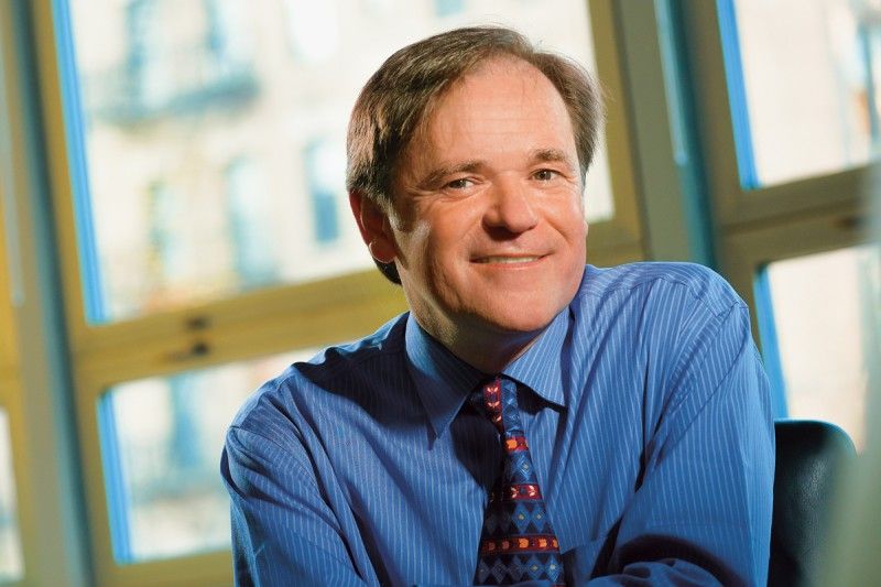 Simon Powell合作论文数Molecular Biology, Weill Cornell Graduate School of Medical Sciences, Cornell University8
 Carl A Morrow合作论文数Univ Queensland, Australian Infect Dis Res Ctr, Brisbane, Qld, Australia6
 Fekret Osman合作论文数Department of Biochemistry, University of Oxford6
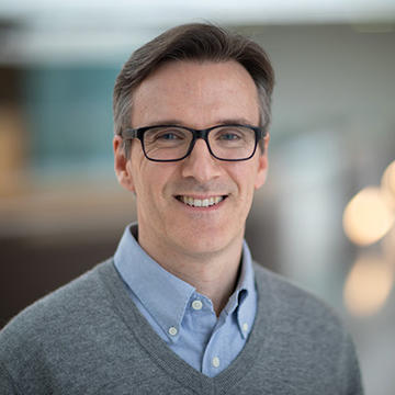 Matthew Whitby合作论文数Department of Biochemistry, University of Oxford6
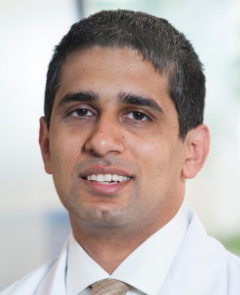 Nadeem Riaz合作论文数Memorial Sloan Kettering Cancer Center5
 Sana Ahmed-Seghir合作论文数4
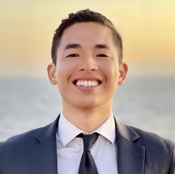 Nguyen Michael O合作论文数Department of Biochemistry, University of Oxford4
 Daniel S. Higginson合作论文数University of North Carolina School of Medicine, Univ. North Carolina Sch. Med.4Sanjeeta Tamang合作论文数3Erika S Buechelmaier合作论文数3
 Anastasiya Kishkevich合作论文数3
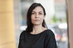 Di Bona Melody合作论文数Nanoscopy, Nanophysics, Istituto Italiano di Tecnologia, Genoa, Italy3
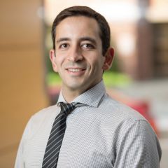 Samuel Bakhoum合作论文数Cell & Developmental Biology, Weill Cornell Graduate School of Medical Sciences, Cornell University3Oehler Judith合作论文数3
 Jorge S. Reis-Filho合作论文数AstraZeneca3Madhulika Srivastava合作论文数3Sara Martin合作论文数2Kyrie S. Olsen合作论文数2Neeman Mohibullah合作论文数2Matthew Deyell合作论文数2Rahul Majumdar合作论文数2
 Christos Andreadis合作论文数2Elena Bulmaga合作论文数2
 Li Yi合作论文数2Rekha Soni合作论文数2
 Pratishtha Rawat合作论文数2
 Christy Hong合作论文数Univ Groningen, Univ Med Ctr Groningen, European Res Inst Biol Ageing, A Deusinglaan 1, NL-9713 AV Groningen, Netherlands2Xiaoqing Rong-Mullins合作论文数2
 Mercedes Duran合作论文数IBGM2Juber Patel合作论文数Mem Sloan Kettering Canc Ctr, 1275 York Ave, New York, NY 10021 USA2
 Albert S Agustinus合作论文数Chemical Biology Program, Memorial Sloan Kettering2
 Yingjie Zhu (朱英杰)合作论文数Department of Pathology, Memorial Sloan Kettering Cancer Center2
 Aman Sharma合作论文数University of Massachusetts-Amherst;University of Massachusetts-Amherst2
 Daniel Bronder合作论文数Department of Radiation Oncology, Memorial Sloan Kettering Cancer Center2
 Pavithran T Ravindran合作论文数Department of Chemical and Biological Engineering, Princeton University, Princeton, NJ 08544, USA2
 Suleman S Hussain合作论文数Dept Radiat Oncol, Mem Sloan Kettering Canc Ctr2
 John Maciejowski合作论文数Molecular Biology, Weill Cornell Graduate School of Medical Sciences, Cornell University2
 Alban Ordureau合作论文数Cell & Developmental Biology, Weill Cornell Graduate School of Medical Sciences, Cornell University2
 Diplas Bill H合作论文数Pediatric Brain Tumor Foundation Institute at Duke, Duke University Medical Center2Izar Benjamin合作论文数Institute of Medical Microbiology, Justus-Liebig-University;E-Mails: bizar@partners.org (B.I.);mikrobio.med.uni-giessen;uni-giessen;uni-giessen;Institute of Medical Microbiology, Justus-Liebig-University2
 Christopher Sifuentes合作论文数2Leeman Jonathan合作论文数Dept Radiat Oncol, Mem Sloan Kettering Canc Ctr2
 Niamh Mcdermott合作论文数2
 Hamzić Edin合作论文数UMR1313 Animal Genetics and Integrative Biology, AgroParisTech;UMR1313 Animal Genetics and Integrative Biology, INRA;Department of Molecular Biology and Genetics, Aarhus University;Department of Molecular Biology and Genetics, Aarhus University2
 Ashley M. Laughney合作论文数2
 Stefano Santaguida合作论文数Department of Experimental Oncology, European Institute of Oncology2
 Himanshi Narang合作论文数Radiation Biology and Health Sciences Division, Bhabha Atomic Research Centre2
 Agnès Viale合作论文数Marie-Josée & Henry R. Kravis Center for Molecular Oncology, Memorial Sloan Kettering Cancer Center2
 Nancy Y. Lee合作论文数Department of Radiation Oncology, Memorial Sloan-Kettering Cancer Center2




