One in three people living with diabetes are affected by diabetic foot ulcers (DFUs) which, if left unmonitored and untreated, reduce quality of life, and may lead to foot amputation. Regular foot screening is suggested by relevant national guidelines. Yet, compliance with screening is poor, mostly due to the inconvenience of the screening process itself. In this work, we describe a preliminary version of a simple low-cost device intended for self-monitoring of foot circulation, to identify the areas of poor perfusion expected to be at the root of the formation of DFUs. This device is based on multispectral imaging of the feet in the visible and near-infrared ranges. The device is tested on the hands and feet of a single subject, where impairment of circulation has been simulated through a brief ligature of a finger/two toes. Multispectral images are captured, and a simple machine-learning-based classifier correctly identifies areas of low perfusion on the hand and shows promising data on toes. Albeit the device and the classifier are still susceptible of significant improvement, this is indicative of the fact that the multispectral images contain relevant information on tissue perfusion.
Amblyopia is a neurodevelopmental disorder that causes irreversible vision loss in one eye. In order to be effective, treatment needs to start as early as possible, ideally in early infancy. The screening for amblyopia assesses the disparity of the child's visual acuity (VA). In pre-verbal children, this screening is performed using a preferential looking test. These testing protocols rely on a subjective estimate by the test operator of the child's attention, and this estimate is subject to errors. Little is understood of the quantitative impact of error rates on screening performance. In this paper, a Monte Carlo simulation to compare the clinical performance of three preferential-looking test protocols for paediatric VA screening tests for amblyopia is described. The inter-protocol differences of “Cardiff Acuity Test” (CAT), “Keeler Acuity Cards for Infants” (KACI) and “Teller Acuity Cards” (TAC) are assessed by iteratively executing through a simulation loop of an examiner testing a test subject using the three set protocols. The measured VA from each protocol and the actual VA have been compared using Bland-Altman statistics. It was determined that CAT and KACI both have a systematic bias, whereby they measure the VA at a greater logMAR value than the smallest testing resolution. KACI bias is greater due to the greater step size used.
A guideline is proposed that comprises the minimum items to be reported in research studies involving an eye tracker and human or non-human primate participant(s). This guideline was developed over a 3-year period using a consensus-based process via an open invitation to the international eye tracking community. This guideline will be reviewed at maximum intervals of 4 years.
Important ophthalmic imaging techniques, such as fundus camera imaging, utilize light sources that need to be focused on an annular section of an eye surface. In order to be performed dynamically, this requires real-time control of the focus plane of the illumination optics. When considering adaptive focusing in ophthalmic instruments, liquid lenses represent the best compromise in terms of tunability, tuning range, compactness, numerical aperture, and speed. In recent years, several compact eye imaging devices using liquid lenses have been described in the literature. These would benefit from low computational complexity algorithms for adaptive local auto-focus on an annular region, to allow the illumination optics to be controlled by low-cost and low-power electronics. In this paper, we propose a novel radial-focus evaluation method based on a revised version of a more traditional Laplacian-based focus estimator. This radial focus evaluation is targeted to focusing annular sections of an image, with the advantage of a drastic reduction of computational complexity.
This paper presents preliminary work on a method to increase the number of detection channels of a commercial 6-channel spectrometric sensor, by employing a set of multiplexed spectrally colored illumination sources. A demonstrator has been built and tested on a set of independent dyes. The results suggest successful enhancement of the number of detection channels. More tests are needed to provide quantitative performance evaluation, and to demonstrate viability in a clinical setting. Clinical Relevance- The technique presented in this paper shows promise to provide a viable method for multispectral sensing in highly-miniaturized systems such as in-vivo sensing capsules.
Screening programs for sight-threatening diseases rely on the grading of a large number of digital retinal images. As automatic image grading technology evolves, there emerges a need to provide a rigorous definition of image quality with reference to the grading task. In this work, on two subsets of the CORD database of clinically grad able and matching non-grad able digital retinal images, a feature set based on statistical and on task-specific morphological features has been identified. A machine learning technique has then been demonstrated to classify the images as per their clinical gradeability, offering a proxy for a rigorous definition of image quality.
Autorefraction consists of the automatic sensing of three parameters - spherical error, cylindrical error, slope of the principal meridian - that describe the deviation of the focusing properties of an ametropic eye with respect to an emmetropic state. Low-cost autorefractors would be highly desirable in resource-poor settings, for the stratification of patients between those who can be treated in the community and those who need to be referred to specialist care. In the present paper, we describe the implementation of an autorefractor based on projecting patterns onto the retina of an eye and observing the projected pattern through an ophthalmoscopic camera configuration coaxial with the projection path. Tunable optics in the coaxial path, combined with appropriate image processing, allows determination of the three parameters. The simplicity and performance of the setup, measured on an eye simulator, shows promise towards clinical use in the community. Further work is needed to confirm the performance in vivo.
Preventing vision loss from glaucoma, the second leading cause of blindness globally, relies on early detection and intervention. A key clinical indicator for early detection is structural change of the optic nerve head (ONH), which impacts the ONHs three-dimensional footprint on the retina. Clinically, such footprint is commonly assessed by subjective observation through a slit lamp, with significant inter- and intra-observer variability. Objective devices that rely on 3D imaging show promise in increasing diagnostic accuracy within clinics but are prohibitively expensive for many. In previous work, we demonstrated the initial feasibility of obtaining 3D surface images by applying stereo vision techniques to a slit lamp fitted with low-cost cameras. In this paper, we present the assessment of the precision and accuracy of our system with ONH targets in an eye phantom. Quantitative reconstructions, referenced to ground truth data, were obtained. Current results, albeit promising, suggest that more work is likely needed to improve accuracy and precision to a clinically significant level.
Background Live teleophthalmology using video conferencing allows real-time, three-way consultation between secondary care, community providers and patients, improving interpretation of slit lamp findings and potentially reducing referrals to secondary care. NHS Forth Valley implemented live teleophthalmology in March 2019. In March 2020, the COVID-19 pandemic created urgency to deliver ophthalmic care while minimising the risk of contracting or spreading the disease. We aim to compare the uptake and two outcomes (number of avoided secondary care referrals; pattern of presenting conditions) of live teleophthalmology consultations in NHS Forth Valley before and during the COVID-19 national lockdown. Methods An NHS secure video conferencing platform connected the video slit lamps of optometrists, or an iPad mounted on a slit lamp and viewing through the eyepieces, to a secondary care ophthalmologist via a virtual live clinic/waiting area. Data about avoiding a secondary care referral were extracted from a post-consultation ophthalmologist survey for 14 months of data. Pre- and during-lockdown intervals were before/after 23 March 2020, when routine eyecare appointments were suspended. Numbers of avoided referrals to secondary care and patterns of presenting conditions were compared for pre- and during-lockdown periods. Results The COVID-19 pandemic markedly increased use of live teleophthalmology in NHS Forth Valley. Surveys were completed for 164 of 250 (66%) teleophthalmology consultations over the study period. Data from 154 surveys were analysed, 78 and 76 for the pre- and during-lockdown periods, respectively. Significantly more during-lockdown (86%) than pre-lockdown (64%; difference 21%, 95% CI 8-34%, p = 0.001) surveys indicated that referrals to secondary care were avoided. Conclusion Survey data from ophthalmologists suggest significantly fewer escalations to secondary care due to teleophthalmology use.
Driven by the global increase in the size and median age of the world population, sight loss is becoming a major public health challenge. Furthermore, the increased survival of premature neonates in low- and middle-income countries is causing an increase in developmental paediatric ophthalmic disease. Finally, there is an ongoing change in health-seeking behaviour worldwide, with consequent demand for increased access to healthcare, including ophthalmology. There is therefore the need to maximise the reach of resource-limited ophthalmology expertise in the context of increasing demand. Yet, ophthalmic diagnostics critically relies on visualisation, through optical imaging, of the front and of the back of the eye, and teleophthalmology, the remote visualisation of diagnostic images, shows promise to offer a viable solution. In this chapter, we first explore the strategies at the core of teleophthalmology and, in particular, real-time vs store-and-forward remote visualisation techniques, including considerations on suitability for different tasks and environments. We then introduce the key technologies suitable for teleophthalmology: anterior segment imaging, posterior segment imaging (retinal imaging) and, briefly, radiographic/tomographic techniques. We highlight enabling factors, such as high-resolution handheld imaging, high data rate mobile transmission, cloud storage and computing, 3D printing and other rapid fabrication technologies and patient and healthcare system acceptance of remote consultations. We then briefly discuss four canonical implementation settings, namely, national service provision integration, field and community screening, optometric decision support and virtual clinics, giving representative examples. We conclude with considerations on the outlook of the field, in particular, on artificial intelligence and on robotic actuation of the patient end point as a complement to televisualisation.
Purpose: Acuity tests for infants and young children use preferential looking methods that require a perceptual match of brightness and color between grey background and target spatial average. As a first step in exploring this matching, this article measures photometric and colorimetric matches in these acuity tests. Methods: The luminance, uniformity, contrast, and color spectra of Teller Acuity Cards, Keeler Acuity Cards for Infants, and Lea Paddles under ambient, warm, and cold lighting, and of grey-emulating patterns on four digital displays, were measured. Five normal adults' acuities were tested at 10 m observationally. Results: Luminance and spectral mismatches between target and background were found for the printed tests (Weber contrasts of 0.3% [Teller Acuity Cards], −1.7% [Keeler Acuity Cards for Infants], and −26% [Lea Paddles]). Lighting condition had little effect on contrast, and all printed tests and digital displays met established adult test luminance and uniformity standards. Digital display grey backgrounds had very similar luminance and color whether generated by a checkerboard, vertical grating, or horizontal grating. Improbably good psychophysical acuities (better than −0.300 logMAR: (logarithm of the minimum angle of resolution)) were recorded from adults using the printed tests at 10 m, but not using the digital test Peekaboo Vision. Conclusions: Perceptible contrast between target and background could lead to an incorrectly measured, excessively good acuity. It is not clear whether the luminance and spectral contrasts described here have clinically meaningful consequences for the target patient group, but they may be avoidable using digital tests. Translational Relevance: Current clinical infant acuity tests present photometric mismatches that may return inaccurate testing results.
In response to 'Quantifying examination distance in ophthalmic assessments.' [1] We read the above correspondence with interest. In ophthalmic care, proximity to the patient is often necessary. In cases where patients are non-mobile, bedside reviews may bring the ophthalmologist even closer, particularly if a portable slit lamp or direct ophthalmoscopy is required. Moreover, in the Coronovirus disease 2019 (COVID-19) pandemic, PPE including a face shield makes biomicroscopic slit lamp examination difficult as there is a physical barrier between the examiner and the oculars. The proximity required may put the ophthalmologist at increased risk of acquiring COVID-19. [2] Social distancing has become a key concept to reduce the spread of COVID-19, with the WHO recommending keeping a 1m distance to others. Teleophthalmology has seen an increased role in service delivery in Scotland. [3] Whilst telemedicine is being used in service redesign, the added benefit with regards to COVID-19 is the increased proximity it affords during examination. Furthermore, by replacing eye-to-eye direct line-of-sight with a digital image, recording or casting visualised signs is straightforward. This allows scrubbing of video to find relevant clinical signs. In the case of paediatric imaging where interpretation is frequently based on a fleeting glimpse, the examiner can now rewind and focus on relevant frames, theoretically gleaning more information from a shorter exam. In addition, where second opinions are required from senior colleagues, the facility to record may reduce the necessity for re-examination by others, further minimising clinician-patient contacts.
Glaucoma is the second leading cause of blindness globally. Stereophotogrammetry-based optic nerve head topographical imaging systems could potentially allow for objective glaucoma assessment in settings where technologies such as optical coherence tomography and the Heidelberg Retinal Tomograph are prohibitively expensive. In the development of such systems, eye phantoms are invaluable tools for both system calibration and performance evaluation. Eye phantoms developed for this purpose need to replicate the optical configuration of the eye, the related causes of measurement artefacts, and give the possibility to present to the imaging system the targets required for system calibration. The phantoms in the literature that show promise of meeting these requirements rely on custom lenses to be fabricated, making them very costly. Here, we propose a low-cost eye phantom comprising a vacuum formed cornea and commercially available stock bi-convex lens, that is optically similar to a gold-standard reference wide-angle schematic eye model and meets all the compliance and configurability requirements for use with stereo-photogrammetry-based ONH topographical imaging systems. Moreover, its modular design, being fabricated largely from 3D-printed components, lends itself to modification for other applications. The use of the phantom is successfully demonstrated in an ONH imager.
Contrast sensitivity is a key visual ability for everyday tasks, as well as a potential indicator of important optical and neurological diseases. Current clinical standards, based on visual discrimination performance on printed charts, present problems that could be bypassed using electronic devices. This work describes the development of new tests for contrast sensitivity, based on the detection of a moving target on a computer screen and in virtual reality headset. It presents preliminary evaluation of these innovations by comparison of their performance, using healthy adults with normal vision and by artificially altering their contrast sensitivity. The results demonstrate consistent correlation between all test modalities explored.
Advancement of techniques for 3D reconstruction of the optic disc could lead to affordable objective detection of glaucoma. Applying computer stereo vision techniques to image pairs is particularly promising. More data, along with the stereo camera calibration parameters and ground truths required for validation, could aid development. This work presents a method to generate, using a virtual environment, synthetic stereo images of optic discs from images in the CORD database and obtain the corresponding stereo camera calibration parameters and ground truths. Our own reconstruction technique was tested using data created using this environment and quantitatively validated.
Early detection and treatment are key in limiting vision loss from glaucoma, the second leading cause of blindness worldwide. Morphological alteration of the optic nerve head (ONH), detectable early in the condition, is a key clinical indicator. The mainstay for evaluation in clinics is the subjective assessment of stereoscopic ONH images. If quantitative diagnostic devices, which extract 3D information and use this to make an objective assessment, could be made affordable, it could mean greater diagnostic capability in primary/community care. A potentially cost-effective solution is to extract, using computer stereo vision, 3D information from stereo images obtained through a slit lamp, a mainstay of eye diagnostics, present in practically all ophthalmology and optometry practices. This work shows 3D ONH reconstruction in an eye phantom through a common slit lamp fitted with low cost cameras. Quantitative reconstructions, in close agreement with ground truths, were obtained.
 Iain Livingstone合作论文数Department of Ophthalmology, Gartnavel General Hospital20
 Andrew Bastawrous合作论文数10
 Nigel Bolster合作论文数9
 Ian Coghill合作论文数7
 Ifor Samuel合作论文数School of Physics and Astronomy, University of St Andrews;Organic Semiconductor Centre, University of St Andrews;Centre for Biophotonics, University of St Andrews7
 Ruth Hamilton合作论文数Yorkhill Hospital, University of Glasgow7
 g guizzetti合作论文数6
 Vittorio Degiorgio合作论文数Department of Electrical, Computer and Biomedical Engineering, University of Pavia6Kirsty C. Jordan合作论文数5
 Matteo Menolotto合作论文数University of Strathclyde5
 Kishan Dholakia合作论文数Centre of Light for Life, University of Adelaide;Department of Physics & Astronomy, University of Adelaide;School of Biological Sciences, University of Adelaide5
 Francesco Mantegazza合作论文数5
 Mario Corti合作论文数C.I.S.E.5stewart jordan合作论文数4R A Black合作论文数4Jay E Self合作论文数4
 Helena Lee合作论文数Leicester Royal Infirmary, The University of Leicester4
 Matt J Dunn合作论文数School of Optometry and Vision Sciences, Cardiff University4
 Iain D. Gilchrist合作论文数4
 Nikola Krstajic合作论文数School of Science and Engineering, University of Dundee;College of Medicine and Biological Information Engineering, Northeastern University4
 Jonathan T. Erichsen合作论文数School of Optometry and Vision Sciences, Cardiff University4
 Lee Mcilreavy合作论文数Eye Research Institute;Medical School, Harvard;Volen Center for Complex Systems;Brandeis University;Eye Research Institute, Brandeis University4
 Paolo Lago合作论文数3
 Tarkeshwar Singh合作论文数3Denize Atan合作论文数Bristol Eye Hosp, Dept Neuroophthalmol, Bristol, Avon, England3
 Jorge Otero-Millan合作论文数Herbert Wertheim School of Optometry & Vision Science, University of California, Berkeley;Center for Innovation in Vision and Optics, University of California, Berkeley3
 Roy Hessels合作论文数Faculty of Social and Behavioural Sciences, Utrecht University;Helmholtz Institute, Utrecht University3
 Praveen Ashok合作论文数School of Physics and Astronomy, University of St Andrews3
 Diederick C. Niehorster合作论文数Department of Psychology, Lund University;Humanities Lab, Joint Faculties of Humanities and Theology, Lund University3Gemma Arblaster合作论文数Division of Ophthalmology and Orthoptics, Health Sciences School, Faculty of Medicine, Dentistry & Health, The University of Sheffield3
 Roberto Piazza合作论文数Condensed Matter Physics, Department of Chemistry, Materials Science and Chemical Engineering, Politecnico di Milano3
 Robert G. Alexander合作论文数Department of Psychology;C. W. Post Campus;Long Island University;Department of Psychology, Long Island University3
 Ignace Hooge合作论文数Helmholtz Institute, Utrecht University;Department of Psychological Function, Utrecht University3
 Andreas Sprenger合作论文数Department of Neurology, University of Lübeck3
 Wilson Sibbett合作论文数University of St Andrews3
 Nikolaos Smyrnis合作论文数Aeginitio Hospital, National University of Athens Medical School3
 Stephen L. Macknik合作论文数barrow neurological institute3
 Ashu Kumar Bansal合作论文数School of Physics and Astronomy, University of St. Andrews3
 Ulrich Ettinger合作论文数Department of Psychiatry, University of Munich3
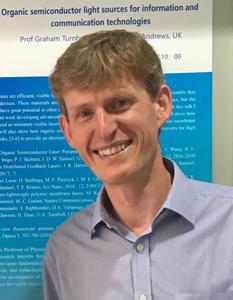 Graham Turnbull合作论文数School of Physics and Astronomy, University of St Andrews3Susana Martinez-Conde合作论文数3
 Marcus Nyström合作论文数Humanities Lab, Lund University3andrea gelmetti合作论文数2Brooke S. Jackson合作论文数2
 Guillermo Vivas-Mateos合作论文数2Maria Katsafadou合作论文数2
 Onyekachukwu Mary-Anne Amiebenomo合作论文数2
 Fadi R. Ghazala合作论文数2
 Thomas F Krauss合作论文数School of Physics, Engineering and Technology, University of York2




