Meckel–Gruber syndrome (MKS) is a recessively inherited, lethal disorder characterized by renal cystic dysplasia, occipital encephalocele, polydactyly and biliary dysgenesis. MKS is genetically heterogeneous with three loci mapped and two identified; MKS1 (17q23) and MKS3 (8q22.1). MKS1 is part of the Finnish disease heritage, while MKS3 has been described exclusively in consanguineous Asian families. Here we aimed to establish molecular diagnostics for MKS, determine the importance of MKS1 and MKS3 in non-consanguineous populations, and study genotype/phenotype correlations. The coding regions of MKS1 and MKS3 were screened for mutations by direct sequencing in 17 families clinically diagnosed with MKS in the US or The Netherlands. The clinical phenotype was compared to genic and allelic effects. Both mutations were identified in ten families; five MKS1 and five MKS3. All but two were compound heterozygotes, consistent with their non-consanguineous nature. The MKS1-Finmajor mutation accounted for 7/10 MKS1 mutations; two novel changes were additionally detected. Seven novel mutations were found in MKS3, including three missense changes. We concluded that MKS1 and MKS3 account for the majority of MKS in non-consanguineous populations of European origin. Polydactyly is usually found in MKS1 but rare in MKS3. Cases with no, or milder, CNS phenotypes were only found in MKS3; hypomorphic missense mutations may be associated with less severe CNS outcomes. This study is consistent with further genetic heterogeneity of MKS, but underlines the value of molecular diagnostics of the known genes to aid family planning decisions.
Leigh syndrome is a frequent, heterogeneous pediatric presentation of mitochondrial oxidative phosphorylation (OXPHOS) disease, manifesting with psychomotor retardation and necrotizing lesions in brain deep gray matter. OXPHOS occurs at the inner mitochondrial membrane through the integrated activity of five protein complexes, of which complex V (CV) functions in a dimeric form to directly generate adenosine triphosphate (ATP). Mutations in several different structural CV subunits cause Leigh syndrome; however, dimerization defects have not been associated with human disease. We report four Leigh syndrome subjects from three unrelated Ashkenazi Jewish families harboring a homozygous splice-site mutation (c.87 + 1G>C) in a novel CV subunit disease gene, USMG5. The Ashkenazi population allele frequency is 0.57%. This mutation produces two USMG5 transcripts, wild-type and lacking exon 3. Fibroblasts from two Leigh syndrome probands had reduced wild-type USMG5 mRNA expression and undetectable protein. The mutation did not alter monomeric CV expression, but reduced both CV dimer expression and ATP synthesis rate. Rescue with wild-type USMG5 cDNA in proband fibroblasts restored USMG5 protein, increased CV dimerization and enhanced ATP production rate. These data demonstrate that a recurrent USMG5 splice-site founder mutation in the Ashkenazi Jewish population causes autosomal recessive Leigh syndrome by reduction of CV dimerization and ATP synthesis.
Rare or novel gene variants in patients with proliferative diabetic retinopathy may contribute to disease development. We performed whole exome sequencing (WES) on patients at the phenotypic extremes of diabetic retinal complications: 57 patients diagnosed with proliferative diabetic retinopathy (PDR) as cases and 13 patients with no diabetic retinopathy despite at least 10years of type 2 diabetes as controls. Thirty-one out of the 57 cases and all 13 controls were from the African American Proliferative Diabetic Retinopathy Study (AA). The rest of the cases were of mixed ethnicities (ME). WES identified 721 candidate genes with rare or novel non-synonymous variants found in at least one case with PDR and not present in any controls. After filtering for genes with null alleles in greater than two cases, 28 candidate genes were identified in our ME cases and 16 genes were identified in our AA cases. Our analysis showed rare and novel variants within these genes that could contribute to the development of PDR, including rare non-synonymous variants in FAM132A, SLC5A9, ZNF600, and TMEM217. We also found previously unidentified variants in VEGFB and APOB. We found that VEGFB, VPS13B, PHF21A, NAT1, ZNF600, PKHD1L1 expression was reduced in human retinal endothelial cells (HRECs) cultured under high glucose conditions. In an exome sequence analysis of patients with PDR, we identified variants in genes that could contribute to pathogenesis. Six of these genes were further validated and found to have reduced expression in HRECs under high glucose conditions, suggestive of an important role in the development of PDR.
Purpose The risk of vision loss from proliferative diabetic retinopathy (PDR) can be reduced with timely detection and treatment. We aimed to identify serum molecular signatures that might help in the early detection of PDR in patients with diabetes. Methods A total of 40 patients with diabetes were recruited at King Khaled Eye Specialist Hospital in Riyadh, Saudi Arabia, 20 with extensive PDR and 20 with mild non-proliferative diabetic retinopathy (NPDR). The two groups were matched in age, gender, and known duration of diabetes. We examined the whole genome transcriptome of blood samples from the patients using RNA sequencing. We built a model using a support vector machine (SVM) approach to identify gene combinations that can classify the two groups. Results Differentially expressed genes were calculated from a total of 25,500 genes. Six genes (CCDC144NL, DYX1C1, KCNH3, LOC100506476, LOC285847, and ZNF80) were selected from the top 26 differentially expressed genes, and a combinatorial molecular signature was built based on the expression of the six genes. The mean area under receiver operating characteristic (ROC) curve was 0.978 in the cross validation. The corresponding sensitivity and specificity were 91.7% and 91.5%, respectively. Conclusions Our preliminary study defined a combinatorial molecular signature that may be useful as a potential biomarker for early detection of proliferative diabetic retinopathy in patients with diabetes. A larger-scale study with an independent cohort of samples is necessary to validate and expand these findings.
Purpose: Despite substantial progress in sequencing, current strategies can genetically solve only approximately 55–60% of inherited retinal degeneration (IRD) cases. This can be partially attributed to elusive mutations in the known IRD genes, which are not easily identified by the targeted next-generation sequencing (NGS) or Sanger sequencing approaches. We hypothesized that copy-number variations (CNVs) are a major contributor to the elusive genetic causality of IRDs. Methods: Twenty-eight cases previously unsolved with a targeted NGS were investigated with whole-genome single-nucleotide polymorphism (SNP) and comparative genomic hybridization (CGH) arrays. Results: Deletions in the IRD genes were detected in 5 of 28 families, including a de novo deletion. We suggest that the de novo deletion occurred through nonallelic homologous recombination (NAHR) and we constructed a genomic map of NAHR-prone regions with overlapping IRD genes. In this article, we also report an unusual case of recessive retinitis pigmentosa due to compound heterozygous mutations in SNRNP200 , a gene that is typically associated with the dominant form of this disease. Conclusions: CNV mapping substantially increased the genetic diagnostic rate of IRDs, detecting genetic causality in 18% of previously unsolved cases. Extending the search to other structural variations will probably demonstrate an even higher contribution to genetic causality of IRDs. Genet Med advance online publication 13 October 2016
We report a Caucasian boy with intractable epilepsy and global developmental delay. Whole-exome sequencing identified the likely genetic etiology as a novel p.K212E mutation in the X-linked gene HSD17B10 for mitochondrial short-chain dehydrogenase/reductase SDR5C1. Mutations in HSD17B10 cause the HSD10 disease, traditionally classified as a metabolic disorder due to the role of SDR5C1 in fatty and amino acid metabolism. However, SDR5C1 is also an essential subunit of human mitochondrial RNase P, the enzyme responsible for 5'-processing and methylation of purine-9 of mitochondrial tRNAs. Here we show that the p.K212E mutation impairs the SDR5C1-dependent mitochondrial RNase P activities, and suggest that the pathogenicity of p.K212E is due to a general mitochondrial dysfunction caused by reduction in SDR5C1-dependent maturation of mitochondrial tRNAs.
Background Next generation sequencing (NGS) offers a rapid and comprehensive method of screening for mutations associated with retinitis pigmentosa and related disorders. However, certain sequence alterations such as large insertions or deletions may remain undetected using standard NGS pipelines. One such mutation is a recently-identified Alu insertion into the Male Germ Cell-Associated Kinase (MAK) gene, which is missed by standard NGS-based variant callers. Here, we developed an in silico method of searching NGS raw sequence reads to detect this mutation, without the need to recalculate sequence alignments or to screen every sample by PCR. Methods The Linux program grep was used to search for a 23 bp “probe” sequence containing the known junction sequence of the insert. A corresponding search was performed with the wildtype sequence. The matching reads were counted and further compared to the known sequences of the full wildtype and mutant genomic loci. (See https://github.com/MEEIBioinformaticsCenter/grepsearch.) Results In a test sample set consisting of eleven previously published homozygous mutants, detection of the MAK-Alu insertion was validated with 100% sensitivity and specificity. As a discovery cohort, raw NGS reads from 1,847 samples (including custom and whole exome selective capture) were searched in ~1 hour on a local computer cluster, yielding an additional five samples with MAK-Alu insertions and solving two previously unsolved pedigrees. Of these, one patient was homozygous for the insertion, one compound heterozygous with a missense change on the other allele (c. 46G>A; p.Gly16Arg), and three were heterozygous carriers. Conclusions Using the MAK-Alu grep program proved to be a rapid and effective method of finding a known, disease-causing Alu insertion in a large cohort of patients with NGS data. This simple approach avoids wet-lab assays or computationally expensive algorithms, and could also be used for other known disease-causing insertions and deletions.
PURPOSE Patients with Usher syndrome type I (USH1) have retinitis pigmentosa, profound congenital hearing loss, and vestibular ataxia. This syndrome is currently thought to be associated with at least six genes, which are encoded by over 180 exons. Here, we present the use of state-of-the-art techniques in the molecular diagnosis of a cohort of 47 USH1 probands. METHODS The cohort was studied with selective exon capture and next-generation sequencing of currently known inherited retinal degeneration genes, comparative genomic hybridization, and Sanger sequencing of new USH1 exons identified by human retinal transcriptome analysis. RESULTS With this approach, we were able to genetically solve 14 of the 47 probands by confirming the biallelic inheritance of mutations. We detected two likely pathogenic variants in an additional 19 patients, for whom family members were not available for cosegregation analysis to confirm biallelic inheritance. Ten patients, in addition to primary disease-causing mutations, carried rare likely pathogenic USH1 alleles or variants in other genes associated with deaf-blindness, which may influence disease phenotype. Twenty-one of the identified mutations were novel among the 33 definite or likely solved patients. Here, we also present a clinical description of the studied cohort at their initial visits. CONCLUSIONS We found a remarkable genetic heterogeneity in the studied USH1 cohort with multiplicity of mutations, of which many were novel. No obvious influence of genotype on phenotype was found, possibly due to small sample sizes of the genotypes under study.
PURPOSE:Next-generation sequencing-based methods are being adopted broadly for genetic diagnostic testing, but the performance characteristics of these techniques with regard to test accuracy and reproducibility have not been fully defined.METHODS:We developed a targeted enrichment and next-generation sequencing approach for genetic diagnostic testing of patients with inherited eye disorders, including inherited retinal degenerations, optic atrophy, and glaucoma. In preparation for providing this genetic eye disease (GEDi) test on a CLIA-certified basis, we performed experiments to measure the sensitivity, specificity, and reproducibility, as well as the clinical sensitivity, of the test.RESULTS:The GEDi test is highly reproducible and accurate, with sensitivity and specificity of 97.9 and 100%, respectively, for single-nucleotide variant detection. The sensitivity for variant detection was notably better than the 88.3% achieved by whole-exome sequencing using the same metrics, because of better coverage of targeted genes in the GEDi test as compared with a commercially available exome capture set. Prospective testing of 192 patients with inherited retinal degenerations indicated that the clinical sensitivity of the GEDi test is high, with a diagnostic rate of 51%.CONCLUSION:Based on quantified performance metrics, the data suggest that selective targeted enrichment is preferable to whole-exome sequencing for genetic diagnostic testing.
Mutations to PKD1 and PKD2 are associated with autosomal dominant polycystic kidney disease (ADPKD). The absence of apparent PKD1/PKD2 linkage in five published European or North American families with ADPKD suggested a third locus, designated PKD3. Here we re-evaluated these families by updating clinical information, re-sampling where possible, and mutation screening for PKD1/PKD2. In the French-Canadian family, we identified PKD1: p.D3782_V3783insD, with misdiagnoses in two individuals and sample contamination explaining the lack of linkage. In the Portuguese family, PKD1: p.G3818A segregated with the disease in 10 individuals in three generations with likely misdiagnosis in one individual, sample contamination, and use of distant microsatellite markers explaining the linkage discrepancy. The mutation PKD2: c.213delC was found in the Bulgarian family, with linkage failure attributed to false positive diagnoses in two individuals. An affected son, but not the mother, in the Italian family had the nonsense mutation PKD1: p.R4228X, which appeared de novo in the son, with simple cysts probably explaining the mother's phenotype. No likely mutation was found in the Spanish family, but the phenotype was atypical with kidney atrophy in one case. Thus, re-analysis does not support the existence of a PKD3 in ADPKD. False positive diagnoses by ultrasound in all resolved families shows the value of mutation screening, but not linkage, to understand families with discrepant data.
Background The retina is a complex tissue comprised of multiple cell types that is affected by a diverse set of diseases that are important causes of vision loss. Characterizing the transcripts, both annotated and novel, that are expressed in a given tissue has become vital for understanding the mechanisms underlying the pathology of disease. Results We sequenced RNA prepared from three normal human retinas and characterized the retinal transcriptome at an unprecedented level due to the increased depth of sampling provided by the RNA-seq approach. We used a non-redundant reference transcriptome from all of the empirically-determined human reference tracks to identify annotated and novel sequences expressed in the retina. We detected 79,915 novel alternative splicing events, including 29,887 novel exons, 21,757 3′ and 5′ alternate splice sites, and 28,271 exon skipping events. We also identified 116 potential novel genes. These data represent a significant addition to the annotated human transcriptome. For example, the novel exons detected increase the number of identified exons by 3%. Using a high-throughput RNA capture approach to validate 14,696 of these novel transcriptome features we found that 99% of the putative novel events can be reproducibly detected. Further, 15-36% of the novel splicing events maintain an open reading frame, suggesting they produce novel protein products. Conclusions To our knowledge, this is the first application of RNA capture to perform large-scale validation of novel transcriptome features. In total, these analyses provide extensive detail about a previously uncharacterized level of transcript diversity in the human retina.
Whole-exome sequencing and autozygosity mapping studies, independently performed in subjects with defective combined mitochondrial OXPHOS-enzyme deficiencies, identified a total of nine disease-segregating FBXL4 mutations in seven unrelated mitochondrial disease families, composed of six singletons and three siblings. All subjects manifested early-onset lactic acidemia, hypotonia, and developmental delay caused by severe encephalomyopathy consistently associated with progressive cerebral atrophy and variable involvement of the white matter, deep gray nuclei, and brainstem structures. A wide range of other multisystem features were variably seen, including dysmorphism, skeletal abnormalities, poor growth, gastrointestinal dysmotility, renal tubular acidosis, seizures, and episodic metabolic failure. Mitochondrial respiratory chain deficiency was present in muscle or fibroblasts of all tested individuals, together with markedly reduced oxygen consumption rate and hyperfragmentation of the mitochondrial network in cultured cells. In muscle and fibroblasts from several subjects, substantially decreased mtDNA content was observed. FBXL4 is a member of the F-box family of proteins, some of which are involved in phosphorylation-dependent ubiquitination and/or G protein receptor coupling. We also demonstrate that FBXL4 is targeted to mitochondria and localizes in the intermembrane space, where it participates in an approximately 400 kDa protein complex. These data strongly support a role for FBXL4 in controlling bioenergetic homeostasis and mtDNA maintenance. FBXL4 mutations are a recurrent cause of mitochondrial encephalomyopathy onset in early infancy.
Leber congenital amaurosis (LCA) is an infantile-onset form of inherited retinal degeneration characterized by severe vision loss(1,2). Two-thirds of LCA cases are caused by mutations in 17 known disease-associated genes(3) (Retinal Information Network (RetNet)). Using exome sequencing we identified a homozygous missense mutation (c.25G>A, p.Val9Met) in NMNAT1 that is likely to be disease causing in two siblings of a consanguineous Pakistani kindred affected by LCA. This mutation segregated with disease in the kindred, including in three other children with LCA. NMNAT1 resides in the previously identified LCA9 locus and encodes the nuclear isoform of nicotinamide mononucleotide adenylyltransferase, a rate-limiting enzyme in nicotinamide adenine dinucleotide (NAD(+)) biosynthesis(4,5). Functional studies showed that the p.Val9Met alteration decreased NMNAT1 enzyme activity. Sequencing NMNAT1 in 284 unrelated families with LCA identified 14 rare mutations in 13 additional affected individuals. These results are the first to link an NMNAT isoform to disease in humans and indicate that NMNAT1 mutations cause LCA.
 Peter C. Harris合作论文数Division of Nephrology & Hypertension, Department of Internal Medicine, Mayo Clinic;Department of Biochemistry and Molecular Biology, Mayo Clinic17
 Eric A. Pierce合作论文数Ocular Genomics Institute, Harvard Medical School;Berman-Gund Laboratory for the Study of Retinal Degenerations, Massachusetts Eye and Ear15
 Vicente E. Torres合作论文数Department of Nephrology and Hypertension, Mayo Clinic;Translational Polycystic Kidney Disease Center, Mayo Clinic14
 Xiaowu Gai合作论文数Department of Zoology;Iowa State University;Department of Zoology, Iowa State University14
 Sandro Rossetti合作论文数13Place Emily合作论文数10
 Marni J. Falk合作论文数Departments of Anesthesiology and Genetics, Case School of Medicine, Cleveland, OH, USA7Kubly Vickie J合作论文数5
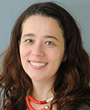 Kinga M. Bujakowska合作论文数Harvard Medical School, Harvard University;Massachusetts Eye and Ear, Mass General Brigham5Daniel Navarro-Gomez合作论文数5
 Joseph D M White合作论文数Baylor University, Baylor University5Farkas Michael H合作论文数Ocular Genomics Institute, Massachusetts Eye and Ear Infirmary4
 Eliot L. Berson合作论文数Department of Ophthalmology, Harvard Medical School, Harvard University4
 Arlene Chapman合作论文数The University of Chicago Medical Center4
 K. Ty Bae合作论文数Department of Diagnostic Radiology, Li Ka Shing Faculty of Medicine, The University of Hong Kong;University of Pittsburgh4Catherine M. Meyers合作论文数3Dan Yi Wang合作论文数3Paul A. Thompson合作论文数3
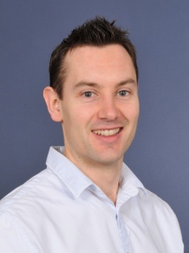 Phillip Miller合作论文数Department of Chemistry, Faculty of Natural Sciences, Imperial College London3
 Katharina Hopp合作论文数3Harper Shyana合作论文数3Taub Daniel G合作论文数3McCormick Elizabeth M合作论文数3Bernard F. King合作论文数Department of Radiology, Mayo Clinic3
 Jared J. Grantham合作论文数University of Kansas Medical Center3
 Douglas C Wallace合作论文数The Center for Mitochondrial and Epigenomic Medicine, Children's Hospital of Philadelphia Research Institute;Department of Pediatrics, Perelman School of Medicine, University of Pennsylvania3Dominique Chauveau合作论文数Centre Hospitalier Universitaire de Toulouse3Carol Weigel Difranco合作论文数3Christopher J. Ward合作论文数Department of Chemistry, University of Bath3
 Janey L. Wiggs合作论文数Department of Ophthalmology, Harvard Medical School, Harvard University;Howe Laboratory, Massachusetts Eye and Ear, Harvard Medical School, Harvard University3Julian Ostrovsky合作论文数3
 Denise D. Walker合作论文数School of Social Work, University of Washington3
 William Bennett合作论文数Legacy Good Samaritan Medical Center3Eliecer Coto合作论文数2Christina M Heyer合作论文数2Keri F Allen合作论文数2Sara Vidoni合作论文数2
 Matteo Gorza合作论文数2Aliete Langsdorf合作论文数2
 Lisa Guay-Woodford合作论文数2
 Holger Prokisch合作论文数2
 Maria E Sousa合作论文数2maria janessian合作论文数2Jeff M Slezak合作论文数2
 Neal Sondheimer合作论文数Department of Molecular Genetics, Temerty Faculty of Medicine, University of Toronto;Division of Clinical and Metabolic Genetics, The Hospital for Sick Children;Synlogic2
 Srinivasan Satish合作论文数2
 Jamie L Sundsbak合作论文数2
 Shane Pankratz合作论文数Health Sciences Center, The University of New Mexico;Comprehensive Cancer Center, The University of New Mexico2
 Tobias Haack合作论文数Institute of Medical Genetics and Applied Genomics, University of Tuebingen;Technische Universität München2




