Mechanisms that maintain genome integrity are crucial for coordinating transcription that drives mammalian forebrain development. Neural progenitor cells and differentiating neurons in the developing forebrain sustain high transcriptional activity and metabolic demand and are therefore vulnerable to DNA damage. Transcription-coupled nucleotide excision repair is a specialized DNA repair pathway that helps to mitigate damage induced by ‘bulky’ adducts such as UV-induced pyrimidine dimers and monoadducts formed by reactive oxygen species. TC-NER factors, which include CSB/CSA, UVSSA-USP7, ELOF1, and STK19, coordinate and assemble the complex to initiate repair. Notably, TC-NER safeguards genome integrity and plays an essential role in neuronal differentiation, synaptogenesis, and neurogenesis. Impaired TC-NER pathway activity manifests in tissue-level pathologies, including neurodegeneration and increased susceptibility to neurological deficits. This is relevant to neurodevelopmental disorders that stem from TC-NER deficiency, such as Cockayne syndrome, Trichothiodystrophy, and Cerebro-Oculo-Facio-Skeletal syndrome. Although deficits in TC-NER have been well established as a contributor to a variety of neurodegenerative disorders, its roles in the developing forebrain across various cell types and neurodevelopmental windows remain poorly defined. In this review, we highlight recent studies investigating mechanisms linking TC-NER deficiency to forebrain developmental phenotypes and summarize knowledge gaps in the field regarding cell-type specificity, regional vulnerability, and therapeutic windows for intervention.
Charcot-Marie-Tooth type 1B (CMT1B) is a demyelination neuropathy caused by over 200 mutations in the myelin protein zero (MPZ) gene. Here, we generated two induced pluripotent stem cell (iPSC) lines from fibroblasts isolated from the skin biopsies of CMT1B patients, each carrying a distinct MPZ mutation (Arg98Cys and Ser63del). The iPSC lines created in this work retained their respective MPZ mutation, exhibited normal karyotypes, expressed pluripotency markers, and demonstrated the ability to differentiate into three germ-layer cell types. These lines offer a valuable tool for exploring and modeling dominant CMT1B disease within a human cellular framework.
Lipid dyshomeostasis and tau pathology are present in frontotemporal lobar degeneration (FTLD) and Alzheimer's disease (AD). However, the relationship between lipid dyshomeostasis and tau pathology remains unclear. We report that GRAM Domain Containing 1B (GRAMD1B), a nonvesicular cholesterol transporter, is increased in excitatory neurons of human neural organoids (HNOs) with the MAPT R406W mutation. Human FTLD, AD cases, and PS19 tau mice also have increased GRAMD1B expression. We show that overexpression of GRAMD1B increases levels of free cholesterol, lipid droplets, and impairs autophagy flux. Modulating GRAMD1B in iPSC-derived neurons also alters key autophagy-related components such as PI3K, phospho-AKT, and p62, as well as phosphorylated tau, and CDK5R1. Blocking GRAMD1B function decreases free cholesterol and lipid droplets. Knocking down GRAMD1B additionally reduces phosphorylated tau, and CDK5R1 expression. Our findings elucidate the role of GRAMD1B in the nervous system and highlight its relevance to FTLD and AD.
Long interspersed nuclear element -1 (L1 or LINE -1) is a highly abundant mobile genetic element in both humans and mice, comprising almost 20% of each genome. L1s are silenced by several mechanisms, as their uncontrolled expression has the potential to induce genomic instability. However, L1s are paradoxically expressed at high levels in differentiating neural progenitor cells. Using in vitro and in vivo techniques to modulate L1 expression, we report that L1s play a critical role in both human and mouse brain development by regulating the rate of neural differentiation in a reverse -transcription -independent manner.
Tuberous sclerosis complex (TSC) is an autosomal dominant disorder caused by mutations in either the TSC1 or TSC2 genes. Though TSC causes the formation of nonmalignant tumors throughout multiple organs, the most frequent causes of mortality and morbidity are due to neurological complications. In two-thirds of cases, TSC occurs sporadically and TSC2 pathogenic variants are approximately three times more prevalent than TSC1 pathogenic variants. Here, we utilized CRISPR-Cas9-mediated homology directed repair in patient induced pluripotent stem cells (iPSCs) to correct two types of TSC2 pathogenic variants generating two isogenic lines. In one line, we corrected a splice acceptor variant (c.2743-1G>A), which causes the skipping of coding exon 23 and subsequent frameshift and introduction of a stop codon in coding exon 25. In the second line, we corrected a missense variant in coding exon 40 within the GTPase-activating protein domain (c.5228G>A, p.R1743Q). The generation of TSC2 patient iPSCs in parallel with their corresponding CRISPR-corrected isogenic lines will be an important tool for disease modeling applications and for developing therapeutics.
Glioblastoma multiforme (GBM) is a highly lethal human cancer thought to originate from a self-renewing and therapeutically-resistant population of glioblastoma stem cells (GSCs). The intrinsic mechanisms enacted by GSCs during 3D tumor formation, however, remain unclear, especially in the stages prior to angiogenic/immunological infiltration. In this study, we performed a deep characterization of the genetic, immune, and metabolic profiles of GBM organoids from several patient-derived GSCs (GBMO). Despite being devoid of immune cells, transcriptomic analysis across GBMO revealed a surprising immune-like molecular program, enriched in cytokine, antigen presentation and processing, T-cell receptor inhibitors, and interferon genes. We find two important cell populations thought to drive GBM progression, Special AT-rich sequence-binding protein 2 (SATB2+) and homeodomain-only protein homeobox (HOPX+) progenitors, contribute to this immune landscape in GBMO and GBM in vivo. These progenitors, but not other cell types in GBMO, are resistant to conventional GBM therapies, temozolomide and irradiation. Our work defines a novel intrinsic immune-like landscape in GBMO driven, in part, by SATB2+ and HOPX+ progenitors and deepens our understanding of the intrinsic mechanisms utilized by GSCs in early GBM formation.
Single-stranded DNA (ssDNA) templates along with Cas9 have been used for gene insertion but suffer from low efficiency. Here, we show that ssDNA with chemical modifications in 10-17% of internal bases (eDNA) is compatible with the homologous recombination machinery. Moreover, eDNA templates improve gene insertion by 2-3 fold compared to unmodified and end-modified ssDNA in airway basal stem cells (ABCs), hematopoietic stem and progenitor cells (HSPCs), T-cells and endothelial cells. Over 50% of alleles showed gene insertion in three clinically relevant loci (CFTR, HBB, and CCR5) in ABCs using eDNA and up to 70% of alleles showed gene insertion in the HBB locus in HSPCs. This level of correction is therapeutically relevant and is comparable to adeno-associated virus-based templates. Knocking out TREX1 nuclease improved gene insertion using unmodified ssDNA but not eDNA suggesting that chemical modifications inhibit TREX1. This approach can be used for therapeutic applications and biological modeling.
Recent studies have shown the importance of the dynamic tumor microenvironment (TME) in high-grade gliomas (HGGs). In particular, myeloid cells are known to mediate immunosuppression in glioma; however, it is still unclear if myeloid cells play a role in low-grade glioma (LGG) malignant progression. Here, we investigate the cellular heterogeneity of the TME using single-cell RNA sequencing in a murine glioma model that recapitulates the malignant progression of LGG to HGG. LGGs show increased infiltrating CD4+ and CD8+ T cells and natural killer (NK) cells in the TME, whereas HGGs abrogate this infiltration. Our study identifies distinct macrophage clusters in the TME that show an immune-activated phenotype in LGG but then evolve to an immunosuppressive state in HGG. We identify CD74 and macrophage migration inhibition factor (MIF) as potential targets for these distinct macrophage populations. Targeting these intra-tumoral macrophages in the LGG stage may attenuate their immunosuppressive properties and impair malignant progression.
Variants in the AUTS2 gene are associated with a broad spectrum of neurological conditions characterized by intellectual disability, microcephaly, and congenital brain malformations. Here, we use a human cerebral organoid model to investigate the pathophysiology of a heterozygous de novo missense AUTS2 variant identified in a patient with multiple neurological impairments including primary microcephaly and profound intellectual disability. Proband cerebral organoids exhibit reduced growth, deficits in neural progenitor cell (NPC) proliferation and disrupted NPC polarity within ventricular zone-like regions compared to control cerebral organoids. We used CRISPR-Cas9-mediated gene editing to correct this variant and demonstrate rescue of impaired organoid growth and NPC proliferative deficits. Single-cell RNA sequencing revealed a marked reduction of G1/S transition gene expression and alterations in WNT-β-catenin signalling within proband NPCs, uncovering a novel role for AUTS2 in NPCs during human cortical development. Collectively, these results underscore the value of cerebral organoids to investigate molecular mechanisms underlying AUTS2 syndrome.
Objective: To characterize the effect of activation of the interferome in human organoids toward understanding human cortical neuronal dysfunction. Background: The mechanisms by which maternal infection and neuroinflammation impact human brain development, function, and repair remain unclear. IFN-g is a proinflammatory cytokine found elevated in serum of pregnant mothers with children diagnosed with autism and in Th1 cells in the cerebrospinal fluid of multiple sclerosis patients. Design/Methods: Nearly three hundred human cerebral organoids derived from iPSCs were used across all analyses. Organoids were continually exposed to IFN-g and compared to controls for up to 80 days in culture. 10X single-cell transcriptomics was performed at three timepoints, along with immunolabeling and pulse EdU experiments. Bioinformatic and Markov Chain analyses were used to determine the molecular, cellular, and systems level effects of IFN-g. Results: IFN-g exposure at low concentration restricts organoid growth across timepoints, diminishes the pool of cycling radial glia progenitors, and disrupts ventricular adherens cytoarchitecture. We find no evidence of cell cycle arrest or apoptosis, but rather an induction of premature neuronal differentiation and acceleration of neuronal maturation, that stems from cell-type-specific transcriptional rewiring by IFN-g, inducing expression of inflammatory and neurogenic genes that persists in all cell lineages. Markov chain modeling demonstrates that IFN-g targets differentiation lineages in a parallel pan-neuronal fashion. Finally, we find that cell-type-specific gene dysregulation induced by IFN-g overlaps with patient derived organoids from genetically linked autism spectrum disorder, and neuronal intrinsic dysregulation observed in cortical neurons in progressive multiple sclerosis. Conclusions: Our results show the feasibility of using a human brain organoid to model the effects of neuroinflammation on human brain development and function. This approach reveals a novel inflammatory mechanistic link with autism and identifies a critical network of neuronal identity and synaptic genes perturbed in multiple sclerosis recapitulated in a preclinical model. Disclosure: Ethan Hollingsworth has nothing to disclose. Mr. Julian has nothing to disclose. Fumihiro Watanabe has nothing to disclose. Mr. Reutershan has nothing to disclose. Miss Julian has nothing to disclose. Ms. Martorell Serra has nothing to disclose. Sofia Lizarraga has nothing to disclose. Dr. Hester has nothing to disclose. Dr. Imitola has received personal compensation in the range of $500-$4,999 for serving as a Consultant for Novartis . The institution of Dr. Imitola has received research support from biogen. Dr. Imitola has a non-compensated relationship as a Board Member with National MS Society that is relevant to AAN interests or activities. Dr. Imitola has a non-compensated relationship as a Committee Member with International Society for Stem Cell Research that is relevant to AAN interests or activities.
Somatic mosaicism is a known cause of neurological disorders, including developmental brain malformations and epilepsy. Brain mosaicism is traditionally attributed to post-zygotic genetic alterations arising in fetal development. Here we describe post-zygotic rescue of meiotic errors as an alternate origin of brain mosaicism in patients with focal epilepsy who have mosaic chromosome 1q copy number gains. Genomic analysis showed evidence of an extra parentally derived chromosome 1q allele in the resected brain tissue from five of six patients. This copy number gain is observed only in patient brain tissue, but not in blood or buccal cells, and is strongly enriched in astrocytes. Astrocytes carrying chromosome 1q gains exhibit distinct gene expression signatures and hyaline inclusions, supporting a novel genetic association for astrocytic inclusions in epilepsy. Further, these data demonstrate an alternate mechanism of brain chromosomal mosaicism, with parentally derived copy number gain isolated to brain, reflecting rescue in other tissues during development.
AUTS2 syndrome is a genetic disorder that causes intellectual disability, microcephaly, and other phenotypes. Syndrome severity is worse when mutations involve 3’ regions (exons 9-19) of the AUTS2 gene. Human AUTS2 protein has two major isoforms, full-length (1259 aa) and C-terminal (711 aa), the latter produced from an alternative transcription start site in exon 9. Structurally, AUTS2 contains the putative “AUTS2 domain” (∼200 aa) conserved among AUTS2 and its ohnologs, fibrosin, and fibrosin-like-1. Also, AUTS2 contains extensive low-complexity sequences and intrinsically disordered regions, features typical of RNA-binding proteins. During development, AUTS2 is expressed by specific progenitor cell and neuron types, including pyramidal neurons and Purkinje cells. AUTS2 localizes mainly in cell nuclei, where it regulates transcription and RNA metabolism. Some studies have detected AUTS2 in neurites, where it may regulate cytoskeletal dynamics. Neurodevelopmental functions of AUTS2 have been studied in diverse model systems. In zebrafish, auts2a morphants displayed microcephaly. In mice, excision of different Auts2 exons (7, 8, or 15) caused distinct phenotypes, variously including neonatal breathing abnormalities, cerebellar hypoplasia, dentate gyrus hypoplasia, EEG abnormalities, and behavioral changes. In mouse embryonic stem cells, AUTS2 could promote or delay neuronal differentiation. Cerebral organoids, derived from an AUTS2 syndrome patient containing a pathogenic missense variant in exon 9, exhibited neocortical growth defects. Emerging technologies for analysis of human cerebral organoids will be increasingly useful for understanding mechanisms underlying AUTS2 syndrome. Questions for future research include whether AUTS2 binds RNA directly, how AUTS2 regulates neurogenesis, and how AUTS2 modulates neural circuit formation.
The high-affinity copper transporter CTR1 is encoded by CTR1 (SLC31A1), a gene locus for which no detailed genotype-phenotype correlations have previously been reported. We describe identical twin male infants homozygous for a novel missense variant NM_001859.4:c.284 G > A (p.Arg95His) in CTR1 with a distinctive autosomal recessive syndrome of infantile seizures and neurodegeneration, consistent with profound central nervous system copper deficiency. We used clinical, biochemical and molecular methods to delineate the first recognized examples of human CTR1 deficiency. These included clinical phenotyping, brain imaging, assays for copper, cytochrome c oxidase (CCO), and mitochondrial respiration, western blotting, cell transfection experiments, confocal and electron microscopy, protein structure modeling and fetal brain and cerebral organoid CTR1 transcriptome analyses. Comparison with two other critical mediators of cellular copper homeostasis, ATP7A and ATP7B, genes associated with Menkes disease and Wilson disease, respectively, revealed that expression of CTR1 was highest. Transcriptome analyses identified excitatory neurons and radial glia as brain cell types particularly enriched for copper transporter transcripts. We also assessed the effects of Copper Histidinate in the patients' cultured cells and in the patients, under a formal clinical protocol. Treatment normalized CCO activity and enhanced mitochondrial respiration in vitro, and was associated with modest clinical improvements. In combination with present and prior studies, these infants' clinical, biochemical and molecular phenotypes establish the impact of this novel variant on copper metabolism and cellular homeostasis and illuminate a crucial role for CTR1 in human brain development. CTR1 deficiency represents a newly defined inherited disorder of brain copper metabolism.
Respiratory system damage is the primary cause of mortality in individuals who are exposed to vesicating agents including sulfur mustard (SM). Despite these devastating health complications, there are no fielded therapeutics that are specific for such injuries. Previous studies reported that SM inhalation depleted the tracheobronchial airway epithelial stem cell (TSC) pool and supported the hypothesis, TSC replacement will restore airway epithelial integrity and improve health outcomes for SM-exposed individuals. TSC express Major Histocompatibility Complex (MHC-I) transplantation antigens which increases the chance that allogeneic TSC will be rejected by the patient’s immune system. However, previous studies reported that Beta-2 microglobulin (B2M) knockout cells lacked cell surface MHC-I and suggested that B2M knockout TSC would be tolerated as an allogeneic graft. This study used a Cas9 ribonucleoprotein (RNP) to generate B2M-knockout TSC, which are termed Universal Donor Stem Cells (UDSC). Whole genome sequencing identified few off-target modifications and demonstrated the specificity of the RNP approach. Functional assays demonstrated that UDSC retained their ability to self-renew and undergo multilineage differentiation. A preclinical model of SM inhalation was used to test UDSC efficacy and identify any treatment-associated adverse events. Adult male Sprague-Dawley rats were administered an inhaled dose of 0.8 mg/kg SM vapor which is the inhaled LD50 on day 28 post-challenge. On recovery day 2, vehicle or allogeneic Fisher rat UDSC were delivered intravenously (n = 30/group). Clinical parameters were recorded daily, and planned euthanasia occurred on post-challenge days 7, 14, and 28. The vehicle and UDSC treatment groups exhibited similar outcomes including survival and a lack of adverse events. These studies establish a baseline which can be used to further develop UDSC as a treatment for SM-induced airway disease.
Abstract Myeloid cells and macrophages have been shown to promote immunosuppression in high-grade gliomas (HGG), however their roles in malignant progression of low-grade glioma (LGG) are poorly understood. Here, we investigated the heterogeneity of the immune microenvironment during glioma progression using a murine model that recapitulates the malignant progression of low to high-grade glioma. To that end, we performed single-cell RNA sequencing on CD45+ immune cells isolated from animals bearing no tumor (NT), LGG, and HGG. We observed an increased infiltration of CD4+ T cells, CD8+ T cells, B cells, and natural killer cells in the tumor microenvironment of LGG, whereas this infiltration was abrogated in HGG. Our study identified two distinct macrophage clusters across all 3 samples, with signatures of bone marrow derived and resident macrophages, respectively. These macrophages showed an immune-activated phenotype (Stat1, Tnf, Cxcl9 and Cxcl10) in LGG, but then evolved to a more immunosuppressive state (Lgals3, Apoc1 and Id2) in HGG, restricting T cell recruitment and activation. In addition, we identified CD74 and macrophage migration inhibition factor (MIF) as potential targets for both these distinct macrophage populations, based on their increased expression in LGG and HGG compared to NT. Targeting these factors during the LGG therapeutic window may inhibit myeloid cells and intra-tumoral macrophages and attenuate their immunosuppressive properties and impair malignant progression.
Human middle temporal gyrus (MTG) is a vulnerable brain region in early Alzheimer’s disease (AD), but little is known about the molecular mechanisms underlying this regional vulnerability. Here we utilize the 10 × Visium platform to define the spatial transcriptomic profile in both AD and control (CT) MTG. We identify unique marker genes for cortical layers and the white matter, and layer-specific differentially expressed genes (DEGs) in human AD compared to CT. Deconvolution of the Visium spots showcases the significant difference in particular cell types among cortical layers and the white matter. Gene co-expression analyses reveal eight gene modules, four of which have significantly altered co-expression patterns in the presence of AD pathology. The co-expression patterns of hub genes and enriched pathways in the presence of AD pathology indicate an important role of cell–cell-communications among microglia, oligodendrocytes, astrocytes, and neurons, which may contribute to the cellular and regional vulnerability in early AD. Using single-molecule fluorescent in situ hybridization, we validated the cell-type-specific expression of three novel DEGs (e.g., KIF5A , PAQR6 , and SLC1A3 ) and eleven previously reported DEGs associated with AD pathology (i.e., amyloid beta plaques and intraneuronal neurofibrillary tangles or neuropil threads) at the single cell level. Our results may contribute to the understanding of the complex architecture and neuronal and glial response to AD pathology of this vulnerable brain region.
36 Variants in the AUTS2 gene are associated with a broad spectrum of neurological 37 conditions characterized by intellectual disability, microcephaly, and congenital brain 38 malformations. Here, we use a human cerebral organoid (CO) model to investigate the 39 pathophysiology of a heterozygous de novo missense AUTS2 variant identified in a 40 patient with multiple neurological impairments including primary microcephaly and 41 profound intellectual disability. Proband COs exhibit reduced growth, deficits in neural 42 progenitor cell (NPC) proliferation and disrupted NPC polarity within ventricular zone43 like regions compared to control COs. We used CRISPR-Cas9-mediated gene editing to 44 correct this variant and demonstrate rescue of impaired organoid growth and NPC 45 proliferative deficits. Single-cell RNA sequencing revealed a marked reduction of G1/S 46 transition gene expression and alterations in WNT-β-Catenin signaling within proband 47 NPCs, uncovering a novel role for AUTS2 in NPCs during human cortical development. 48 Collectively, these results underscore the value of COs to uncover molecular 49 mechanisms underlying AUTS2 syndrome. 50 51
Neurosphere cultures have been used to propagate and study the intrinsic properties of neural stem cells (NSCs) for more than two decades but this method has many limitations. It is well known that neurospheres fuse in culture, but the long‐term biological consequences of this phenomena are not well characterized. We leveraged the fusion behavior of human neurospheres to improve upon this technique with our Neurosphere‐derived organoid‐like aggregates (NEDAS) culture method, allowing the fusion of human NSCs at high density, which were maintained in orbital shaker conditions for 8‐12 weeks without passing leading to the formation of 3D organoid‐like aggregates without the use of Matrigel. NEDAS organoids proliferate and self‐organize into neural rosettes, expressing PAX6 and SOX2 in ventricular zone (VZ)‐like proliferative areas. Outside these rosettes, we identified corridors of migratory radial glial progenitors expressing Phospo‐vimentin, CRYAB. In addition to DLX2, CXCR4 + progenitors. Further, we found immature neurons within cortical‐like areas highly enriched for DCX and TUJ1, in addition to GABA+ and excitatory VGLUT1+ neurons. Here, we provide a protocol to generate NEDAS, additionally, we present a protocol for immunostaining of NEDAS organoids for confocal imaging. This protocol may be useful to dissect the self‐organization and morphogenetic programs of populations of human NSCs offering an advantageous alternative to the conventional neurospheres method, generating more cell type diversity, within tissue‐like aggregates over extended periods of time without dissociation or passing. NEDAS may be a complementary method to cerebral organoids protocols from IPSCs. © 2021 Wiley Periodicals LLC.
Phosphatase and tensin homologue (PTEN) regulates cell growth and survival through inhibition of the mammalian target of rapamycin (MTOR) signalling pathway. Germline genetic variation of PTEN is associated with autism, macrocephaly and PTEN hamartoma tumour syndromes. The effect of developmental PTEN somatic mutations on nervous system phenotypes is not well understood, although brain somatic mosaicism of MTOR pathway genes is an emerging cause of cortical dysplasia and epilepsy in the paediatric population. Here we report two somatic variants of PTEN affecting a single patient presenting with intractable epilepsy and hemimegalencephaly that varied in clinical severity throughout the left cerebral hemisphere. High-throughput sequencing analysis of affected brain tissue identified two somatic variants in PTEN. The first variant was present in multiple cell lineages throughout the entire hemisphere and associated with mild cerebral overgrowth. The second variant was restricted to posterior brain regions and affected the opposite PTEN allele, resulting in a segmental region of more severe malformation, and the only neurons in which it was found by single-nuclei RNA-sequencing had a unique disease-related expression profile. This study reveals brain mosaicism of PTEN as a disease mechanism of hemimegalencephaly and furthermore demonstrates the varying effects of single- or bi-allelic disruption of PTEN on cortical phenotypes.
 Brian K. Kaspar合作论文数11
 Summer Fair合作论文数Steve and Cindy Rasmussen Institute for Genomic Medicine, Nationwide Children’s Hospital8
 Dominic Julian合作论文数The Steve and Cindy Rasmussen Institute for Genomic Medicine, Abigail Wexner Research Institute at Nationwide Children's Hospital7
 Bedrosian Tracy A合作论文数Wexner Med Ctr, Ohio State Univ7
 Katherine E Miller合作论文数The Steve and Cindy Rasmussen Institute for Genomic Medicine, Abigail Wexner Research Institute at Nationwide Children's Hospital6
 Jaime Imitola合作论文数Center for Neurologic Diseases, Brigham and Women's Hospital, 77 Avenue Louis Pasteur, HIM 714, Boston, MA 02115, USA6Watanabe Fumihiro合作论文数College of Medicine Wexner Medical Center, The Ohio State University;Hertie-Institute for Clinical Brain Research, University of Tübingen;Department of Neurology and Immunobiology, Yale School of Medicine;Department of Neurology, Thomas Jefferson University;Hospital Universitari Vall d'Hebron, Universitat Autònoma de Barcelona;Hospital Universitari Vall d'Hebron, Universitat Autònoma de Barcelona5John C Thompson合作论文数4
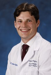 Ethan W Hollingsworth合作论文数University of California Irvine4
 Thomas Beach合作论文数Civin Laboratory for Neuropathology, Banner Sun Health Research Institute;Evelyn F. McKnight Brain Institute, The University of Arizona4Rao Meghan合作论文数4
 Geidy Serrano合作论文数Civin Laboratory for Neuropathology, Banner Sun Health Research Institute;Arizona State University4
 Elaine R. Mardis合作论文数Departments of Pediatrics, College of Medicine, The Ohio State University;Institute for Genomic Medicine, Nationwide Children's Hospital4
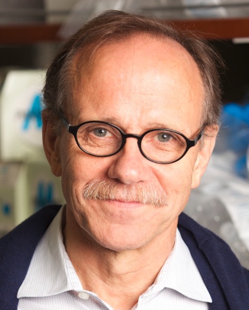 Rusty Gage合作论文数Laboratory of Genetics, Salk Institute for Biological Studies;Kavli Institute for Brain and Mind, Salk Institute for Biological Studies;Department of Neurosciences, University of California, San Diego4Michael B. Weinstein合作论文数4Hongjun Fu合作论文数4
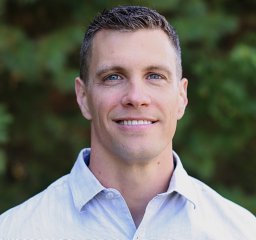 Craig A. Mcelroy合作论文数College of Medicine, Ohio State University4Diana Acosta合作论文数3
 J. Westfall合作论文数3
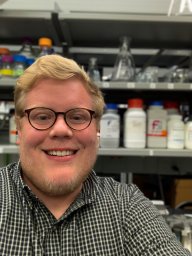 Cody Morrison合作论文数3Annalisa M. Hartlaub合作论文数Nationwide Childrens Hosp3
 Meisam Naeimi Kararoudi合作论文数Department of Pediatrics, The Ohio State University College of Medicine3Liangping Li合作论文数3
 Yuzhou Chang合作论文数Ohio State Univ, Coll Med, Dept Biomed Informat, Columbus, OH 43210 USA3Soo H. Kim合作论文数3
 Cankun Wang合作论文数Ohio State Univ, Coll Med, Dept Biomed Informat, Columbus, OH 43210 USA3
 Nathalie L Maitre合作论文数Nationwide Childrens Hosp, Abigail Wexner Res Inst3
 Mehboob Ali合作论文数The Research Institute at Nationwide Children's Hospital3
 Prajwal Rajappa合作论文数Departments of Neurosurgery and.3Ye Liu合作论文数3
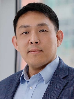 Qin Ma (马勤)合作论文数Division of Bioinformatics and Computational Biology, Comprehensive Cancer Center, The Ohio State University3
 Irina Buhimschi合作论文数Perinatal Research Laboratory, Department of Obstetrics and Gynecology, College of Medicine, University of Illinois at Chicago3
 Richard K. Wilson合作论文数Institute for Genomic Medicine, Nationwide Children's Hospital;Ohio State University College of Medicine3
 Ryan Roberts合作论文数College of Medicine and Public Health, The Ohio State University3Seth A. Faith合作论文数Department of Immunology, University of Pittsburgh School of Medicine3
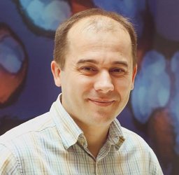 Carlos J. Miranda合作论文数Department of Medicine, Hôpital Notre-Dame3
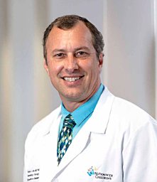 Dean A. Lee合作论文数Division of Hematology and Oncology, Nationwide Children's Hospital;Center for Childhood Cancer, Nationwide Children's Hospital;College of Medicine, The Ohio State University3
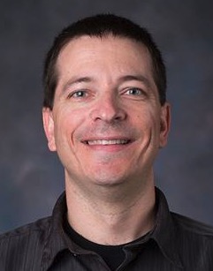 Dan Koboldt合作论文数Institute for Genomic Medicine, Nationwide Children's Hospital;The Ohio State University3
 Lamya Shihabuddin合作论文数Montai Therapeutics;Shihabuddin BioTech Consulting3Seng H. Cheng合作论文数3Liza Rizo合作论文数2Wesley Schwind合作论文数2
 Ryan Rutherford合作论文数2Matthew J Murtha合作论文数2Theresa Mihalic Mosher合作论文数2Angela Minard-Smith合作论文数2Esley Heizer合作论文数2
 Emir Turkes合作论文数2James R Fitch合作论文数2




