Heart Failure and Left Ventricular Assist Devices as Destination Therapy Heart failure (HF) is a worldwide epidemic, exacting an economic toll of $30 billion dollars annually. In addition to the significant allocation of healthcare resources, HF has a profound effect on the individual, often limiting quality of life and functional capacity. Improved medical therapies, particularly for ischemic heart disease, continue to decrease mortality from myocardial infarction. Additionally, application of mechanical assist devices to patients with heart failure has led to further advancement in therapeutic strategies. Unintentionally, more patients are surviving to develop heart failure shifting the burden of this disorder to an older population. The diagnosis of other comorbidities, such as malignancy, increases with age. Therefore, it stands to reason that a patient with end-stage HF receiving a left ventricular assist device (LVAD) as destination therapy (DT) may develop a malignancy. In this setting, a host of considerations arise for optimal treatment. Concurrently a critical reexamination of ethical principles is required as prior interpretations may no longer be applicable to evolving clinical strategies and technology. In this edition of ASAIO J, Loyaga-Rendon et al.1 present an article entitled “Cancer in end-stage heart failure supported by left ventricular assist device.” This series of patients were diagnosed with cancer after LVAD implantation. Although the number of patients is too small for definitive conclusions, a number of fascinating and important questions are introduced. How does informed consent differ for patients with cancer? Is cancer a contraindication for LVAD therapy, or in some circumstances an indication LVAD-DT? What are the priority goals for surgical resection of cancer and how do we monitor progress towards clinical goals? How do we integrate prognosis for severe HF in cancer into the therapeutic plan? What are the ethical principles regarding withdrawal of LVAD-DT? Ethical Issues Prognosis and Clinical Outcomes LVAD use as a bridge to cardiac transplantation (LVAD-BTT) has shifted into destination therapy (LVAD-DT) with mechanical support as the therapeutic endpoint.2 End-stage HF with 1 year mortality up to 90% improved with LVAD-DT to 73% 2 year survival. Despite improved longevity and quality of life with LVAD-DT for HF patient, the financial feasibility remains an unresolved dilemma.3 The debate on whether all patients who would benefit from LVAD-DT should receive this therapy is beyond the scope of this commentary. However identifying LVAD-DT patients in which further intervention(s) influences clinical outcomes is fiscally responsible. Cancer is a leading cause of death with increased prevalence in the elderly. Surgical resection is frequently required for any chance at curing cancer and long-term survival. Anatomical staging is essential to formulate a prognosis for each case. Particularly relevant is lung cancer, in that LVAD implantation requires surgical access to the thorax and typically abdominal compartments. Repeat procedures are more difficult because of scarring and bleeding associated with reoperation. Pulmonary lesions often require surgical biopsy to establish diagnosis and prognosis. Video-assisted thoracoscopic surgery (VATS) is the technique of choice. VATS in the left thorax of a patient with LVAD-DT may not be technically feasible. Any malignancy in the mediastinum may also present challenges. LVAD-DT exclusion criteria currently includes patients who are not expected to survive at least 2 years despite LVAD-DT. Clinicians must now account for a second disease process, which may independently have a prognosis of less than 2 years. Treatment algorithms for cancer seem obvious. Superficial skin cancer (except melanoma) should be excised under local or regional anesthesia. Likewise hematologic cancers should be treated with chemotherapy per accepted guidelines. Solid tumors should be treated per oncologic guidelines with the consideration of VAD position in relation to the tumor and potential complications that may arise during radiation therapy or surgical resection. In the Loyaga-Rendon et al.1 study, the renal cell carcinoma patient would have likely undergone a robotically assisted nephrectomy at our institution. The esophageal cancer patients are more complex, but the LVAD per se would not have prevented the procedure (Table 1).Table 1: Centers for Medicare and Medicaid Services Criteria for Left Ventricular Assist Devices as Destination TherapyInformed Consent Patient autonomy is a foundation of medical ethics. However dissemination and patient understanding of the information that constitutes “informed” consent is not clearly defined. It is not unusual for HF patients to be acutely decompensated during LVAD-DT initiation. The patient may be intubated and mechanically ventilated. Shared decision-making has been advocated for elderly patients considered for LVAD implantation.4 Consent for cancer surgery differs in a subtle yet ethically critically important manner. Patients are usually not acutely ill with potentially imminent death without immediate intervention. Most importantly, the patient makes a decision for himself or herself as opposed to a surrogate attempting to answer for the patient. Patient and healthcare teams should discuss advanced directives and possible therapy withdrawal contingency plans for complications that could arise. Device Termination LVAD-DT patients are at constant risk for infectious or thrombotic complications. Catastrophic neurological complications are the most feared adverse event in this patient population. Any emboli will result in infarction of the organ supply by that arterial vasculature. Organs with disproportionately greater perfusion are at the greatest risk. The brain does not tolerate infarction of any size without significant functional deficits. It is imperative that the patient’s wishes be ascertained, particularly for common complications. Improved quality of life is a major goal of LVAD-DT. Stroke is a common complication, which precludes the patient returning to a meaningful quality-of-life. Deactivation of the LVAD-DT device is now viewed ethically as withdrawal of medically futile support.5 Technology and Clinical Issues Clinical Challenges: Cardiopulmonary Monitoring Pulseless patients are difficult to monitor. Most general surgeons or anesthesiologists providing care for LVAD-BTT and LVAD-DT patients are very apprehensive and understandably reluctant to engage. Virtually all cardiothoracic surgeons completed general surgery residency, whereas very few general surgeons are trained cardiothoracic surgeons. Likewise cardiac anesthesiologists have advanced training. Monitoring the patient is a unique challenge, often requiring an individualized plan that diverges from the American Society of Anesthesiologists’ standard monitoring. The gold standard monitors of blood pressure, pulse oximetry, and transesophageal echocardiography (TEE) depend upon pulsatility. Continuous flow LVAD, depending on the speed (rpm), may have an intermittent pulse or no pulse. The native LV must contract with sufficient stroke volume to contribute pulsatility to the arterial catheter signal. Significant reduction in cardiac stroke-work occurs with even modest continuous flow LVAD support,6 and a pulse may be present with partial support. When present, to measure pulse oximetry the pulse must be strong enough to produce a signal-to-noise ratio sufficient to discriminate oxyhemoglobin from deoxyhemoglobin. The anesthesiologists or intensivists have used and relied on these monitors for every patient they have ever encountered. Thus the inability in providing interpretable physiological data to guide anesthetic or intensive care unit (ICU) management renders traditional techniques impossible. TEE has a role, particularly to evaluate the right ventricle. However, TEE would be contraindicated in the esophageal cancer patients in the Loyaga-Rendon et al.1 study. The current anesthesia LVAD-DT literature is sparse. Case series of low to intermediate surgical complexity were performed without deviation from standard anesthetic technique.7,8 There were no procedures performed in the thorax or mediastinum, which would be required for the esophageal cancer patients in the Loyaga-Rendon et al.1 study. Clinical Challenges: Making a Mathematical Variable a Mathematical Constant An alternate approach to thinking about the LVAD-DT monitoring problem may simplify development of a management plan. Regional anesthesia is preferred so that the patient’s mental status monitors hemodynamic adequacy. Surgical procedures under general anesthesia require modification in anesthetic or ICU care. Oxygen delivery (ḊO2) is the overriding goal. ḊO2 is governed by arterial oxygen content (CaO2) and cardiac output (CO) expressed mathematically as: CaO2 is determined by two major variables (hemoglobin and arterial oxygen saturation) in the equation: Although the dissolved oxygen content (0.003 × PaO2) is mathematically insignificant, it is imperative physiologically. When substituting the CaO2 parameters into the ḊO2 equation: The clinical reality is that there are only four parameters that can be manipulated: Hemoglobin Arterial oxygen saturation Dissolved oxygen Cardiac output Anemia and dyshemoglobinemia decrease ḊO2. Most medical centers do not have a hyperbaric facility making dissolved oxygen impractical as a therapeutic target. Five mechanisms govern cardiac output: preload, afterload, contractility, heart rate, and rhythm. Anesthetic or ICU care primarily consists of pharmacological interventions to support CO and correction of anemia. Clinical translation into the simplest paradigm, the continuous flow LVAD-DT has a fixed CO (i.e., LVAD outflow) thus maintaining ḊO2 by changing a physiological variable (CO) into a mathematical constant. LVAD management during surgical procedures may allow significant clinical flexibility. Decreasing LVAD rotary speed can be used to induce controlled hypotension thereby decreasing blood loss or increased to augment CO. Primary monitoring modalities (in addition to electrocardiogram, arterial mean pressure, and PaCO2) would be ḊO2, O2, and serum lactate.9 Adequate ḊO2 can be measured through oxygen consumption ( O2), achieved clinically by real-time SVO2 measurement with an oximetric PA catheter. Unlike SpO2, which requires an adequate pulsatile signal, SVO2 does not. Literature reports show a correlation between high SVO2 and improved outcomes in goal-directed therapy algorithms.10–12 The intervention threshold of SVO2 ≤~65% or serum lactate ≥2.0 mmol/L seems to be a reasonable, conservative goal although some authors advocate that central venous saturation (SCVO2) is superior to.13–15 Miniaturization and Robotic-Assisted Thoracic Surgery The current generation of LVADs is markedly smaller and delivers continuous rather than pulsatile flow. Because device therapy is not limited by access or availability compared with the limited number of donor hearts available, the number of patients receiving LVAD-DT now exceeds LVAD-BTT. The HeartMate II (Thoratec Corporation, Pleasanton, CA), a second generation LVAD, demonstrated improved outcomes.15 Miniaturization of ventricular assist devices continues to improve device performance. The HeartWare HVAD (Heartware, Framingham, MA) is a third generation device, which is small enough to be implanted in the left thoracic cavity while maintaining capacity to increase flow up to approximately 10 L/minute.16 Advances in technology and novel therapeutic strategies are not limited to the ventricular assist device. The surgical strategy of implanting the LVAD through thoracotomy is a significant advance of surgical technique, which then makes robotic-assisted thoracic surgery (RATS) feasible. RATS has more favorable patient outcomes, at least for pulmonary resection, compared with traditional VATS or thoracotomy17,18 There is a trend towards widespread use of RATS for a variety of surgical procedures including LVAD-DT. Disadvantages associated with RATS include acquisition expense, requisite technical expertise, and requirement for single lung ventilation, which complicates anesthetic management. One potential outcome in LVAD-DT candidates with a concomitant cancer could lead to a paradoxical ethical dilemma. If a patient with moderate–severe HF and a surgically resectable cancer (for cure) may benefit from LVAD-DT, is it ethical to deny LVAD-DT? Hemostasis: To Clot or Not to Clot The pertinent clinical question for LVAD-DT patients is how to manage the perioperative bleeding with the risk of thrombosis and device failure, which is increased secondary to the hypercoagulopathy associated with malignancy. Patients with LVAD-DT require anticoagulation therapy to prevent thrombosis within the device itself or the inflow or outflow cannula. Continuous flow devices with magnetically suspended impeller wheels decrease, but do not eliminate, the requirement for anticoagulation compared with pulsatile LVAD. The patient with cancer and LVAD-DT is a complex clinical challenge. The hypercoagulable state of cancer exacerbates the thrombotic risk and confounds appropriate dosing of anticoagulation medicines. Clinically even small thrombotic emboli can have a devastating neurological outcome. Alternatively anticoagulation therapy inhibits hemostasis resulting in increased perioperative bleeding and transfusion of blood product. In our opinion, accepting surgical bleeding and maintaining adequate anticoagulation should be the overriding goal. There is a paucity of literature to make an evidence-based decision regarding anticoagulation therapy and LVAD-DT with cancer at this time. Summary In summary, patients with LVADs will continue to increase as the implantation of these devices as destination therapy becomes more accepted, particularly in the geriatric population.19 We congratulate Loyaga-Rendon et al.1 for their report in this issue of ASAIO J on cancer in LVAD-DT patients. Cancer increases in prevalence with age thus more patients will present for treatment of various malignancies. For superficial and hematological malignancies (not near driveline site), no significant additional perioperative precautions are necessary. For solid tumors amenable to surgical resection for cure, the surgeon, HF cardiologist, intensivist, and anesthesiologist should collaborate in preoperative planning so appropriate precautions can be considered and implemented. However, they should be treated as any other patient who has a malignancy except where medically or surgically impossible (e.g., surgical resection of tumor or malignancy that could harm or jeopardize the device). For moderate- to high-risk surgical procedures, we recommend focusing on ḊO2 and O2. Clinically the primary challenge is balancing the anticoagulation therapy and risk of bleeding the perioperative period. Unquestionably, there are economic and ethical implications, and these should be considered by the VAD team and discussed with the patient, including device termination in response to a catastrophic complication. But as a society if we are willing to pay for the VAD implant, we should be willing to deal with these patients subsequent medical needs and give them the same dignity and therapeutic options as any other patient with a life-threatening illness, be it driveline infection, pneumonia, or renal cell cancer.
BACKGROUND:Anesthesiologists routinely perform high-risk procedures that are associated with permanent disability or death. Critical perioperative events require that the anesthesiologist perform procedures that are only used intermittently. Teaching these procedures is complicated by their infrequency and pressure to maximize operating room efficiency; therefore we created an annual 1-day anesthesiology skills lab as an innovative method of residency education.METHODS:Anesthetized pigs served as the educational platform for first-year anesthesiology residents to gain hands-on experience performing regional, thoracic, vascular, and difficult airway techniques. A 20-question test was administered pre- and post-lab to assess the effectiveness of our educational intervention. Participants evaluated the quality of the educational experience (1-poor to 5-excellent).RESULTS:First-year anesthesiology residents participated (n=70, 2008-2012). Residents' pre-test scores measured 50.3±2.6% (range 20-80). Following lab participation, their test scores signi ficantly improved to a mean of 84.2±1.9% (range 45-100, p<0.0001). Participants increased their knowledge and skills (mean 4.96±0.02), considered the skills lab to be a worthwhile educational experience (mean 4.99±0.01), and agreed that the lab should be repeated in the future (mean 5.00).CONCLUSIONS:In vivo labs confers educational benefits which are both perceived and self-reported by the participants and objectively demonstrated by marked improvements in their post-test scores. Animal models provide high-fidelity tactile learning, real-time physiological changes, and potential for complications which builds residents' familiarity and confidence with anesthesiology-related procedures and crisis events that would otherwise involve potential patient risk. Our lab is an enriching and well-received educational tool which promotes our goal of improving patient safety.
Restrictive ventricular septal defect (rVSD) presents with little/no hemodynamic aberrations despite a patent septal defect. Clinically, these patients are observed with the hope that the defect will functionally close over time without the need for surgical repair and development of heart failure. Without evidence supporting a definitive therapeutic strategy, rVSD patients may have increased risk of a poor outcome. We tested the hypothesis that rVSD results in subclinical RV diastolic dysfunction and molecular remodeling. Five pigs underwent surgical rVSD creation. Echocardiography, hemodynamics, myocyte contractility experiments, and proteomics/Western blot were performed 6-weeks post-rVSD and in controls. *p<0.05. LV and RV hemodynamics in rVSD were comparable to controls. The tricuspid valve early/late diastolic inflow velocity ratio (TV E/A ratio) decreased from 1.6±0.05 in controls to 1.0±0.08* in rVSD, indicating RV diastolic dysfunction. rVSD RV myocytes showed abnormalities in contraction (departure velocity (Vd) −51%*, Vd time +55%*) and relaxation (return velocity (Vr) −50%*, Vr time +62%*). Mitochondrial proteins (fatty acid, TCA cycle) increased 2-fold*, indicating heightened RV work. Desmin protein upregulated 285%* in rVSD RV myocardium, suggesting cytoskeletal remodeling. rVSD causes RV diastolic dysfunction, myocyte functional impairment, and mitochondrial/cytoskeletal protein upregulation in our model. Desmin upregulation may hinder sarcomeric organization/relaxation, representing a key subclinical early marker for future RV dysfunction. TV E/A measurements are a non-invasive modality to assess rVSD patients for diastolic dysfunction. Translational research applications may lead to fundamental changes in the clinical management of rVSD by providing evidence for early repair of the defect.
Trauma is one of the leading causes of death in the world. Thoracic trauma, particularly to the heart or great vessels, accounts for 20–25 percent of the trauma mortality. Although most significant injuries to the cardiac or great vessel structures are immediately fatal, some very common traumatic clinical scenarios can result in excellent outcomes with appropriate diagnosis and rapid institution of treatment [1]. Hypovolemia has been implicated as a primary factor in traumatic fatalities, [1] a clinical problem that is integral to an anesthesiologist's resuscitative attempts. When compared with out-of-hospital cardiopulmonary arrest, survival following traumatic cardiopulmonary arrest may be similar [1] and reversible problems should be considered. It is imperative that anesthesiologists understand trauma of the heart and great vessels so that appropriate and expeditious care can be provided. Supplemental material can be accessed via the Internet at the Web sites shown in Table 18.1 (see also Chapter 17).
The cytoskeletal protein desmin is upregulated in ischemic heart failure (HF). To test the hypothesis that wall stress mechanisms influence desmin upregulation we examined its transmural distribution in HF. HF was induced in sheep (coronary embolization). LV EF and end‐systolic and ‐diastolic areas (ESA, EDA) were measured at baseline and 4‐ or 12mos later. Desmin content was measured in myocardium via histochemistry and image analysis. Multivariate regression with beta coefficient (β) assessed which parameter exerted the greatest influence on the outcome. *p=0.05. EF decreased from 55% to 24%*. ESA and EDA increased 235%* and 123%* at 12mos. Desmin content was greatest in the LV endocardium (+30% compared to epicardium), increased with HF progression (145%* and 259%* at 4‐ and 12‐mos), inversely correlated to EF (r=‐0.61*), and better predicted EF (β=‐1.18*) and ESA (β=0.89*) than meso‐ or epicardial. Transmural desmin better predicted cardiac dysfunction/remodeling than parallel measurements of fibrosis (r=0.32*) or myocyte hypertrophy (r=0.36*). Desmin upregulation shows a transmural endo‐>epicardial gradient in HF. Endocardial desmin content better correlates to worsening HF than meso‐ or epicardial desmin, suggesting that intra‐myocyte remodeling, likely related to mechanical stretch, are useful predictors of LV function and disease progression. Funding provided by AHA, FAER, NHF, and SCA.
This report describes a reproducible swine model for creating muscular ventricular septal defects (VSDs). The model not only facilitates the development and modification of hybrid techniques for closing muscular VSDs, but also serves as a teaching tool that allows operators to become accustomed to the specific technical requirements necessary when using the hybrid approach to perform perventricular VSD device closure. The authors’ institutional experience using this novel animal model is presented.
Aims: Fibrosis and myocyte hypertrophy are classical remodeling parameters in heart failure (HF): however, an intriguing possibility is that myocytes undergo intracellular remodeling which decrease compliance, contributing to diastolic dysfunction. The most obvious candidates are cytoskeletal proteins. The cytoskeletal protein desmin reinforces the sarcomeres, enabling force generation. As a contributor to sarcomere performance, desmin may represent a better appraisal of dysfunction than fibrosis or myocyte hypertrophy.Main methods: HF was induced in sheep via coronary microembolization, Echocardiography was performed at baseline, 4-, and 12-months in HE Desmin, fibrosis, and myocyte hypertrophy from infarcted LV posterior and noninfarcted LV anterior walls were measured using Western blot, immunohistochemistry, and digital image analysis. Multivariate regression analysis was performed, providing structure/function mechanisms. *p<0.05.Key findings: EF decreased from 55% to 24%*. LV end-diastolic area (LVEDA) increased 123%* at month-12. Fibrosis increased only in posterior LV whereas myocyte hypertrophy increased in both LV posterior and LV anterior regions but only at month-12. Desmin content progressively increased 121%* at month-4 and 182%* at month-12 in both LV posterior and anterior walls. Multivariate linear regression (beta coefficient standardization) demonstrated that desmin was a much better predictor of EF (beta=-0.38*) and LVEDA (beta=0.58*) than fibrosis or myocyte hypertrophy.Significance: Desmin, fibrosis, and myocyte hypertrophy are temporally and spatially heterogeneous in HE Desmin content more accurately correlated with remodeling than fibrosis or myocyte hypertrophy, suggesting that intra-myocyte responses, likely related to mechanical stretch, are better predictors of LV function and may represent novel targets for therapeutic intervention. (C) 2008 Elsevier Inc. All rights reserved.
Introduction: Myocyte hypertrophy is an adaptive-to-maladaptive feature of ventricular remodeling. Protein kinase Akt has been implicated in triggering hypertrophy via the phosphorylation and consequent inactivation of the constitutively active glycogen synthase kinase-3β (GSK-3β), a negative regulator of hypertrophy. Increases in Akt and GSK-3β phosphorylation have been demonstrated in failing human myocardium; however, regional Akt and GSK-3β phosphorylation mechanisms have not been elucidated. Specifically, are Akt and GSK-3β phosphorylation a global cardiac response or region-specific in hypertrophic and/or infarcted myocardium? We test the hypothesis that Akt and GSK-3β phosphorylation result in hypertrophy in an ovine model of CHF. Methods: CHF was induced in sheep via selective LCx microembolization. 12-24 months post-microembolization, myocardium was collected from CHF sheep and healthy controls (n = 3/group) from distinct myocardial regions: infarcted LV (perfused by the LCx), noninfarcted LV (perfused by the LAD), noninfarcted RV (perfused by the RCA). Protein microarray with densitometry was used to quantify Akt and GSK-3β phosphorylation. Regional prevalence of myocyte hypertrophy and fibrosis were determined by immunohistochemistry and digital image analysis. ∗p < 0.05 significant. Results: Infarcted LV myocardium demonstrated increased hypertrophy, fibrosis, and kinase phosphorylation. Akt and GSK-3β phosphorylation increased 77% and 84%∗, respectively, compared to controls. This region also underwent marked increases in both fibrosis and myocyte hypertrophy in CHF (852%∗ and 252%∗, respectively). In noninfarcted LV, Akt and GSK-3β phosphorylation were unchanged despite significant increases in fibrosis and hypertrophy (74%∗ and 282%∗) in this region. In contrast, RV Akt and GSK-3β phosphorylation decreased in CHF compared to controls (32%∗ and 20%∗, respectively) while fibrosis and hypertrophy remained unchanged. Conclusion: Hypertrophy and fibrosis were increased in both infarcted and noninfarcted regions of the CHF LV. If the Akt/GSK-3β cascade is responsible for ventricular remodeling in this model, we would expect enhanced phosphorylation globally within the LV; however, increased Akt/GSK-3β phosphorylation was limited to infarcted LV myocardium. Therefore, we conclude that the putative hypertrophic pathway Akt/GSK-3β is not the mechanism for these remodeling phenomena.
The regulation of myocardial electrolyte concentrations is critical to proper cardiac function. Myocardial ischemia is associated with deranged ion transport. Left ventricular assist device (LVAD) therapy improves myocyte bioenergetics in chronic heart failure (CHF), which may manifest as electrolyte alterations; however, rapid electrolyte shifts may place critically ill patients at risk for arrhythmias upon initiation of LVAD support. We examine the effect of incremental increases in LVAD support on acute changes in myocardial arteriovenous electrolytes in CHF. CHF was induced in sheep via coronary microembolization. Four months later, sheep underwent acute LVAD implantation. LVAD support was incrementally increased (0%, 25%, 50%, 75% support). Paired arterial and coronary sinus blood samples were obtained at each increment and analyzed for K+, Ca2+, and Na+ concentrations. Arteriovenous electrolyte concentrations (mmol/l) were inverted in CHF before LVAD support: K+ (–0.08), Ca2+ (−0.04), and Na+ (0.04). These imbalances were corrected within 20 minutes and with as little as 25% LVAD support: K+ (0.06), Ca2+ (0.012), and Na+ (–0.80). The arteriovenous differences further widened as LVAD support was increased. In conclusion, LVAD support in CHF induces acute alterations in myocardial electrolytes. Rapid shifts myocardial arteriovenous electrolyte balances during LVAD support may in part explain the incidence of post-LVAD arrhythmias observed clinically in humans.
Currently used methods of sedation for fiberoptic intubation such as benzodiazepines, propofol, or opioids have their limitations. Dexmedetomidine (DEX) is a selective α-2 adrenergic agonist that has been used clinically for its sympatholytic, analgesic, and sedative properties. We report on 4 patients with particularly difficult airways who underwent successful awake fiberoptic intubation with DEX. Dexmedetomidine was used to provide a moderate level of conscious sedation without causing respiratory distress or hemodynamic instability during fiberoptic intubation.
To the Editor: A 63-year-old woman presented with double vision and intermittent headaches. A cerebral angiogram showed a 27-mm globular wide-based basilar apex aneurysm. Preoperative cardiac catheterization revealed severe stenosis involving the left anterior descending (LAD) (80%) and the right coronary artery (90%). The LAD lesion required surgical repair rather than stenting. The plan was to proceed with the combined surgical approach under deep hypothermic cardiac arrest. The patient was monitored with arterial, central venous, and pulmonary arterial catheters, as well as electroencephalogram, brainstem auditory evoked potentials, bladder/nasal temperature, intracranial pressure via a lumbar drain, and transesophageal echocardiography. Anesthesia was induced with fentanyl, sodium thiopental, and vecuronium. Anesthesia was maintained with isoflurane and IV infusions of fentanyl and midazolam. We administered mannitol and hyperventilated to a Paco2 of 30 mm Hg, allowed the temperature to decrease to 34°C, and drained spinal fluid to maintain a cerebral perfusion pressure >60 mm Hg. Craniotomy and exposure of the aneurysm preceded median sternotomy. Aprotinin was administrated for antifibrinolysis. The patient was placed on cardiopulmonary bypass, with arterial blood pressure maintained at approximately 60 mm Hg. Dexamethasone and sodium thiopental were used for additional cerebral protection. Distal coronary anastamoses were performed while cooling to a nasopharyngeal temperature of 18°C. Once the patient was deeply hypothermic, bypass was stopped and the neurosurgery team dissected and clipped the aneurysm within 39 min. Cardiopulmonary bypass resumed, and the proximal coronary anastomosis were completed during rewarming. The patient was easily weaned off bypass with one defibrillation. Protamine was administered. The total bypass time was 107 min. Craniotomy closure followed chest closure. An intraoperative cerebral angiogram showed no residual aneurysm. The patient did well until the fourth postoperative day, when an undiagnosed pneumothorax from a central line placed postoperatively led to a prolonged hypoxic event, with a poor neurological outcome unrelated to the surgery. This is a unique case that required the cooperation among many teams including cardiac and neuroanesthesia, nursing, cardiac surgery, neurosurgery, and critical care. The anesthetic technique was unique in order to involve the two procedures. Except for the postoperative hypoxic event that eventually took the life of the patient, our example would be good for future for similar cases. Bachar Hachwa, MD Department of Anesthesia The Ohio State University [email protected] Michele Walker, MD Anesthesiologist Grant Hospital Columbus, Ohio Ryan Dalton, MD Mark Gerhardt, PhD, MD Sergio D. Bergese, MD Department of Anesthesia The Ohio State University Columbus, Ohio
Context: Epidural anesthesia for labor pain is frequently complicated by maternal hypotension. Objective: To test whether continuous epidural infusion (CEI) of local anesthetic, without bolus administration, lowers the incidence of hypotension in parturient patients. Methods: In a single-blind clinical study, subjects were randomly assigned to CEI-only (10 mL/h of 0.2% ropivacaine hydrochloride without bolus) or control (10 mL of 0.2% ropivacaine hydrochloride per hour with 10-mL bolus) epidural dosing groups. The incidence of hypotension (20% decrease in systolic blood pressure or mean arterial pressure (MAP), systolic blood pressure lower than 100 mm Hg, or MAP lower than 65 mm Hg) was recorded for 2 hours after dosing. Statistical analysis included a 2 X 2 chi(2) analysis, the Fisher exact test, and paired two-tailed t tests.Results: Fifty subjects were studied, with 25 randomly assigned to each study group (CEI-only vs control). Baseline blood pressure was not different between groups (CEI-only, 127 [11]/77 [8.7] mm Hg; control, 131 [14]/78 [2]). The incidence of hypotension was lower in the CEI-only group than in the control group (5 [20%] vs 15 [60%]; P=.009), with intervention required in 1(20%) of 5 CEI-only subjects and 7 (47%) of 15 control subjects. Sensory block reached the T10 dermatome in 54.4 (18) minutes in the CEI-only group and 38 (24) minutes in the control group (P=.04). Pain scores and maternal and fetal pulse rates were not different between groups. Analgesic supplementation (250 mu g of epidural fentanyl) was used more frequently in the CEI-only group (72% vs 32%; P=.01), without adverse effects.Conclusions: Continuous epidural infusion of 0.2% ropivacaine hydrochloride without bolus administration reduces the incidence of hypotension by 67% and is safer than traditional bolus dosing for routine labor. This method requires further study in high-risk patients, including those with preeclampsia and cardiovascular disease.
A 57-year-old male with a documented history of obstructive sleep apnea with loud snoring received deep intravenous sedation with midazolam, fentanyl, ketamine, and propofol infusion and a left interscalene brachial plexus nerve block for a left biceps tendon repair.Loud snoring during the case was noted.On the second postoperative day, he was observed to have significant uvular edema.After due consideration of the various elements in the differential diagnosis, it was concluded that negative pressure trauma from deep snoring during the sedation was the most likely etiology.
This article focuses on regional anesthesia for orthopedic procedures of the lower extremity.
Shepherd, Kimberly R. MD; Miller, Robert DO; Samuels, Philip MD; Gerhardt, Mark A. MD, PhD Author Information
Regional anesthesia (RA) is the anesthetic of choice for all foot and ankle surgery. Advances in anesthetic equipment and techniques have made peripheral nerve blocks the perfect anesthetic technique for these patients, who should be educated about them in their surgeon's office. The anesthetic alternative of choice is, in the authors' opinion, a neuraxial (i.e. spinal or subarachnoid) technique, rather than a general anesthesia (GA). GA has a higher morbidity and complication rate compared to RA. Performance of a peripheral nerve block, or PNB, requires proper training, equipment, and support personnel in order to handle any and all complications, including general anesthesia.
 Monreal Gretel合作论文数7
 Alistair Phillips合作论文数4T Reilley合作论文数3
 Sergio D Bergese合作论文数Dept Anesthesiol, Ohio State Univ3
 Debra A. Schwinn合作论文数Palm Beach Atlantic University3Gretel Monreal合作论文数3Darryl M. Atwell合作论文数3Thomas E. Reilley合作论文数3
 Lynn Chesnut合作论文数3Bonita L. Funk合作论文数2Antolin Flores合作论文数2
 Madan Kwatra合作论文数Department of Pharmacology and Cancer Biology, Duke University School of Medicine2Miller Robert J合作论文数2
 Lisa M Nicholson合作论文数2El-Moalem Habib合作论文数2
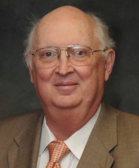 Duane D. Miller合作论文数Neuroscience Institute, University of Tennessee Health Science Center;Department of Pharmaceutical Sciences, College of Pharmacy, University of Tennessee Health Science Center2
 Cynthia Moreton Kuhn合作论文数Sanford School of Public Policy, Duke University2Karl J. Romstedt合作论文数2
 Loren Eugene Wold合作论文数Department of Pharmacology, Physiology, and Therapeutics, University of North Dakota School of Medicine2
 John Anthony Bauer合作论文数Division of Pharmacology/College of Pharmacy and OSU Heart and Lung Research Institute, The Ohio State University2William D. Roberts合作论文数2Yangmee Shin合作论文数2Dennis R. Feller合作论文数2
 David Mark Stafford Smith (Mark Stafford Smith)合作论文数Commonwealth Scientific and Industrial Research Organisation2Elliott Bennett-Guerrero合作论文数2Thomas D. Mcsweeney合作论文数2
 Mark Galantowicz合作论文数Nationwide Children’s Hospital, The Ohio State University2Kevin P. Landolfo合作论文数2Kathrine Doyle合作论文数2
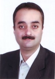 Babak Khabiri合作论文数Department of Anesthesiology, The Ohio State University Medical Center2Marc W. Harrold合作论文数2John V. Booth合作论文数2Michael B. Howie合作论文数2V. Bergdall合作论文数1JV Booth合作论文数1Kimberly R. Shepherd合作论文数1Philip Samuels合作论文数1Ryan Dalton合作论文数1PN PATIL合作论文数1John Bauer合作论文数1Ac Cook合作论文数1Tn Barnett合作论文数1Bl Funk合作论文数1Kp Landolfo合作论文数1
 Mandar Joshi合作论文数Google1
 Jonathan Green合作论文数Division of Neuroscience and Experimental Psychology, School of Biological Sciences, Faculty of Biology, Medicine and Health, The University of Manchester1Patil Popat N合作论文数1Smith M S合作论文数1Youtz Dane J合作论文数1




