Background: A hallmark feature in individuals with a Fontan circulation (FTN) is reduced exercise tolerance, possibly the result of slower oxygen kinetics. However, the kinetics of oxygen uptake (VO2) in FTN during the six-minute walk test (6MWT) have not been studied. Hypothesis: We tested the hypothesis that VO2 kinetics would be slower in FTN than control (CTL). Methods: 18 adolescents and emerging adults between the ages of 12-23 years old were recruited with FTN (m/f = 9/9; age = 16 ± 4yrs) and compared to similarly age- and sex-matched 14 healthy CTLs (m/f = 7/7, age = 16 ± 4yrs). Each participant performed a 6MWT using a wearable metabolic system (COSMED K5). VO2 kinetics were modeled using mono-exponential curve fitting (Origin, 2024b). Analyses were made using unpaired t-tests with P < 0.05 indicating significance. Results: During walk testing, the highest VO2 achieved was significantly lower in FTN (24 ± 7 ml/kg/min) compared to CTL (32 ± 8 ml/kg/min, P = 0.0040) and FTN also covered a significantly shorter distance (529 ± 79 m) than CTL (645 ± 46 m, P = 0.0001), despite both groups reporting similar perceived exertion scores (FTN: 6 ± 3 vs CTL: 5 ± 2, P = 0.2420). Resting VO2 was not different between FTN (6 ± 2 ml/kg/min) and CTL (5 ± 1 ml/kg/min, P = 0.9305). VO2 amplitude of Phase II was significantly lower in FTN (13 ± 5 ml/kg/min) compared to CTL (21 ± 6 ml/kg/min, P = 0.0007). The VO2 time constant, tau, of the exercise phase was significantly delayed in FTN (44 ± 11 s) compared to CTL (28 ± 8 s, P = 0.0002). Similarly, the tau of the recovery phase was slow in FTN compared to CTL (63 ± 17 s vs 51 ± 9 s, P = 0.0236). Conclusion: VO2 kinetics during the 6MWT in FTN are delayed. Funding: Saskatchewan Centre for Patient Oriented Research, Saskatchewan Health Research Foundation, Heart and Stroke Foundation of Canada, Scottish Rite Charitable Foundation of Canada, Canadian Institute of Health Research) This abstract was presented at the American Physiology Summit 2026 and is only available in HTML format. There is no downloadable file or PDF version. The Physiology editorial board was not involved in the peer review process.
AIMS:Cardiovascular disease (CVD) is a leading cause of mortality in childhood cancer survivors (CCS) that may be related to the cardiotoxic effects of radiation or chemotherapy and concomitant reductions in cardiorespiratory fitness. Therefore, we sought to compare cardiorespiratory fitness (peak oxygen uptake, V̇O2peak) between CCS and age-matched non-cancer controls (CON). Secondary outcomes included haemodynamics and resting cardiac function. METHODS AND RESULTS:Embase, Scopus, MEDLINE, CINAHL, and SPORTDiscus databases were searched from inception to June 2023 for eligible studies. Cross-sectional studies with V̇O2peak measured in CCS and CON were included. Differences in outcomes and pooled estimates for each outcome were estimated from a fixed-effects meta-analysis and between-group differences were reported as a weighted mean difference (WMD). Of 2026 studies identified, 18 reported V̇O2peak (CCS: n = 786, 44% female, mean age: 16 years, time post-therapy: 5.8 years; CON: n = 1379, 50% female, mean age: 16 years). V̇O2peak was lower in CCS [WMD: -7.08 mL/kg/min, 95% confidence interval (CI): -7.75 to -6.42, I2: 79%, n = 2165] with no difference for peak exercise heart rate (WMD: -1.4 b.p.m., 95% CI: -3.0 to 0.2, I2: 63%, n = 741). Resting left ventricular ejection fraction (WMD: -1.61%, 95% CI: -2.60 to -0.62, I2: 49%, n = 222) and systolic blood pressure were lower (WMD: -3.8 mmHg, 95% CI: -5.7 to -1.9, I2: 25%, n = 184) while resting heart rate was higher in CCS (WMD: 4.9 bpm; 95% CI: 1.8-7.9, I2: 55%, n = 262). CONCLUSION:Childhood cancer survivors have a marked reduction in cardiorespiratory fitness (7.1 mL/kg/min lower than CON) that may have important prognostic implications for their future risk of CVD and mortality.
RATIONALE: Patients with heart failure with preserved ejection fraction (HFpEF) have exercise intolerance that impacts quality of life. Impairments in peripheral oxygen (O 2 ) delivery and extraction (i.e., lower arterio-venous O 2 difference [a-vO 2 diff] and/or muscle O 2 diffusive conductance [DMO 2 ]) contribute prominently to exercise intolerance in patients with HFpEF. Exercise improves functional capacity in patients with HFpEF, however whole-body exercise is associated with symptoms of dyspnea and fatigue. Small muscle mass exercise that reduces cardiopulmonary limitations while maximizing peripheral adaptations has been proposed as an alternative training strategy for patients with HFpEF. The objective of this investigation was to quantify peripheral O 2 transport and utilization during single leg knee extensor exercise (SKLE) before and after 16-weeks of SLKE or cycle exercise training in patients with HFpEF. We tested the hypothesis that small muscle mass training would elicit greater peripheral adaptations compared to whole-body aerobic exercise in patients with HFpEF. METHODS: 34 patients with HFpEF (77% females; age: 71±7 y) were randomly assigned to either SLKE (n=19) or cycle training with cardiac unloading with sublingual nitroglycerin (CYCLE, n=15) for 16-weeks. The SLKE group performed one endurance (55-70% baseline peak power output [PPO]) and two interval (85-95% baseline PPO) training sessions per week, for 30 minutes per leg. CYCLE performed two endurance (1-20 bpm < maximal steady state heart rate [HR]) and two interval (≥95% peak HR) exercise training sessions per week, for 30 minutes each. Sublingual nitroglycerin (0.4mg) was taken before and during each training session to lower cardiac filling pressure during exercise in the CYCLE group. Before (PRE) and after (POST) the training intervention, participants completed an invasive incremental SLKE test to exhaustion during which time leg blood flow (LBF; Doppler ultrasound), leg a-vO 2 diff (PvO 2 , mmHg and SvO 2 , % [femoral venous catheter], SaO 2 , [pulse O 2 saturation] and venous hemoglobin concentration [spectrophotometry]), leg [Formula: see text]O 2 (direct Fick) and DMO 2 were calculated. Data were compared using two-way (Training×Type) ANOVA. RESULTS: Baseline hemodynamics were not different between SLKE and CYCLE. PPO increased after training (SLKE: 3±4 Watts, P< 0.001; CYCLE: 5±4 Watts, P=0.017). SLKE training increased peak LBF (PRE: 1877±662 vs. POST: 2115±748 mL/min, P=0009) and peak leg [Formula: see text]O 2 (PRE: 214.7±102.9 vs. POST: 256.9±121.8 mL/min, P=0.014), with no effect on peak leg a-vO 2 diff (PRE: 11.1±2.3 vs. POST: 11.3±2.5%, P=0.426). Conversely, CYCLE training did not improve peak LBF (PRE: 1935±593 vs. POST: 1976±508 mL/min, P=0.680) but did increase peak leg a-vO 2 diff (PRE: 10.5±2.2 vs. POST: 11.8±1.8%, P< 0.001) and tended to increase peak leg [Formula: see text]O 2 , though with more variability (PRE: 207.0±85.1 vs. POST: 231.5±71.4 mL/min, P=0.094). DMO 2 was improved in both SLKE (PRE: 4.9±2.7 vs. POST: 6.1±3.5 mL/min/mmHg, P=0.003) and CYCLE (PRE: 4.2±2.2 vs. POST: 5.5±2.1 mL/min/mmHg, P=0.001). CYCLE training reduced the slope of the relationship between LBF and leg [Formula: see text]O 2 (PRE: 9.2±2.1 vs. POST: 8.1±1.4, P=0.002), whereas SLKE did not (PRE: 8.7±2.2 vs. POST: 8.4±1.8, P=0.177). CONCLUSION: Both whole body and small muscle mass exercise training were effective in improving leg [Formula: see text]O 2 , though the mechanism of improvement was different. SLKE improved both LBF and DMO 2 while CYCLE exercise increased DMO 2 and leg O 2 extraction. Future studies should evaluate the combination of small muscle mass and whole-body exercise training to improve overall exercise capacity in patients with HFpEF. Funding: NIH 1P01HL137630 (BDL); 1F32HL137285 (CMH) This abstract was presented at the American Physiology Summit 2026 and is only available in HTML format. There is no downloadable file or PDF version. The Physiology editorial board was not involved in the peer review process.
INTRODUCTION:Hypertension (HT) is a leading modifiable risk factor for cardiovascular disease, but resting blood pressure (BP) measurements often miss hypertensive episodes during daily activities, affecting 10-15% of adults. A hypertensive response to exercise (HRE), characterized by abnormally high systolic BP (SBP) increases, is associated with future arterial HT and cardiovascular events, even in normotensive individuals and athletes. Despite its clinical significance, definitions of HRE vary widely, leading to inconsistent incidence estimates. The aim of this study was to collect available values of HRE and investigate definitions for HRE. EVIDENCE ACQUISITION:This systematic review followed PRISMA guidelines, conducting a comprehensive search of MEDLINE and Embase from 1974 to 2024. The search included studies on normotensive adults and athletes with or without HRE, focusing on BP cutoffs across exercise modalities and intensities. Eligible study designs included original research studies of any design, while reviews, case reports, and meta-analyses were excluded. Data extraction and synthesis involved multiple reviewers to ensure accuracy, with results presented in narrative and tabular formats. EVIDENCE SYNTHESIS:A total of 25 studies with 15,391 participants (weighted mean age 50 years, 28.3% female, 5.4% athletes) were analyzed, encompassing various study designs, including cross-sectional, case-control, cohort, and longitudinal studies. Exercise test protocols included treadmill (14 studies), bicycle ergometry (seven), shuttle/runs (three), and hand-grip strength (one), with most studies utilizing peak exercise intensities and automated BP measurements. Cut-offs for HRE varied, with most studies using SBP thresholds of ≥210 mmHg for men and ≥190 mmHg for women, though some studies proposed higher thresholds or included diastolic BP criteria. Definitions and methodologies for HRE were heterogeneous, reflecting variability across studies. Age, sex, fitness level, and test protocols significantly influence BP response, yet these factors are mostly omitted in the definition of HRE, with older adults and postmenopausal women showing exaggerated responses. Athletes exhibit higher peak SBP during exercise due to increased peak exercise cardiac output and augmented muscular strength enabling the athletes to push peak SBP higher, but thresholds applied are often the same as for non-athletes, underscoring the need for fitness-specific cut-offs. Variations in test protocols, measurement methods, and reliance on legacy cut-offs, which have been reproduced over decades further complicate consensus on standardized thresholds. CONCLUSIONS:Standardized, phenotype-specific criteria are essential to improve diagnostic accuracy and guide clinical recommendations in HRE.
Background Breast cancer survivors (BCS) have an increased risk of developing cardiovascular disease risk factors (CVDRF). However, the role of cardiopulmonary fitness, echocardiography and treatment regimen e.g. anthracycline, HER2-targeted therapy (AC/H) or endocrine therapy (ET) are uncertain. Methods This single-center, retrospective study included BCS without visceral metastases, who had been treated with either AC/H or ET and assessed by echocardiography, cardio-pulmonary exercise testing (CPET) and the H2FPEF score at baseline. We included BCS with a left ventricular ejection fraction ≥50 % and absence of CVDRF at baseline. The primary outcome was the incidence of CVDRF. Results A total of 112 BCS were included (mean age of 54.6 ± 9.9 years, BC stage I-III). After a median follow-up of 21 months new-onset arterial hypertension was the most common CVDRF observed (n = 17). New onset hypertension was related to higher baseline resting systolic blood pressure (127.4 ± 9.2 mmHg vs. 117.3 ± 13.1 mmHg, p = 0.002) and H2FPEF scores (1.2 ± 0.8 vs. 0.8 ± 1.1, p = 0.043). Echocardiographic and CPET findings associated with new-onset arterial hypertension included greater left ventricular mass index (77.3 ± 24.2 g/m2 vs. 65.6 ± 15.5 g/m2, p = 0.007), higher peak systolic blood pressure (193.1 ± 19.5 mmHg vs. 173.3 ± 21.2 mmHg, p = 0.017) and ventilatory power (6.4 ± 1.3 mmHg vs. 5.7 ± 1.2 mmHg, p = 0.022). Treatment regimen had no influence on the development of CVDRF. Conclusions The most common CVDRF among BCS is hypertension, underscoring the importance of monitoring this outcome irrespective of breast cancer treatment regimens. The H2FPEF score, CPET and echocardiography may help identify BCS at risk of developing hypertension. Strain and biomarkers were not available, impeding detection of cardiotoxicity.
Patients with heart failure with preserved ejection fraction (HFpEF) often have multiple cardiac and/or non-cardiac comorbidities that may contribute to exercise intolerance, the hallmark symptom in HFpEF. Exercise training is one of the most effective treatments to improve exercise tolerance but it is unclear whether individual comorbidities or the comorbidity burden are associated with altered exercise training effects in HFpEF. To evaluate the association between baseline comorbidities and the change in peak oxygen consumption (VO2) in patients with HFpEF. This is a pooled analysis of the two largest randomized controlled exercise training trials performed in HFpEF to date. In the OptimEx-Clin trial, 180 patients were randomized (1:1:1) to 12 months of high-intensity interval training (3× per week), moderate continuous training (5× per week) or usual care (UC). In the Ex-DHF trial, 322 patients were randomized (1:1) to 12 months of endurance and resistance training (3× per week) or UC. For the present analysis, all exercise training groups and both UC groups were combined into one exercise and one UC group. Peak V̇O2 was defined as the highest 30-sec average during symptom-limited incremental cardiopulmonary exercise testing. Comorbidity burden was evaluated by a simple score assigning 1 point to each assessed comorbidity (arterial hypertension, hyperlipidaemia, obesity, coronary heart disease, atrial fibrillation, diabetes mellitus, chronic kidney disease, cancer, anaemia, sleep apnoea, cerebrovascular disease, depression, chronic obstructive pulmonary disease, primary valve disease, peripheral vascular disease, congenital heart disease). The associations between baseline comorbidities and change in peak VO2 after 12 months were analysed using linear regression analyses adjusted for sex, age at inclusion and baseline peak VO2. All analyses were performed using R Statistical Software with significant levels of α=0.05. A total of 400 patients with available peak VO2 measurements at baseline and 12 months (60.7% women; mean age: 70 years; median no. of comorbidities: 4 [range: 0-10; IQR: 2-5]) were included in this analysis. Change in peak VO2 at 12 months was significantly higher following exercise training vs. UC (mean difference, 1.22 mL/kg/min [95% CI, 0.58-1.86], P<0.001). There was no significant interaction between any of the investigated comorbidities and treatment group for the change in peak VO2 (Figure 1). Comorbidity burden was also not significantly associated with the change in peak VO2 (P-interaction = 0.99) with mean between-group differences of 1.26 mL/kg/min (95%CI, -0.25-2.77) for ≤2 comorbidities, 1.37 mL/kg/min (95%CI, 0.44-2.31) for 3-4 comorbidities and 1.27 mL/kg/min (95%CI, 0.19-2.36) for ≥5 comorbidities. In patients with HFpEF, exercise training significantly improved peak VO2 over 12 months, regardless of individual comorbidities and overall comorbidity burden.
Endurance exercise training (ET) is an effective treatment in heart failure with preserved ejection fraction (HFpEF), but the efficacy of resistance training in this patient population has been only scarcely evaluated. In this multicenter, randomized trial, we evaluated the effects of combined endurance and resistance training over 12 months in patients with HFpEF. The primary endpoint was a modified Packer score, including all-cause mortality, hospitalizations classified as potentially related to heart failure or exercise and changes in peak oxygen consumption ( V̇O_2 ), diastolic function (E/e′), New York Heart Association (NYHA) class and global self-assessment (GSA). In total, 322 patients (mean age, 70 years; 192 females (59.6 V̇O_2 (mean difference, 1.3 ml kg−1 min−1 (95 V̇O_2 and NYHA class, as compared to UC. ISRCTN registration: ISRCTN86879094 . In a multicenter, randomized trial, patients with heart failure with preserved ejection fraction who underwent a regimen of combined endurance and resistance exercise training over the course of 1 year did not show a statistically significant improvement in the modified Packer score—the primary efficacy endpoint—as compared to patients who received usual care, but they did show improvements in secondary endpoints for maximal oxygen consumption and NYHA heart failure class.
Heart failure (HF) is a significant global health issue, categorized by left ventricular ejection fraction, being either reduced (HFrEF < 0.40) or preserved (HFpEF > 0.50), or in the middle of this range. Although the overall incidence of HF remains stable, HFpEF cases are increasing, representing about 50% of all HF cases. Outcomes for HFpEF are similar to those for HFrEF, leading to substantial health-care resource use. Despite extensive research over the past 2 decades, the prognosis and mortality rates for HFpEF remain high. A key feature of HFpEF is exercise intolerance, characterized by severe exertional dyspnea and fatigue, which significantly impacts quality of life. The underlying mechanisms of exercise intolerance are not fully understood due to the complex pathophysiology and multisystem involvement. Obesity is a common comorbidity in HFpEF, especially in North America, leading to worsening symptoms, hemodynamics, and mortality rates. Increased adiposity leads to inflammation, hypertension, dyslipidemia, and insulin resistance, and impairing cardiac, vascular, pulmonary, and skeletal muscle function. Therefore, managing obesity is crucial in treating HFpEF. In this review we explore the pathophysiologic mechanisms of HFpEF, emphasizing obesity’s role, and we discuss current management strategies while identifying areas needing further research.
Since COVID-19, cardiac rehabilitation (CR) programs are moving towards fewer in-person sessions, therefore the need for home-based support is paramount for patient well-being and fitness benefits. Moreover, reducing the barriers for participation and ensuring better CR accessibility is important. Our research group recently developed a home-based online rehabilitation support program "My Heart Coach" which provides exercise support to people throughout their CR program. We determined aerobic endurance (6-minute walk test [6MWT] distance) and heart rate (HR) and pulmonary oxygen uptake (VO2) during and following (i.e., recovery kinetics) 6MWT in patients undertaking combined center- and home-based CR. Twenty-seven patients (64±11 years, 5 female) attended assessments at baseline (BL) and after 12 weeks of combined in-person CR (exercise training 1 day/week for 8 weeks) with online MHC support. At each assessment, 6MWT was performed following American Thoracic Society guidelines1. HR and VO2 were continuously measured at rest, during, and immediately following (for 5 min) 6MWT. Rate of perceived exertion (RPE; Borg 6-20 scale) was recorded following 6MWT. Exercise HR and VO2 were determined in the last two-minutes of the 6MWT, and recovery kinetics for both variables were determined using non-linear modelling (OriginLab, 2023b) to yield the mean response time (MRT = time delay to onset of the exponential recovery + time constant of the recovery curve; Figure 1) during seated rest. The effect of the intervention was evaluated using two-tailed paired t-tests and differences considered significant when p<0.05. At baseline, patients walked 452±117m at an RPE of 13±2 and this improved by 63±46m after the intervention (515±131m, p<0.01) with a similar RPE (14±2, p=0.13). Resting VO2 was higher following the intervention (p<0.01) with no change in resting HR (p=0.71; Figure 2A/B). Pulmonary VO2 during 6MWT was higher after the 12-week intervention (BL: 12.4±3.2 ml/kg/min; 12wk: 14.5±4.3 ml/kg/min; p<0.01); however, exercising HR was not different between time-points (BL: 103±19 bpm; 12wk: 107±18 bpm; p=0.21; Figure 2C/D). The MRT for HR (BL: 54±23 s; 12wk: 8±16 s; p=0.35; Figure 2E) and VO2 (BL: 73±25 s; 12wk: 81±38 s; p=0.16; Figure 2F) were not significantly changed by the intervention despite greater distance covered (i.e., higher speed) at a higher exercising VO2. A 12-week blended CR program is an effective intervention to improve aerobic endurance during a 6MWT. Our finding of a higher VO2, but not HR, during exercise despite a similar VO2 recovery kinetics suggests favorable peripheral adaptations that result in greater skeletal muscle oxygen utilization after CR participation.
Duchenne muscular dystrophy is a muscle-wasting, progressive, X-linked inherited disease in young male individuals, who—aside from peripheral muscular impairment—may also suffer from severe cardiac complications. In women who are muscular dystrophy carriers (MDCs), muscular symptoms and cardiac complications are less severe or even absent. While male individuals with muscular dystrophy are not usually able to perform strenuous exercise, women who are MDCs can exercise at mild, or even high, intensity. However, the impact of participating in elite sports, particularly endurance sports with high cardiopulmonary exercise strain, on female athletes who are MDCs is uncertain. Herein, we describe two rare cases of female elite athletes who are MDCs who participated in endurance sports. We describe their clinical presentation, kinetics of cardiac biomarkers and peripheral muscle enzymes during acute exercise, and cardiac manifestations in the context of sports eligibility, including an interdisciplinary shared decision-making approach to whether to continue participating in sports. This approach focuses on pathophysiology and genetics in dystrophinopathies, with a particular focus on genetic carrier status. While the primary concern is risk stratification for sudden cardiac death and its prevention, the potential risk of early onset of myocardial dysfunction or even heart failure also needs to be considered in MDCs. To optimize exercise recommendations, these complex and rare cases of athletes require an interdisciplinary approach, including experts in sports cardiology, sports medicine, radiology, and genetics, and should be included in a long-term international sports cardiology registry.
Early breast cancer (EBC) remains the most common invasive cancer in North American women. With improvements in detection, systemic therapies and supportive care, long-term survival in early stage disease now approaches 90%. In EBC survivors, fat pool distribution, specifically visceral and intermuscular fat (IMF) is associated with increased cardiovascular disease risk regardless of pre-existing risk factors or cancer treatments. We previously showed that 1 year after trastuzumab initiation, visceral fat and IMF volumes increased significantly while muscle volume decreased in HER2+ EBC patients. However, the longer-term effects of such exposures on body composition remain unknown. We hypothesized that over time, deleterious changes would progress with increasing fat and decreasing skeletal muscle in EBC patients exposed to trastuzumab-based chemotherapy. A follow-up MRI study was conducted with prior participants of the MANTICORE (Multidisciplinary Approach to Novel Therapies In Cardio-Oncology REsearch) trial. Enrolment was limited to those without recurrent disease; 54 patients consented to participate. Eight scans were not included (tissues outside field of view, files unevaluable) thus 46 scans were included. The identical MRI protocol was used at all timepoints with five slices centered at the 3rd lumbar vertebra. Sum of volumes of muscle, IMF and visceral fat were measured using custom semi-automated software. Bones and organs were removed from the visceral fat pool. Repeated measures of ANOVA with appropriate post-hoc correction for multiple comparisons were performed at baseline, 3-month, 12-month, 24-month and 5 years. Participants were 58.6 ± 8.6 years of age and median 5.5 years had elapsed since trastuzumab completion. All were living independently with no cardiac events occurring in the intervening years. Ten had initiated cardiometabolic pharmacotherapy (beta-blockade, ACEI/ARB, diuretic or diabetes). Muscle mass increased over the 5 years (main effect of time, p=0.002); however, was lower at 1 year versus 3 months (Fig 1A). IMF also increased over 5 years (main effect of time, p=0.005) but the pattern was different with increases observed as early as 3-months (Fig 1B). Visceral fat increased up to 2 years then plateaued (main effect of time, p= 0.038; Fig 1C). To our knowledge, this is the first and longest prospective study assessing changes in body composition in EBC using MRI both during and years after trastuzumab-based chemotherapy. Our data suggests that there may be ‘targetable’ timepoints for interventions such as pharmacotherapy, nutrition and exercise-based rehabilitation aiming to improve the quality and quantity of life in EBC survivors.Figure 1
Over 50,000 hematopoietic stem cell transplants (HSCTs) are performed worldwide annually. Patients receiving HSCT for relapsed disease have commonly received prior chemotherapy protocols resulting in reduced physiologic capacity. We have previously shown that treatment-related alterations to muscle/fat distribution and muscle quality significantly reduces physical functioning (VO2peak). Sarcopenic obesity is an independent risk factor of prolonged hospitalization and inferior overall survival in HSCT. In APOLLO, we are utilizing novel MRI techniques studying lean and fat mass in relation to physical performance and quality of life. to characterize muscle and fat distribution in baseline MRI scans of patients with hematologic diseases participating in the APOLLO study compared to sex-matched normal population. In the initial 11 participants, quantitative fat and water separated images were used to measure intermuscular fat and muscle volumes from mid-thigh and abdomen were performed. The volumes of muscle, intermuscular, visceral, and subcutaneous fat was measured using custom semi-automated software to identify the boundaries of muscle and the distinct fat groups. Four females (age 59.5 ± 8) and 7 males (age 67.1 ± 7.5) were included. All were pretreated with a range of 1 – 4 lines of chemotherapy. Compared to our sex-matched healthy control cohort, both females and males had relatively less total thigh muscle volume (5% and 23%) and greater thigh intramuscular fat fraction (17% and 37%). Mean subcutaneous thigh fat in females was 21% lower vs 29% higher in men compared to controls. Abdominal MRI demonstrated 23% less abdominal muscle in women with no difference in males from norms. Abdominal intermuscular fat was 19% greater in women and 52% greater in men. Visceral fat was 21% greater than normal in females and 91% greater in men. In these preliminary results, prior to undergoing HSCT we observed abnormal patterns and potentially deleterious changes to muscle and fat pools in thigh and abdominal regions. High-intensity drug doses alongside severe deconditioning with prolonged bedfast states would only exacerbate poor body composition and frailty. The high morbidity and mortality rates of HSCT may well be associated with low muscle/ high fat volume distributions, not evaluable without advanced imaging. This work has implications for pre-habilitation in people with high-risk hematologic malignancies, targeted post-HSCT rehabilitation, nutritional studies and other interventions specifically aimed at improving body composition.
Atrial fibrillation (AF) is a common comorbidity in individuals with heart failure with preserved ejection fraction (HFpEF) that contributes to increased morbidity and mortality. However, the impact of AF on key HFpEF physiologic features, including exercise tolerance (peak oxygen uptake, VO2peak) hemodynamic responses, and peripheral oxygen extraction remains unclear. To compare VO2peak, peak exercise hemodynamics and arterio-venous oxygen difference (a-vO2diff) in patients with HFpEF with or without AF. The study cohort included patients referred to a multi-disciplinary unexplained dyspnea clinic with an established HFpEF diagnosis after detailed clinical and hemodynamic evaluation. Patients were sub-grouped based on whether they were in AF (HFpEF-AF; n=88) or sinus rhythm (HFpEF-SR; n=625) at the time of the evaluation. Peak VO2, exercise hemodynamics and a-vO2diff were assessed from maximal cardiopulmonary exercise testing with simultaneous echocardiography (CPETecho). AF and SR sub-groups were compared using ANCOVA with adjustment for age and sex. The HFpEF-AF group was slightly older (76±6yrs vs 73±8yrs, P<0.001), with similar (P>0.2 for all) body mass index (28.2±4.8kg/m2 vs 29.6±11.2kg/m2), resting left-ventricular (LV) ejection fraction (61±8 vs 62±8) and LV mass index (92±36g/m2 vs 93±25g/m2) but higher indexed left-atrial volume (47±15mL/m2 vs 32±13mL/m2, P<0.001) and median [interquartile range] NT-proBNP levels (1200ng/L [805, 1750] vs 290ng/L [140, 530], P<0.001). The HFpEF-AF group also had a higher proportion of males (42% vs 55%, P=0.023), but similar prevalence of hypertension (75% vs 79%, P=0.43) and diabetes (22% vs 22%, P=0.91). Both groups showed comparable peak effort during CPETecho (peak respiratory exchange ratio: HFpEF-AF=1.06±0.11 vs HFpEF-SR=1.07±0.12, P=0.25). Peak VO2 was 9% lower in HFpEF-AF (P=0.027), accompanied by a 10% reduction in peak cardiac output (P=0.007), despite a 12% higher heart rate (P<0.001). Stroke volume was 19% lower in HFpEF-AF, related to smaller left ventricular end-diastolic volume (12% lower, P=0.001), which decreased further during exercise (5% decrease vs. 3% increase in HFpEF-SR, interaction P<0.001). In contrast, the a-vO2diff was not different between groups (P=0.94). HFpEF-AF and HFpEF-SR also showed similar resting (17.8±4.1mmHg vs 17.2±4.1 mmHg, P=0.29) and peak exercise mean pulmonary artery pressures (mPAP; 32.2±6.6mmHg vs 32.7±7.0mmHg, P=0.48), but the mPAP-CO slope tended to be 14% higher in HFpEF-AF (Fig. 1, P=0.086). Patients with HFpEF in AF have a lower peak oxygen uptake and reduced end-diastolic volume, stroke volume and cardiac output reserve compared to patients with HFpEF in sinus rhythm. These findings highlight differences in HFpEF exercise physiology associated with AF, suggesting a potential need for tailored therapeutic approaches to optimize their functional outcomes.Fig. 1Impact of AF on exercise responsesFig. 2Impact of AF on LV responses
Hematopoietic stem cell transplantation (HSCT) is a common therapy for many hematologic malignancies. While advances in transplant practice have improved cancer-specific outcomes, multiple and debilitating long term physical and psychologic effects remain. Patients undergoing allogeneic bone marrow transplantation (allo-BMT) are often critically ill at initial diagnosis and with necessary sequential treatments become increasingly frail and deconditioned. Despite modern treatment regimens and support, cardiovascular disease remains a leading cause of non-relapse mortality among allo-BMT survivors. Well-established multi-disciplinary care models such as cardiac rehabilitation offer holistic care including exercise training, nursing support, physical/occupational therapy, psychosocial support and nutritional education. HSCT patients may be excluded from conventional outpatient physical rehabilitation programs due to prolonged pancytopenia and frequent hospital admissions. In Canada, dedicated cancer-specific rehabilitation programs are available only at major tertiary academic centers. The primary aim of this study will evaluate the feasibility and acceptability of a multimodal care navigation (nursing, exercise, nutrition) intervention with content delivery facilitated by a supportive care web-based ‘app’ extending from diagnosis to 1 year in the allogeneic bone marrow transplant population. Adult patients scheduled for allo-BMT will receive support from exercise specialist, nursing support and dietician expertise alongside a supportive care ‘app’ with additional in-person or virtual cardiac rehabilitation support. To our knowledge, no research team is taking such a holistic, multidisciplinary approach to address the debilitating physiologic and psychological consequences of allo-BMT. We expect the findings to inform the optimal timing and patient preferences to develop studies examining risk-specific, individualized interventions (including exercise, pharmacotherapy, combination treatments) to reduce or prevent symptoms and dysfunction. We expect this innovative program to identify ways to benefit innumerable patients with hematologic and other malignancies. Ultimately, we hope to transform supportive care in hematopoietic stem cell transplantation. Clinicaltrials.gov ID: NCT05579678.
During standard cardiovascular magnetic resonance (CMR) the horizontal long-axis cine image (i.e., 4-chamber) is captured which includes a cross-section of the descending aorta. The aortic cross-section can be used to assess aortic stiffness (distensibility; ∆area/pressure) or circumferential strain (percentage vascular deformation). We examined whether descending aortic strain from traditional CMR is sensitive to age- and disease-related (heart failure with preserved ejection fraction; HFpEF) arteriosclerosis. We recruited 83 participants into three groups: (1) 34 young individuals (age: 22 ± 3 years; body mass index (BMI): 24.3 ± 2.8 kg/m2); (2) 19 older individuals (age: 69 ± 5 years; BMI: 26.9 ± 4.7 kg/m2) and (3) 26 patients with HFpEF (age: 69 ± 6 years; BMI: 35.8 ± 6.1 kg/m2). All participants were studied in the same 3 T scanner (Phillips, Achieva). Descending aortic cross-sectional area and circumferential strain were measured using cvi42 software. Blood pressure was measured via a brachial oscillometric cuff. Data were compared via ANOVA. All data are reported as means ± standard deviation. Compared to the young group (71 ± 5 mmHg), mean arterial pressure was higher in the older (83 ± 9 mmHg, P < 0.001) and HFpEF groups (86 ± 10 mmHg, P < 0.001). Minimum and maximum aortic areas were greater in the older and HFpEF groups (both, P < 0.01). Peak descending aortic strain (young: 11.4% ± 2.2%; older: 4.8% ± 1.6%; HFpEF 3.8% ± 1.6%) and absolute distension were lower (all, P < 0.02) in the older and HFpEF groups compared to the young. Peak descending aortic strain and strain rates are sensitive to age and may provide a novel assessment of arterial stiffness for longitudinal studies that utilize or have utilized CMR.
AIMS:Exercise training (ET) is an effective therapy in heart failure with preserved ejection fraction (HFpEF), but the influence of different ET characteristics is unclear. We aimed to evaluate the associations between ET frequency, duration, intensity [% heart rate reserve (%HRR)] and estimated energy expenditure (EEE) with the change in peak oxygen consumption (V̇O2) over 3 months of moderate continuous training (MCT, 5×/week) or high-intensity interval training (HIIT, 3×/week) in HFpEF. METHODS AND RESULTS:ET duration and heart rate (HR) were recorded with a smartphone application. EEE was calculated using the HR data during ET and the individual HR-V̇O2 relationships during cardiopulmonary exercise testing. Differences between groups and associations between ET characteristics and peak V̇O2 change were assessed with linear regression analyses. Peak V̇O2 improved by 9.2 ± 13.2% after MCT and 8.7 ± 15.9% after HIIT (P = 0.67). The average EEE of 1 HIIT session was equivalent to ∼1.42 MCT sessions and when adjusted for EEE, the mean difference between MCT and HIIT was -0.1% (P = 0.98). For both MCT and HIIT, peak V̇O2 change was positively associated with ET frequency (MCT: R2 = 0.103; HIIT: R2 = 0.149) and duration/week (MCT: R2 = 0.120; HIIT: R2 = 0.125; all P < 0.05). Average %HRR was negatively associated with peak V̇O2 change in MCT (R2 = 0.101; P = 0.034), whereas no significant association was found in HIIT (P = 0.234). Multiple regression analyses explained ∼1/3 of the variance in peak V̇O2 change. CONCLUSION:In HFpEF, isocaloric HIIT and MCT seem to be equally effective over 3 months. Within each mode, increasing ET frequency or duration/week may be more effective to improve peak V̇O2 than increasing ET intensity.
Heart failure with preserved ejection fraction (HFpEF) is a complex clinical syndrome characterized by reduced peak oxygen uptake (VO2peak) secondary to central and peripheral limitations. Obesity is a well-established risk factor for HFpEF and has garnered considerable interest as a therapeutic target, particularly in the context of the obese-HFpEF phenotype. However, the impact of obesity on VO2peak and its Fick determinants in HFpEF remains unclear. To understand the impact of obesity on VO2peak and it’s central and peripheral determinants in individuals with HFpEF. Patients referred to a multi-disciplinary unexplained dyspnea clinic with an established HFpEF diagnosis were included (n=448), and sub-grouped based on body mass index (BMI, ≥30 kg/m2 or <30) into HFpEF with- (HFpEFObese, n=138; BMI=34.3±3.7 kg/m2; 68% female; 71±9 yrs) or without obesity (HFpEFNon-Obese, n=310; BMI=24.9±3.0 kg/m2; 60% female; 74±9 yrs.). Patients underwent maximal cardiopulmonary exercise testing with simultaneous echocardiography (CPETecho) to assess VO2peak, exercise hemodynamics (cardiac output, CO; stroke volume, SV; heart rate, HR, mean pulmonary artery pressure, mPAP) and the arterio-venous oxygen difference (a-vO2diff). HFpEF sub-groups were compared using ANCOVA with adjustment for age and sex. Comparison was made using absolute values and values adjusted for bodyweight (peak VO2) or body surface area (BSA; CO, SV) . Pooled HFpEF sub-groups were also compared using ANCOVA (adjusted for age and sex) to controls (CON, n=153; BMI: 23.1±2.7 kg/m2, 41% female; 62±5 yrs) without HFpEF to elucidate the impact of HFpEF, versus HFpEF plus obesity on VO2peak and its determinants. HFpEFObese and HFpEFNon-Obese had comparable absolute VO2peak, peak exercise HR and a-vO2diff, while HFpEFObese had higher CO at peak exercise secondary to a larger SV. Resting and exercise mPAP and mPAP-CO slopes were higher in HFpEF vs CON (P<0.001 for all; Fig. 2), with no differences seen between HFpEF obesity phenotypes (P>0.50 for all). In contrast, bodyweight-adjusted peak VO2 was markedly lower in HFpEFObese (Fig. 1B), despite comparable peak exercise cardiac index and stroke volume index. Regardless of HFpEF sub-group, peak VO2, central (CO, HR, mPAP) and peripheral factors (a-vO2diff) were markedly impaired in HFpEF versus CON (Fig. 1). Obese HFpEF patients demonstrate a higher stroke volume reserve while maintaining oxygen extraction. However, these adaptations are insufficient to sustain weight-adjusted peak VO2 at the levels of their non-obese counterparts. This suggests that obesity induces adaptive cardiac and peripheral muscle remodeling. Accordingly, HFpEF patients achieving weight reduction should be supported with adjuvant exercise training programs to preserve cardiac and peripheral muscle performance as the load of excess body mass is alleviated, thereby enhancing exercise tolerance.Fig 1.Differences in VO2 determinantsFig 2.Differences in mPAP and CO
While interpreting the mechanisms underlying exercise limitation in cardiopulmonary exercise testing (CPET), oxygen pulse (O2pulse) is often assumed to be a surrogate for stroke volume (SV). However, in the rearranged Fick’s equation, O2pulse represents the product of SV and arteriovenous oxygen difference (a-vO2diff). Consequently, interpreting O2pulse exclusively as a reflection of SV can be misleading, as it disregards one of its key determinants. To assess the effort response of the determinants of O2pulse (SV and a-vO2Diff) by simultaneous CPET and echocardiography (CPETecho) or invasive CPET (iCPET). This observational study analysed the O2pulse in CPETecho and iCPET examinations conducted between July/15 and May/24. SV in CPETecho was measured using the ultrasound velocity time integral of the left ventricular outflow tract, and with VO2, a-vO2diff was calculated. For iCPET, a-vO2diff was directly measured using arterial and mixed-venous blood gas analyses, while SV was calculated. During CPETecho, measurements were taken at rest, intermediate, and peak exercise, whereas during iCPET, at rest, at 3, 6, 9 minutes, and at peak exercise. Variables were compared using one-way ANOVA with post hoc Tukey tests, with statistical significance set at p < 0.05. CPETecho: A total of 1,866 exams were analyzed (54.1% male; age: 65.4±13.9 y). The main indication was heart failure with preserved ejection fraction (HFpEF, 39.7%), valvular heart disease (35.5%), myocardial ischemia (6.3%), and others (18.5%). O2pulse increased significantly from rest to intermediate and peak (4.0±1.9, 9.0±3.3, and 10.9±3.9 mL/beat, respectively; p<.0001; Figure 1). SV increased from rest to intermediate load (70.2±18.1 vs. 84.5±20.9 mL/beat; p<.0001), with no further significant increase from intermediate to peak exercise (84.5±20.9 vs. 85.1±21.3 mL/beat; p=.07). In contrast, a-vO2 diff increased progressively from rest to intermediate and peak (p<.001). iCPET: A total of 68 exams were analyzed, primarily conducted in suspected HFpEF (age: 64.1±10.6 y). O2pulse increased progressively from rest to peak (p<.0001; Figure 2). SV significantly increased from rest to the 6-minute measurement, with no further significant changes thereafter. In contrast, a-vO2diff showed a continuous progressive increase from rest to peak exercise. Analysis of the determinants of O2pulse (SV and a-vO2diff) using both CPETecho and iCPET demonstrated that the increase in O2pulse from intermediate to peak exercise is primarily driven by a rise in a-vO2diff, while SV remains relatively constant. Therefore, in cardiac patients, changes in O2pulse should not be assumed to reliably reflect changes in SV alone but rather represent the combined effects of SV and peripheral oxygen extraction. For a definitive interpretation of the O2pulse response during exercise, performing measurement of either SV or a-vO2diff is essential.Figure 1.Variables measured by CPETechoFigure 2.Variables measuredf by iCPET
Background:Cardiovascular disease risk factors (CVDRF) are linked to increased morbidity in cardiomyopathies (CMP), but whether new onset CVDRF differ among CMPs is unknown. In addition, whether the acute exercise response during cardiopulmonary exercise testing (CPET) differs among CMPs remains unclear. Methods:This single-center, retrospective study analyzed patients with arrhythmogenic, hypertrophic, and dilated (DCM) cardiomyopathy without CVDRF at baseline. Resting echocardiography and CPET were performed, and exercise response was assessed depending on sex and CMP. After a median follow-up of 19.5 months, CPET and echocardiography were analyzed in relation to the development of new CVDRF. Results:A total of 104 CMPs were included (median age 53.0 years). New-onset CVDRF was rare (11.5 %) and driven by arterial hypertension (8.7 %) but did not differ among CMPs. DCM displayed significantly lower resting left ventricular ejection fraction (40.5 %, interquartile range, IQR, 11.5 %, p < 0.001), diastolic function (E/e' 9.3, IQR 5.5, p < 0.001), and had the lowest peak systolic blood pressure (170.0 mmHg, IQR 52.5 mmHg, p = 0.011), predicted peak oxygen consumption (82.0 %, IQR 39.8 %, p = 0.003), oxygen pulse (101.0 %, IQR 28.8 %, p = 0.030) as well as lower ventilatory (VP, 5.5 mmHg, IQR 1.4 mmHg, p = 0.033) and circulatory (CP, 4096.0 mL/kg/min x mmHg, IQR 2299.3 mL/kg/min x mmHg, p = 0.015) power compared to the other groups. Lower VP (5.0 mmHg, IQR 1.3 mmHg, p = 0.003) and CP (3660.0 mL/kg/min x mmHg, IQR 3298.0 mL/kg/min x mmHg, p = 0.004) were observed for females. Conclusions:Arterial hypertension was the most common CVDRF among CMPs, underscoring the importance of monitoring this outcome. Exercise limitations differ between CMPs and should be interpreted depending on sex.
 Richard B. Thompson合作论文数Department of Biomedical Engineering, Faculty of Engineering, University of Alberta93
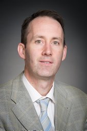 Ian Paterson合作论文数77
 Michael D Nelson合作论文数School of Health and Exercise Sciences, University of British Columbia;Faculty of Rehabilitation Medicine, University of Alberta and Mazankowki Alberta Heart Institute;Cedars-Sinai Heart Institute;Centre for Translational Physiology, University of Otago;Research Institute for Sport and Exercise Sciences, Liverpool John Moores University;University of British Columbia, Liverpool John Moores University68
 Andre La Gerche合作论文数52
 Darren Warburton合作论文数School of Kinesiology, Faculty of Education, The University of British Columbia;Physical Activity Promotion and Chronic Disease Prevention Unit, The University of British Columbia50
 Corey R. Tomczak合作论文数Faculty of Rehabilitation Medicine, University of Alberta50
 Edith Pituskin合作论文数49
 Jessica M. Scott合作论文数NASA Johnson Space Center45
 Dalane W. Kitzman合作论文数Congdon Heart and Vascular Center, Atrium Health Wake Forest Baptist;Clinical and Translational Science Institute, Wake Forest School of Medicine;Department of Medicine, Wake Forest School of Medicine38Rhys I Beaudry合作论文数36
 John Mackey合作论文数Department of Oncology, Faculty of Medicine & Dentistry, University of Alberta;Pacylex Pharmaceuticals Inc;SMHeartCard Inc;Illumisonics;J2 Endeavours Inc34
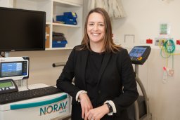 Howden Erin J合作论文数Schools of Human Movement Studies, University of Queensland33Steve Foulkes合作论文数32
 Satyam Sarma合作论文数Medicine (Cardiology),, Baylor College of Medicine31Martin Halle合作论文数30
 Wesley J Tucker合作论文数Point Loma Nazarene University, Arizona State University30
 Lee Jones合作论文数Department of Medicine, Memorial Sloan Kettering Cancer Center27
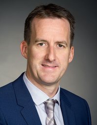 Michael Stickland合作论文数Division of Pulmonary Medicine, Department of Medicine, Faculty of Medicine & Dentistry, University of Alberta;G.F. MacDonald Centre for Lung Health, Covenant Health26
 Amy Kirkham合作论文数Faculty of Kinesiology and Physical Education, University of Toronto25Ben T. A. Esch合作论文数24
 Steve F Fraser合作论文数Discipline of Exercise Sciences, RMIT University23
 Jason Dyck合作论文数Department of Pediatrics, Faculty of Medicine & Dentistry, University of Alberta;Cardiovascular Research Centre, University of Alberta22Dylan Taylor合作论文数22
 Rachel Skow合作论文数rachel.skow@ualberta.ca.21
 Peter H. Brubaker合作论文数Department of Health and Exercise Science, Wake Forest University20
 Gavin Oudit合作论文数Department of Medicine, Faculty of Medicine & Dentistry, University of Alberta;PEARKO Therapeutics Inc.20
 Robert C Welsh合作论文数Division of Adult Psychiatry, Department of Psychiatry, School of Medicine, University of Utah Health19Wayne Tymchak合作论文数17Kelvin Chow合作论文数17
 Stephen Foulkes合作论文数Baker Department of Cardiometabolic Health (S.J.F., E.J.H., A.L.G.),, University of Melbourne16
 Antill Yoland C合作论文数Department of Hematology and Medical Oncology, Peter MacCallum Cancer Centre16Stewart R. Petersen合作论文数16
 T. Jake Samuel合作论文数College of Nursing and Health Innovation, University of Texas at Arlington15Dp Humen合作论文数15Haennel Robert G合作论文数15
 Koon Teo合作论文数Division of Cardiology, Faculty of Health Sciences, McMaster University15
 Sandra Mandic合作论文数15Joseph Pagano合作论文数14
 Benjamin Levine合作论文数Department of Internal Medicine, University of Texas Southwestern Medical Center;Institute for Exercise and Environmental Medicine, University of Texas Southwestern Medical School14Justin A. Ezekowitz合作论文数14
 Margaret L. Mcneely合作论文数Department of Rehabilitation Medicine, Cross Cancer Institute14
 Abraham Joy合作论文数Department of Bioengineering, College of Engineering, Northeastern University14
 Kerry Courneya合作论文数Faculty of Kinesiology, Sport, and Recreation, College of Health Sciences, University of Alberta;Cancer Research Institute of Northern Alberta, University of Alberta;Department of Oncology, Faculty of Medicine and Dentistry, University of Alberta;Behavioral Medicine Laboratory and Fitness Center, University of Alberta;Cross Cancer Institute Edmonton14June Cheng Baron合作论文数14H. Arthur Quinney合作论文数14James P MacNamara合作论文数13Claessen Guido合作论文数13
 Kenneth J Riess合作论文数13
 Neil Eves合作论文数Centre for Heart, Lung & Vascular Health, School of Health and Exercise Sciences, Faculty of Health and Social Development, The University of British Columbia13




