ABSTRACTBackgroundPerineural spread of cutaneous head and neck squamous cell carcinoma is most effectively managed by surgical resection with a clear central margin at the affected nerve. For ophthalmic nerve disease, there is no clear consensus regarding the extent of spread which is resectable via an orbit‐preserving neurectomy, either in regard to oncological outcome or technical feasibility.MethodsCadaveric anatomical study of 10 human orbits, with endoscopic and exoscopic dissection of the frontal division of the ophthalmic nerve, to quantify the maximal extent of resection via orbit‐preserving techniques and to discern the advantages and disadvantages of exoscopic versus endoscopic visualization aids.ResultsMean achievable resection length measured from the supraorbital ridge was 40.9 mm (n = 10). No branching patterns were identified which would preclude neurectomy as an oncologically sound option.ConclusionsOrbit‐preserving resection of the ophthalmic nerve is technically feasible to a distance of approximately 4 cm, encompassing the majority of Williams Zone 1.
ABSTRACTBackgroundStandardized surgical approaches to advanced pre‐auricular cutaneous squamous cell carcinomas (cSCC) are lacking.MethodsFifty–four patients who underwent lateral temporal bone resection (LTBR) for pre‐auricular cSCC were grouped into “Levels” of increasing disease spread. Surgical approaches to achieve negative‐margin resection were designed for each Level and replicated on cadaveric specimens.ResultsLevel 1 extended to the external auditory canal, requiring LTBR ± superficial parotidectomy. Level 2 involved the retromandibular space ± temporomandibular joint, necessitating partial mandibulectomy, in addition to the above. Level 3 and 4 involved the deep parotid, being situated either away from (> 5 mm) or close (≤ 5 mm) to the anterior carotid sheath (ACS), respectively. These tumors require radical parotidectomy, with incorporation of the ACS for Level 4. Level 5 involved the ACS at the skull base and should be treated non‐surgically.ConclusionThis Level‐based system will hopefully lead to further prospective studies and improvements in outcomes for advanced pre‐auricular cSCC.
Objective: To establish reference intervals using a new point-of-care thromboelastometry device in dogs for the extrinsically activated test (EX-test), intrinsically activated test (IN-test), fibrin polymerization test (FIB-test), ecarin test (ECA-test), and tissue plasminogen activator test (TPA-test) and to investigate the effects of storage time on the results. Design: Prospective clinical study in 2022. Setting: University teaching hospital. Animals: Forty-eight healthy privately or university-owned dogs were prospectively enrolled and included on the basis of normal physical examination and normal baseline laboratory results (CBC, biochemistry profile, prothrombin time, and activated partial thromboplastin time [aPTT]). Interventions: After a 30-minute storage time, the EX-test, IN-test, FIB-test, ECA-test, and TPA-test were performed on citrated blood samples. To determine the effect of storage time, 11 samples had the EX-test, FIB-test, and IN-test repeated 90 and 150 minutes after sample collection. Measurements and Main Results: Ten thromboelastometry parameters were evaluated for each test. Reference intervals were calculated using the robust method for parametric data, and the robust Box-Cox transformed or nonparametric methods were used for nonparametric data. Increasing storage time resulted in more hypocoagulable tracings. A correlation was found between the IN-test and aPTT (r = 0.62, P < 0.0001). Other weak to moderate correlations were seen between thromboelastometry parameters and platelet count and hematocrit. Conclusions and Clinical Importance: The development of reference intervals for the thromboelastometry device allows for the clinical use of this technology. Analyzing samples after a prolonged storage time of more than 30 minutes may result in erroneous results. Results may also be affected by an abnormal hematocrit or platelet count.
Background The treatment of traumatic haemorrhagic shock has been transformed through better haemorrhage control, use of tranexamic acid and use of blood products. The improved survival seen from these strategies has stimulated an interest in pre-hospital transfusion. Objectives To determine if the clinical effectiveness of resuscitation with red blood cells and lyophilised plasma was superior to 0.9% saline for improving tissue perfusion and reducing mortality in adults with haemorrhagic shock following major trauma. Design A multi-centre, allocation concealed, open-label, parallel group, randomised controlled trial (with internal pilot). Setting The trial was conducted in four civilian pre-hospital critical care services who operated within the National Health Service (NHS) England Major Trauma Networks. Participants Adults (aged ≥16 years) who had sustained traumatic injuries, were attended by a pre-hospital emergency medical team and were hypotensive (systolic blood pressure <90 mmHg or absence of radial pulse) as a consequence of traumatic haemorrhage were eligible for inclusion. The exclusion criteria were known or apparently <16 years, blood administered on scene prior to arrival of the RePHILL team, traumatic cardiac arrest where (1) the arrest occurred prior to arrival of the team and/or (2) the primary cause is not hypovolaemia, refusal of blood product administration, known Jehovah’s Witness, pregnancy, isolated head injury without evidence of external haemorrhage, prisoners in the custody of HM Prison and Probation Service. Interventions Participants were randomised to receive up to either two units each of red blood cells and lyophilised plasma or up to 1 L 0.9% saline. Treatment was administered through the intravenous or intraosseous route. Main outcome measures The primary outcome was a composite of episode mortality and/or impaired lactate clearance. The secondary outcomes included the individual components of the primary outcome. Results From 6 December 2016 to 2 January 2021, pre-hospital medical teams randomised 432 participants to red blood cell/lyophilised plasma (n = 209) or 0.9% saline (n = 223) out of a target sample size of 490. Most participants were white (62%), males (82%), median age 38 (interquartile range 26 to 58), involved in a road traffic collision (62%) with severe injuries (median injury severity score 36, interquartile range 25 to 50). Prior to randomisation participants had received on average 430 ml crystalloid fluids and tranexamic acid (90%). The primary outcome occurred in 128/199 (64.3%) of participants randomised to red blood cell/lyophilised plasma and 136/210 (64.8%) randomised to 0.9% saline [adjusted risk difference –0.025% (95% confidence interval –9.0% to 9.0%), p = 0.996]. The event rates for the individual components of the primary outcome, episode mortality and lactate clearance were not statistically different between groups [adjusted average differences −3% (−12% to 7%); p = 0.57 and −5% (−14% to 5%), p = 0.33, respectively]. Limitations Recruitment stopped prematurely due to disruption caused by the COVID-19 pandemic. Future work Identify the characteristics of patients who may benefit from pre-hospital blood products and whether alternative transfusion regimens are superior to standard care. Conclusions The trial did not demonstrate that pre-hospital red blood cell/lyophilised plasma resuscitation was superior to 0.9% saline for trauma-related haemorrhagic shock. Trial registration This trial is registered as ISRCTN62326938. Funding This award was funded by the National Institute for Health and Care Research (NIHR) Efficacy and Mechanism Evaluation Programme (NIHR award ref: 14/152/14) and is published in full in Efficacy and Mechanism Evaluation; Vol. 11, No. 2. See the NIHR Funding and Awards website for further award information.
BACKGROUND:Textbook outcomes are composite outcome measures that reflect the ideal overall experience for patients. There are many of these in the elective surgery literature but no textbook outcomes have been proposed for patients following emergency laparotomy. The aim was to achieve international consensus amongst experts and patients for the best Textbook Outcomes for non-trauma and trauma emergency laparotomy. METHODS:A modified Delphi exercise was undertaken with three planned rounds to achieve consensus regarding the best Textbook Outcomes based on the category, number and importance (Likert scale of 1-5) of individual outcome measures. There were separate questions for non-trauma and trauma. A patient engagement exercise was undertaken after round 2 to inform the final round. RESULTS:A total of 337 participants from 53 countries participated in all three rounds of the exercise. The final Textbook Outcomes were divided into 'early' and 'longer-term'. For non-trauma patients the proposed early Textbook Outcome was 'Discharged from hospital without serious postoperative complications (Clavien-Dindo ≥ grade III; including intra-abdominal sepsis, organ failure, unplanned re-operation or death). For trauma patients it was 'Discharged from hospital without unexpected transfusion after haemostasis, and no serious postoperative complications (adapted Clavien-Dindo for trauma ≥ grade III; including intra-abdominal sepsis, organ failure, unplanned re-operation on or death)'. The longer-term Textbook Outcome for both non-trauma and trauma was 'Achieved the early Textbook Outcome, and restoration of baseline quality of life at 1 year'. CONCLUSION:Early and longer-term Textbook Outcomes have been agreed by an international consensus of experts for non-trauma and trauma emergency laparotomy. These now require clinical validation with patient data.
Examination of gait patterns has been used to determine severity, intervention triage and prognostic measures for many health conditions. Methods that generate detailed gait data for clinical use are typically logistically constrained to a formal gait laboratory setting. This has led to an interest in portable analysis systems for near clinical or community-based assessments. The following study assessed with the wearable accelerometer/gyroscopic, gait analysis system (LEGSYS+TM) and the standard of static motion capture camera (MOCAP) analysis during a treadmill walk at three different walking speeds in healthy participants (n = 15). To compare each speed, 20 strides were selected from the MOCAP data and compared with the LEGSYS+ strides at the same time point. Both scatter and bland-Altman plots with accompanying linear regression analysis for each of the parameters. Each stride parameter showed minimal or a consistent difference between the LEGSYS+ and MOCAP, with the phase parameters showing inconsistencies between the systems. Overall, LEGSYS+ stride parameters can be used in the clinical setting, with the utility of phase parameters needing to be taken with caution.
Abstract Background Direct assessment of microcirculatory function remains a critical care research tool but approaches for analysis of microcirculatory videomicroscopy clips are shifting from manual to automated algorithms, with a view to clinical application in the intensive care unit. Automated analysis software associated with current sidestream darkfield videomicroscopy systems is demonstrably unreliable; therefore, semi-automated analysis of captured clips should be undertaken in older generations of software. We present a method for capture of microcirculatory clips using current version videomicroscope hardware and resizing of clips to allow compatibility with legacy analysis software. The interobserver reliability of this novel approach is examined, in addition to a comparison of this approach with the current generation of automated analysis software. Results Resizing microcirculatory clips did not significantly change image quality. Assessment of bias between observers for manual analysis of resized clips; and between manually analysed clips and automated software analysis was undertaken by Bland–Altman analysis. Bias was demonstrated for all parameters for manual analysis of resized clips (total vessel density = 6.8, perfused vessel density = 6.3, proportion of perfused vessels = − 8.79, microvascular flow index = − 0.08). Marked bias between manual analysis and automated analysis was also evident (total vessel density = 16.6, perfused vessel density = 16.0, proportion of perfused vessels = 1.8). The difference between manual and automated analysis was linearly related to the magnitude of the measured parameter. Conclusions Poor reliability of automated analysis is a significant hurdle for clinical translation of microcirculatory monitoring. The method presented here allows capture of microcirculatory clips using current hardware that are backwards compatible with older versions of manual analysis software. We conclude that this approach is appropriate for research applications in the intensive care unit, however the time delay to results limits utility for clinical translation.
BACKGROUND:In our experience, the anterior carotid sheath forms an important plane of dissection when excising temporal bone region cancers. However, its anatomical composition, relationships, and radiological appearance remains unclear.METHODS:Eight sides of cadaveric heads were dissected. Anatomical findings were correlated with a high-resolution baseline T1 MRI.RESULTS:The anterior carotid sheath was formed by the tensor-vascular-styloid fascia, stylopharyngeal fascia, buccopharyngeal fascia (BPF), and longus capitis fascia (LCF), and appeared as a hypointense line on MRI. Not previously described, the glossopharyngeal nerve pierced the sheath 9.0 mm (SD 2.1 mm) below the skull base and traveled through its LCF and BPF layers to exit near the pharynx.CONCLUSION:Multiple fascial layers formed the anterior carotid sheath at the skull base, and this was radiologically identifiable. Further studies are required to validate findings and investigate the role this fascial plane has in forming an effective barrier to spread of malignancy.
Objective: The objective of this review was to identify quantitative biomechanical measurements of human tissues, the methods for obtaining these measurements, and the primary motivations for conducting biomechanical research. Introduction: Medical skills trainers are a safe and useful tool for clinicians to use when learning or practicing medical procedures. The haptic fidelity of these devices is often poor, which may be because the synthetic materials chosen for these devices do not have the same mechanical properties as human tissues. This review investigates a heterogeneous body of literature to identify which biomechanical properties are available for human tissues, the methods for obtaining these values, and the primary motivations behind conducting biomechanical tests. Inclusion criteria: Studies containing quantitative measurements of the biomechanical properties of human tissues were included. Studies that primarily focused on dynamic and fluid mechanical properties were excluded. Additionally, studies only containing animal, in silico, or synthetic materials were excluded from this review. Methods: This scoping review followed the JBI methodology for scoping reviews and the Preferred Reporting Items for Systematic Reviews and Meta-Analyses extension for Scoping Reviews (PRISMA-ScR). Sources of evidence were extracted from CINAHL (EBSCO), IEEE Xplore, MEDLINE (PubMed), Scopus, and engineering conference proceedings. The search was limited to the English language. Two independent reviewers screened titles and abstracts as well as full-text reviews. Any conflicts that arose during screening and full-text review were mediated by a third reviewer. Data extraction was conducted by 2 independent reviewers and discrepancies were mediated through discussion. The results are presented in tabular, figure, and narrative formats. Results: Data were extracted from a total of 186 full-text publications. All of the studies, except for 1, were experimental. Included studies came from 33 countries, with the majority coming from the United States. Ex vivo methods were the predominant approach for extracting human tissue samples, and the most commonly studied tissue type was musculoskeletal. In this study, nearly 200 unique biomechanical values were reported, and the most commonly reported value was Young’s (elastic) modulus. The most common type of mechanical test performed was tensile testing, and the most common reason for testing human tissues was to characterize biomechanical properties. Although the number of published studies on biomechanical properties of human tissues has increased over the past 20 years, there are many gaps in the literature. Of the 186 included studies, only 7 used human tissues for the design or validation of medical skills training devices. Furthermore, in studies where biomechanical values for human tissues have been obtained, a lack of standardization in engineering assumptions, methodologies, and tissue preparation may implicate the usefulness of these values. Conclusions: This review is the first of its kind to give a broad overview of the biomechanics of human tissues in the published literature. With respect to high-fidelity haptics, there is a large gap in the published literature. Even in instances where biomechanical values are available, comparing or using these values is difficult. This is likely due to the lack of standardization in engineering assumptions, testing methodology, and reporting of the results. It is recommended that journals and experts in engineering fields conduct further research to investigate the feasibility of implementing reporting standards. Review registration: Open Science Framework https://osf.io/fgb34
It is generally accepted that the human abdominal wall comprises skin, subcutaneous tissues, muscles and their aponeuroses, and the parietal peritoneum. Understanding these layers and their mechanical properties provides valuable information to those designing procedural skills trainers, supporting surgical procedures (hernia repair), and engineering-based work (in silico simulation). However, there is little literature available on the mechanical properties of the abdominal wall in layers or as a composite in the context of designing a procedural skills trainer. This work characterizes the tensile properties of the human abdominal wall by layer and as a partial composite. Tissues were collected from fresh-never-frozen and fresh-frozen cadavers and tested in uniaxial tension at a rate of 5 mm/min until failure. Stress–strain curves were created for each sample, and the values for elastic moduli, ultimate tensile strength, and strain at failure were obtained. The experimental outcomes from this study demonstrated variations in tensile properties within and between tissues. The data also suggest that the tensile properties of composite abdominal walls are not additive. Ultimately, this body of work contributes to a deeper comprehension of these mechanical properties and will serve to enhance patient care, refine surgical interventions, and assist with more sophisticated engineering solutions.
OBJECTIVE:The objective of this scoping review is to identify instruments that measure the physical haptic fidelity of procedural skills trainers. INTRODUCTION:Procedural skills trainers have demonstrated beneficial outcomes for clinicians when used to practice and rehearse procedures. Despite this, several design flaws currently limit the widespread implementation of such trainers. One notable deficit in current trainer designs is haptic fidelity. Identifying measurements of haptic fidelity may maximize the benefit of using certain training devices as well as guiding future design. INCLUSION CRITERIA:This review will consider studies that assess the high fidelity haptics of procedural skills training devices in adult physicians above the level of an intern physician. Studies that do not include physicians will be excluded. METHODS:The review will follow the JBI methodology for scoping reviews and will be reported in line with the Preferred Reporting Items for Systematic Reviews and Meta-Analyses extension for Scoping Reviews (PRISMA-ScR). Both published and unpublished studies will be searched for in MEDLINE (PubMed), Scopus, Web of Science Core Collection, Cochrane Central Register of Controlled Trials (CENTRAL), Embase (Ovid), CINAHL (EBSCOhost), ProQuest Dissertations and Theses (ProQuest), and Google Scholar. There will be no date, setting, or geographical limits, but only studies in English will be included. REVIEW REGISTRATION:Open Science Framework osf.io/pvazu/.
Background:This study aimed to compare sublingual microcirculatory parameters between anesthetized pigs and conscious adult humans using sidestream darkfield videomicroscopy.The overarching aim of the work was to validate the pig as an ex-perimental model of changes in microcirculatory function following traumatic haem-orrhagic shock and resuscitation. Methods:Fourteen large white pigs and 14 humans were recruited for the study.Sublingual sidestream darkfield videomicroscopy clips were captured in anesthetized pigs and conscious humans.Clips underwent manual analysis in Automated Vascular Analysis 3.2 software.The total vessel density(TVD),perfused vessel density(PVD),proportion of perfused vessels(PPVs)and microvascular flow index(MFI)were quan-tified.An independent samples t test was used for between species comparison of microcirculatory parameters. Results and Conclusions:Conscious humans had a significantly lower TVD,PVD and MFI than anesthetized pigs.No significant difference in PPVs was observed between the species.Perfusion of the microcirculation is a critical determinant of tissue meta-bolic function and viability.Whilst it may not be surprising that some inter species differences in the sublingual microcirculatory anatomy were identified between pig and human subjects,it is interesting to report the insignificant difference in PPVs.This direct microcirculatory measure represents a relative change which should hold translatable value across species.We therefore conclude the pig is a suitable model for microcirculatory research and may be a suitable species to investigate changes in microcirculatory perfusion following perturbations in cardiovascular homeostasis,for example during traumatic haemorrhagic shock and resuscitation.
To examine the relationship between sublingual microcirculatory measures and frailty index in those attending a kidney transplant assessment clinic.
Department of Surgery, University Hospitals Birmingham NHS Foundation Trust, Birmingham, UK NIHR Global Health Unit on Global Surgery, Institute of Translational Medicine, University of Birmingham, Birmingham, UK Department of Major Trauma, East Midlands Major Trauma Centre, Queen’s Medical Centre, Nottingham, UK Department of Surgery, Division of Trauma and Acute Care Surgery, Los Angeles County & USC Medical Center, Los Angeles, California, USA The Center for Translational Injury Research, McGovern Medical School at The University of Texas Health Science Center at Houston, Houston, Texas, USA Department of Surgery, University Hospitals Sussex NHS Foundation Trust, Brighton, UK School of Biomedical Sciences, The University of Queensland, Brisbane, Australia Department of Surgery, Salford Royal NHS Foundation Trust, Salford, UK Department of Surgery, Division of Acute Care Surgery, University of Alabama at Birmingham, Birmingham, USA
Introduction: The lateral skull base is one of the most anatomically complex regions of the human body, owing to the proximity of vital neurovascular structures. Knowledge of anatomical relations is essential for the skull base surgeon in operative planning and in preventing inadvertent damage and surgical morbidity. Furthermore, fascial planes in this region can provide safe routes of access and act as barriers to spread of malignancy. Although the fascial layers and spaces of the head and neck have been well described in literature, a study from a lateral approach has not been conducted.
Background Contention exists amongst anatomists, clinicians and surgeons about how much anatomical knowledge medical students need, although what is taught should be aligned with current surgical practice. The aim of this study was to explore the scope of recent advances in applied anatomy as highlighted in the ANZ Journal of Surgery in each of the surgical specialties. Methods The 2018 volume of the ANZ Journal of Surgery was narrowed to 254 articles by applying the search term ‘anatomy’. The main topic was extracted from each paper. The content of the paper was assessed for ‘novel description’ or ‘novel application’ of anatomical knowledge and classified accordingly. Results Most papers with an anatomical focus were from general surgery, which focused on surgical techniques, outcomes and management. Vascular surgery had the highest percentage of papers with a novel description and application of anatomy. Although cardiothoracic and paediatric surgery had no papers with a novel description of anatomy, novel applications of anatomy were a focus in each speciality. Conclusion The trend towards novel applications of anatomical knowledge in all surgical specialties should encourage medical schools to shape their anatomy curricula in tandem with such advances as they evolve. The high proportion of novel applications and descriptions of anatomy in general surgery indicates continued growth as a benchmark of anatomical understanding. Vascular surgery's proportion of novel application and description of anatomy may change the way students will learn vascular anatomy to incorporate endovascular, radiologically based approaches.
Objectives: A dysfunctional microcirculation is universal in shock and is often dissociated from global hemodynamic parameters. Persistent microcirculatory derangements reflect ongoing tissue hypoperfusion and organ injury. The initial microcirculatory dysfunction and subsequent resolution could potentially guide therapy and predict outcomes. We evaluated the microcirculation early in a heterogenous shocked population. Microcirculatory resolution was correlated with measures of tissue perfusion and global hemodynamics. The relationship between the microcirculation over 24 h and outcome were evaluated. Design: We prospectively recruited patients with all forms of shock, based on global hemodynamics and evidence of organ hypoperfusion. Setting: A 30-bed adult intensive care unit (ICU). Patients: Eighty-two shocked patients. Measurements and Main Results: Following the diagnosis of shock, patients underwent a sublingual microcirculation examination using Sidestream Dark Field Imaging. The median age of patients was 66 years old (interquartile range [IQR] 54-71), with an Acute Physiology and Chronic Health Evaluation II of 27 (IQR 20-32). Microcirculatory parameters included Percentage Perfused Vessels (PPV), De Backer Score, and a heterogeneity index in patients with septic shock, according to the second consensus guidelines Additional parameters collected: temperature, heart rate and arterial pressure, cumulative fluid balance, and vasopressor use. Arterial blood samples were taken at the time of microcirculatory assessments, providing HCO3, lactate concentrations, PaO2, and PaCO2 measurements. A statistically significant improvement in PPV and the heterogeneity index was demonstrated. This improvement was mirrored by biomarkers of perfusion; however, the global hemodynamic parameter changes were not significantly different over the 24-h period. The early microcirculatory improvement was not predictive of an improvement in acute kidney injury, length of stay, ICU, or hospital mortality. Conclusions: Early sequential evaluation of the microcirculation in shocked patients, demonstrated statistically significant improvement in the PPV and microvascular heterogeneity with standard care. These improvements were mirrored by biomarkers of organ perfusion; however, the changes in global hemodynamics were not as pronounced in this early phase. Early improvement in the microcirculation did not predict clinical outcome.
BackgroundCompared with similarly injured patients of a younger age, elderly patients have worse outcomes from acute injury. One factor adversely affecting outcomes is sarcopenia, which has been assessed in healthy elderly populations through established clinical and radiological criteria. However, in the acute care setting, no such criteria have been established. Sarcopenia has been opportunistically assessed via radiographic means but there is as of yet no gold standard. The purpose of this review is to summarize the radiological methods used to diagnose sarcopenia in the acute care setting, and suggest ways in which these methods may lead to a consensus definition of sarcopenia and its relationship to patient outcomes.MethodsA systematic survey of medical databases was conducted, with 902 unique publications identified. After screening and application of inclusion and exclusion criteria, data regarding study population, outcome, imaging modality, and criteria for assessment of sarcopenia were extracted from 20 studies. Quality was assessed with the Newcastle-Ottawa Scale.ResultsCT was the imaging modality for 18 of the studies, with total psoas muscle cross-sectional area at the level of L3 and L4 being the dominant method for assessing sarcopenia. Adjustment for body morphology most commonly used patient height or L4 vertebral body area. The majority of articles found radiographically assessed sarcopenia to be significantly correlated to outcomes such as mortality, length of hospital stay, morbidity, and in-hospital complicationsConclusionsEstablishing a consistent definition would strengthen its applicability and generalizability to admission and discharge planning.Level of evidenceSystematic review, level III.
Augmented and virtual reality are transforming the practice of healthcare by providing powerful and intuitive methods of exploring and interacting with digital medical data, as well as integrating data into the physical world to create natural and interactive virtual experiences. These immersive technologies use lightweight stereoscopic head-mounted displays to place users into simulated and realistic three-dimensional digital environments, unlocking significant benefits from the seamless integration of digital information with the healthcare practitioner and patient's experience. This review article explores some of the current and emerging technologies and applications in surgery, their benefits and challenges around immersion, spatial awareness and cognition, and their reported and projected use in learning environments, procedure planning and perioperative contexts and in the surgical theatre. The enhanced access to information, knowledge, and experience enabled by virtual and augmented reality will improve healthcare approaches and lead to better outcomes for patients and the wider community.
Context: Survival rates after severe injury are improving, but complication rates and outcomes are variable. Objective: This cohort study addressed the lack of longitudinal data on the steroid response to major trauma and during recovery. Design: We undertook a prospective, observational cohort study from time of injury to 6 months postinjury at a major UK trauma centre and a military rehabilitation unit, studying patients within 24 hours of major trauma (estimated New Injury Severity Score (NISS) > 15). Main outcome measures: We measured adrenal and gonadal steroids in serum and 24-hour urine by mass spectrometry, assessed muscle loss by ultrasound and nitrogen excretion, and recorded clinical outcomes (ventilator days, length of hospital stay, opioid use, incidence of organ dysfunction, and sepsis); results were analyzed by generalized mixed-effect linear models. Findings: We screened 996 multiple injured adults, approached 106, and recruited 95 eligible patients; 87 survived. We analyzed all male survivors <50 years not treated with steroids (N = 60; median age 27 [interquartile range 24–31] years; median NISS 34 [29–44]). Urinary nitrogen excretion and muscle loss peaked after 1 and 6 weeks, respectively. Serum testosterone, dehydroepiandrosterone, and dehydroepiandrosterone sulfate decreased immediately after trauma and took 2, 4, and more than 6 months, respectively, to recover; opioid treatment
 David N Naumann合作论文数28
 Morrison Jonathan James合作论文数26
 Jan O Jansen合作论文数Royal Army Medical Corps24
 Emrys Kirkman合作论文数Defence Science and Technology Laboratory20Tom Woolley合作论文数17
 Jon C Clasper合作论文数Royal Ctr Def Med, Acad Dept Mil Surg & Trauma14
 Jowan G Penn-Barwell合作论文数Selly Oak Hospital12Allwyn M. Cota合作论文数12
 Jon Bishop合作论文数11
 Sam David Hutchings合作论文数Department of Critical Care, Kings College Hospital11Todd E. Rasmussen合作论文数11Nigel Tai合作论文数10
 John Breeze合作论文数Queen Elizabeth Hospital Birmingham, University Hospitals Birmingham NHS Foundation Trust10
 Janet M Lord合作论文数Institute of Inflammation and Ageing, University of Birmingham10
 John B. Holcomb合作论文数Department of Surgery, Heersink School of Medicine, The University of Alabama at Birmingham;Decisio Health, Inc.10Peter Mahoney合作论文数9
 Smith Iain M合作论文数9
 Charles Anton Fries合作论文数9Sarah Watts合作论文数9
 Adam Brooks合作论文数8Joseph DuBose合作论文数Department of Surgery and Perioperative Care, Dell Medical School, University of Texas at Austin7
 Antonio Belli合作论文数Institute of Inflammation and Ageing, College of Medical and Dental Sciences, University of Birmingham7Douglas M. Bowley合作论文数Royal Centre for Defence Medicine7Rm Charnley合作论文数6A W Lambert合作论文数6Crombie Nicholas合作论文数6
 Philippa M Bennett合作论文数Academic Department of Military Surgery and Trauma, Royal Centre for Defence Medicine (RCDM),6Watts Sarah A合作论文数6
 Henrietta Poon合作论文数Academic Department of Military Surgery and Trauma, Birmingham, UK henrietta.poon@nhs.net.6
 Somayyeh Mossadegh合作论文数6
 Robert T. Russell合作论文数Department of Surgery D-4314 Medical Center, Vanderbilt University Medical Center6
 Arul Ramasamy合作论文数Blast Biomechanics Group, Imperial College6
 Jon Hazeldine合作论文数6
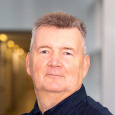 Paul Harrison合作论文数Institute of Inflammation and Ageing, College of Medical and Dental Sciences, University of Birmingham6N R M Tai合作论文数5Ryan Homes合作论文数5
 David Sharp合作论文数Neurology, Department of Brain Sciences, Faculty of Medicine, Imperial College London5Dretzke Janine合作论文数5Rickard Rory F合作论文数Academic Department of Military Surgery and Trauma, Royal Centre for Defence Medicine;Department of Plastic Surgery, Derriford Hospital;Department of Surgery, University of Cape Town;Derriford Hospital, University of Cape Town5David Baxter合作论文数5Sharrock A E合作论文数5
 Matthew Martin合作论文数Madigan Army Medical Center;Legacy Emanuel Medical Center5Tim J. Hodgetts合作论文数5
 Mark Anthony Foster合作论文数Department of Molecular Biology and Skaggs Institute for Chemical Biology, The Scripps Research Institute5
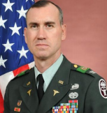 Lorne H. Blackbourne合作论文数Army Institute of Surgical Research5Raushan Lala合作论文数4
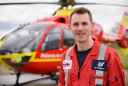 James M Hancox合作论文数West Midlands Ambulance Service NHS Foundation Trust4H Doughty合作论文数4




