Achieving bone union remains a significant clinical dilemma. The use of osteoinductive agents, specifically bone morphogenetic proteins (BMPs), has gained wide attention. However, multiple side effects, including increased incidence of cancer, have renewed interest in investigating alternatives that provide safer, yet effective bone regeneration. Here we demonstrate the robust bone healing capabilities of the main megakaryocyte (MK) growth factor, thrombopoietin (TPO), and second-generation TPO agents using multiple animal models, including mice, rats, and pigs. This bone healing activity is shown in two fracture models (critical-sized defect [CSD] and closed fracture) and with local or systemic administration. Our transcriptomic analyses, cellular studies, and protein arrays demonstrate that TPO enhances multiple cellular processes important to fracture healing, particularly angiogenesis, which is required for bone union. Finally, the therapeutic potential of thrombopoietic agents is high since they are used in the clinic for other indications (eg, thrombocytopenia) with established safety profiles and act upon a narrowly defined population of cells.
Autosomal dominant osteopetrosis type II (ADO2) is a heritable bone disease of impaired osteoclastic bone resorption caused by missense mutations in the chloride channel 7 (CLCN7) gene. Clinical features of ADO2 include fractures, osteomyelitis of jaw, vision loss, and in severe cases, bone marrow failure. Currently, there is no effective therapy for ADO2, and patients usually receive symptomatic treatments. Theoretically, bone marrow transplantation (BMT), which is commonly used in recessive osteopetrosis, could be used to treat ADO2, although the frequency of complications related to BMT is quite high. We created an ADO2 knock-in (p.G213R mutation) mouse model on the 129 genetic background, and their phenotypes mimic the human disease of ADO2. To test whether BMT could restore osteoclast function and rescue the bone phenotypes in ADO2 mice, we transplanted bone marrow cells from 6-8 weeks old male WT donor mice into recipient female ADO2 mice. Also, to determine whether age at the time of transplant may play a role in transplant success, we performed BMT in young (12-week-old) and old (9-month-old) ADO2 mice. Our data indicate that ADO2 mice transplanted with WT marrow achieved more than 90% engraftment up to 6 months post-transplantation at both young and old ages. The in-vivo DXA data revealed that young ADO2 mice transplanted with WT marrow had significantly lower whole body and spine areal bone mineral density (aBMD) at month 6 post-transplantation compared to the ADO2 control mice. The old ADO2 mice also displayed significantly lower whole body, femur, and spine aBMD at months 4 and 5 post-transplantation compared to the age-matched control mice. The in-vivo micro-CT data showed that ADO2 experimental mice transplanted with WT marrow had significantly lower BV/TV at months 2 and 4 post-transplantation compared to the ADO2 control mice at a young age. In contrast, ADO2 control and experimental mice displayed similar BV/TV values for all post-transplantation time points at old age. In addition, serum CTX was significantly higher at month 2 post-transplantation in both young and old ADO2 experimental mice compared to the ADO2 control mice. Serum P1NP levels in young ADO2 experimental mice were significantly higher at baseline and month 2 post-transplantation compared to the ADO2 control mice. These data suggest that BMT may provide, at least, some beneficial effect at both young and adult ages.
Withdrawal: "Megakaryocytes Enhance Mesenchymal Stromal Cells Proliferation and Inhibit Differentiation", by Arbi M. Emmakah, Hussain E. Arman, Marta B. Alvarez, Paul J. Childress, Joseph P. Bidwell, William S. Goebel, Tien-Min Gabriel Chu, and Melissa A. Kacena, J Cell Biochem. The above article, published as accepted article online on 19 July 2017 in Wiley Online Library () has been withdrawn by agreement between authors, the journal's Editor in Chief, Prof. Dr. Christian Behl, and Wiley Periodicals LLC. The withdrawal has been agreed because final approval for the publication of the article could not be received from the authors.
Megakaryocytes (MKs) support bone formation by stimulating osteoblasts (OBs) and inhibiting osteoclasts (OCs). Aging results in higher bone resorption, leading to bone loss. Whereas previous studies showed the effects of aging on MK-mediated bone formation, the effects of aging on MK-mediated OC formation is poorly understood. Here we examined the effect of thrombopoietin (TPO) and MK-derived conditioned media (CM) from young (3-4 months) and aged (22-25 months) mice on OC precursors. Our findings showed that aging significantly increased OC formation in vitro. Moreover, the expression of the TPO receptor, Mpl, and circulating TPO levels were elevated in the bone marrow cavity. We previously showed that MKs from young mice secrete factors that inhibit OC differentiation. However, rather than inhibiting OC development, we found that MKs from aged mice promote OC formation. Interestingly, these age-related changes in MK functionality were only observed using female MKs, potentially implicating the sex steroid, estrogen, in signaling. Further, RANKL expression was highly elevated in aged MKs suggesting MK-derived RANKL signaling may promote osteoclastogenesis in aging. Taken together, these data suggest that modulation in TPO-Mpl expression in bone marrow and age-related changes in the MK secretome promote osteoclastogenesis to impact skeletal aging.
Osteoblast number and activity decreases with aging, contributing to the age-associated decline of bone mass, but the mechanisms underlying changes in osteoblast activity are not well understood. Here, we show that the age-associated bone loss critically depends on impairment of the ability of megakaryocytes (MKs) to support osteoblast proliferation. Co-culture of osteoblast precursors with young MKs is known to increase osteoblast proliferation and bone formation. However, co-culture of osteoblast precursors with aged MKs resulted in significantly fewer osteoblasts compared to co-culture with young MKs, and this was associated with the downregulation of transforming growth factor beta. In addition, the ability of MKs to increase bone mass was attenuated during aging as transplantation of GATA1low/low hematopoietic donor cells (which have elevated MKs/MK precursors) from young mice resulted in an increase in bone mass of recipient mice compared to transplantation of young wild-type donor cells, whereas transplantation of GATA1low/low donor cells from old mice failed to enhance bone mass in recipient mice compared to transplantation of old wild-type donor cells. These findings suggest that the preservation or restoration of the MK-mediated induction of osteoblast proliferation during aging may hold the potential to prevent age-associated bone loss and resulting fractures.
Limitations in scaffold material properties, such as sub-optimal degradation time, highlight the need for alternative approaches to engineer de novo tissues. One emerging solution for fabricating tissue constructs is scaffold-free tissue engineering. To facilitate this approach, three-dimensional (3D) bioprinting technology (Regenova Bio 3D Printer) has been developed to construct complex geometric shapes from discrete cellular spheroids without exogenous scaffolds. Optimizing spheroid fabrication and characterizing cellular behavior in the spheroid environment are important first steps prior to printing larger constructs. Here, we characterized spheroids of immortalized mouse bone marrow stromal cells (BMSCs) that were differentiated to the osteogenic lineage. Immortalized BMSCs were seeded in low attachment 96-well plates in various numbers to generate self-aggregated spheroids either under the force of gravity or centrifugation. Cells were cultured in control or osteogenic media for up to 28 days. Spheroid diameter, roundness and smoothness were measured. Cell viability, DNA content and alkaline phosphatase activity were assessed at multiple time points. Additionally, expression of osteogenic markers was determined using real time qPCR. Spheroids formed under gravity with 20 K, 30 K and 40 K cells had average diameters of 498.5 ± 8.3 μm, 580.0 ± 32.9 μm and 639.2 ± 54.0 μm, respectively, while those formed under 300G centrifugation with the same numbers of cells had average diameters of 362.3 ± 3.5 μm, 433.1 ± 6.4 μm and 491.2 ± 8.0 μm. Spheroids formed via centrifugation were superior to those formed by gravity, as evidenced by better roundness and smoothness and double the retention of DNA (cellular) content. Cells in spheroids exhibited a robust osteogenic response to the differentiation medium, including higher mRNA expression of alkaline phosphatase, collagen type I, and osteocalcin than those cultured in control medium, as well as greater alkaline phosphatase activity. The optimal spheroid fabrication technique from this study was to aggregate 40K cells under 150-300G centrifugation. In future investigations, these spheroids will be 3D printed into larger tissue constructs.
Spaceflight results in reduced mechanical loading of the skeleton, which leads to dramatic bone loss. Low bone mass is associated with increased fracture risk, and this combination may compromise future, long-term, spaceflight missions. Here, we examined the systemic effects of spaceflight and fracture surgery/healing on several non-injured bones within the axial and appendicular skeleton. Forty C57BL/6, male mice were randomized into the following groups: (1) Sham surgery mice housed on the earth (Ground + Sham); (2) Femoral segmental bone defect surgery mice housed on the earth (Ground + Surgery); (3) Sham surgery mice housed in spaceflight (Flight + Sham); and (4) Femoral segmental bone defect surgery mice housed in spaceflight (Flight + Surgery). Mice were 9 weeks old at the time of launch and were euthanized approximately 4 weeks after launch. Micro-computed tomography (μCT) was used to evaluate standard bone parameters in the tibia, humerus, sternebra, vertebrae, ribs, calvarium, mandible, and incisor. One intriguing finding was that both spaceflight and surgery resulted in virtually identical losses in tibial trabecular bone volume fraction, BV/TV (24–28% reduction). Another important finding was that surgery markedly changed tibial cortical bone geometry. Understanding how spaceflight, surgery, and their combination impact non-injured bones will improve treatment strategies for astronauts and terrestrial humans alike.
Emerging evidence demonstrates that megakaryocytes (MK) play key roles in regulating skeletal homeostasis and hematopoiesis. To test if the loss of MK negatively impacts osteoblastogenesis and hematopoiesis, we generated conditional knockout mice where Mpl, the receptor for the main MK growth factor, thrombopoietin, was deleted specifically in MK (Mplf/f;PF4cre). Unexpectedly, at 12 weeks of age, these mice exhibited a 10-fold increase in platelets, a significant expansion of hematopoietic/mesenchymal precursors, and a remarkable 20-fold increase in femoral midshaft bone volume. We then investigated whether MK support hematopoietic stem cell (HSC) function through the interaction of MK with osteoblasts (OB). LSK cells (Lin-Sca1+CD117+, enriched HSC population) were co-cultured with OB+MK for 1 week (1wk OB+MK+LSK) or OB alone (1wk OB+LSK). A significant increase in colony-forming units was observed with cells from 1wk OB+MK cultures. Competitive repopulation studies demonstrated significantly higher engraftment in mice transplanted with cells from 1wk OB+MK+LSK cultures compared to 1wk OB+LSK or LSK cultured alone for 1 week. Furthermore, single-cell expression analysis of OB cultured±MK revealed adiponectin as the most significantly upregulated MK-induced gene, which is required for optimal long-term hematopoietic reconstitution. Understanding the interactions between MK, OB, and HSC can inform the development of novel treatments to enhance both HSC recovery following myelosuppressive injuries, as well as bone loss diseases, such as osteoporosis.
Spaceflight results in bone loss like that associated with osteoporosis or decreased weight-bearing (for example, high-energy trauma such as explosive injuries and automobile accidents). Thus, the unique spaceflight laboratory on the International Space Station presents the opportunity to test bone healing agents during weightlessness. We are collaborating with NASA and the US Army to study bone healing in spaceflight. Given the unique constraints of spaceflight, study design optimization was required. Male mice were selected primarily because their femur is larger than females', allowing for more reproducible surgical outcomes. However, concern was raised regarding male mouse aggression. In addition, the original spaceflight study design included cohousing nonoperated control mice with mice that had undergone surgery to create a segmental bone defect. This strategy prompted the concern that nonoperated mice would exhibit aggressive behavior toward vulnerable operated mice. We hypothesized that operated and nonoperated male mice could be cohoused successfully when they were cagemates since birth and underwent identical anesthetic, analgesic, preoperative, and postoperative conditions. Using quantitative behavioral scoring, body weight, and organ weight analyses (Student t test and ANOVA), we found that nonoperated and operated C57BL/6 male mice could successfully be housed together. The male mice did not exhibit aggressive behavior toward cagemates, whether operated or nonoperated, and the mice did not show evidence of stress, as indicated by veterinary assessment, or change in body or proportional organ weights. These findings allowed our mission to proceed (launched February 2017) and may inform future surgical study designs, potentially increasing housing flexibility.
Department of Orthopaedic Surgery, Indiana University School of Medicine, Indianapolis, Indiana Department of Microbiology and Immunology, Indiana University School of Medicine, Indianapolis, Indiana Department of Medicine, Indiana University School of Medicine, Indianapolis, Indiana Department of Biomedical and Applied Sciences, Indiana University School of Dentistry, Indianapolis, Indiana Department of Orthopaedics and Rehabilitation, Yale University School of Medicine, New Haven, Connecticut Department of Physical Therapy, Indiana University School of Health and Rehabilitation Sciences, Indianapolis, Indiana Department of Pediatrics, Indiana University School of Medicine, Indianapolis, Indiana
Combining anticatabolic agents with parathyroid hormone (PTH) to enhance bone mass has yielded mixed results in osteoporosis patients. Toward the goal of enhancing the efficacy of these regimens, we tested their utility in combination with loss of the transcription factor Nmp4 because disabling this gene amplifies PTH-induced increases in trabecular bone in mice by boosting osteoblast secretory activity. We addressed whether combining a sustained anabolic response with an anticatabolic results in superior bone acquisition compared with PTH monotherapy. Additionally, we inquired whether Nmp4 interferes with anticatabolic efficacy. Wild-type and Nmp4(-/-) mice were ovariectomized at 12 weeks of age, followed by therapy regimens, administered from 16 to 24 weeks, and included individually or combined PTH, alendronate (ALN), zoledronate (ZOL), and raloxifene (RAL). Anabolic therapeutic efficacy generally corresponded with PTH + RAL = PTH + ZOL > PTH + ALN = PTH > vehicle control. Loss of Nmp4 enhanced femoral trabecular bone increases under PTH + RAL and PTH + ZOL. RAL and ZOL promoted bone restoration, but unexpectedly, loss of Nmp4 boosted RAL-induced increases in femoral trabecular bone. The combination of PTH, RAL, and loss of Nmp4 significantly increased bone marrow osteoprogenitor number, but did not affect adipogenesis or osteoclastogenesis. RAL, but not ZOL, increased osteoprogenitors in both genotypes. Nmp4 status did not influence bone serum marker responses to treatments, but Nmp4(-/-) mice as a group showed elevated levels of the bone formation marker osteocalcin. We conclude that the heightened osteoanabolism of the Nmp4(-/-) skeleton enhances the effectiveness of diverse osteoporosis treatments, in part by increasing hyperanabolic osteoprogenitors. Nmp4 provides a promising target pathway for identifying barriers to pharmacologically induced bone formation.
Early stages of chronic kidney disease (CKD) are characterized by development of progressive anemia as well as concurrent marked elevation of the phosphaturic hormone fibroblast growth factor 23 (FGF23). As kidney function declines, FGF23 further increases and anemia worsens, due to either inadequate production of renal erythropoietin (EPO) or incidence of hypoferremia. Moreover, in CKD, anemia and elevated FGF23 levels are associated with left ventricular hypertrophy (LVH), CKD progression, and mortality. Treatment of CKD-related anemia involves iron repletion and erythropoietin (EPO) administration. EPO is one of the most extensively used medications in CKD, but its administration is associated with increased risks of cardiovascular disease and mortality. Although FGF23 levels increase early in CKD, the pathophysiological regulation of FGF23 is still not completely understood. Phosphate, 1,25-dihydroxyvitamin D (1,25D), parathyroid hormone, and calcium affect FGF23 production; however, these factors are still within normal ranges when bone and circulating FGF23 increase. Recent studies demonstrate intriguing associations between hypoxia, iron deficiency, and FGF23 upregulation. Indeed, in the settings of normal and impaired kidney function, iron deficiency potently increases bone Fgf23 expression. However, other anemia-related factors, including EPO, could potentially contribute to elevated FGF23 production. As both EPO therapy and FGF23 are associated with adverse outcomes in CKD, we explored the hypothesis that EPO is a previously unrecognized regulator of this phosphaturic hormone. Collectively, our pre-clinical findings suggest that modulating EPO exposure in CKD patients may lower FGF23 and thereby decrease its adverse effects. To examine whether exogenous EPO stimulates FGF23 in vivo, wild-type C57BL/6 mice at 6-8 weeks of age were injected with increasing doses of recombinant human EPO (25-250 U/g of body weight). A 3-day regimen induced a dose-dependent, 40-fold maximal increase in whole bone Fgf23 mRNA expression (Figure 1A), paralleled by increased serum total FGF23 as measured with an ELISA that detects both C-terminal FGF23 fragments (‘cFGF23’) and bioactive intact FGF23 (‘iFGF23’) (Figure
Networking between hematopoietic stem cells (HSCs) and cells of the hematopoietic niche is critical for stem cell function and maintenance of the stem cell pool. We characterized calvariae-resident osteomacs (OMs) and their interaction with megakaryocytes to sustain HSC function and identified distinguishing properties between OMs and bone marrow (BM)-derived macrophages. OMs, identified as CD45+F4/80+ cells, were easily detectable (3%-5%) in neonatal calvarial cells. Coculture of neonatal calvarial cells with megakaryocytes for 7 days increased OM three- to sixfold, demonstrating that megakaryocytes regulate OM proliferation. OMs were required for the hematopoiesis-enhancing activity of osteoblasts, and this activity was augmented by megakaryocytes. Serial transplantation demonstrated that HSC repopulating potential was best maintained by in vitro cultures containing osteoblasts, OMs, and megakaryocytes. With or without megakaryocytes, BM-derived macrophages were unable to functionally substitute for neonatal calvarial cell-associated OMs. In addition, OMs differentiated into multinucleated, tartrate resistant acid phosphatase-positive osteoclasts capable of bone resorption. Nine-color flow cytometric analysis revealed that although BM-derived macrophages and OMs share many cell surface phenotypic similarities (CD45, F4/80, CD68, CD11b, Mac2, and Gr-1), only a subgroup of OMs coexpressed M-CSFR and CD166, thus providing a unique profile for OMs. CD169 was expressed by both OMs and BM-derived macrophages and therefore was not a distinguishing marker between these 2 cell types. These results demonstrate that OMs support HSC function and illustrate that megakaryocytes significantly augment the synergistic activity of osteoblasts and OMs. Furthermore, this report establishes for the first time that the crosstalk between OMs, osteoblasts, and megakaryocytes is a novel network supporting HSC function.
Journal of Cellular BiochemistryVolume 118, Issue 8 p. 1947-1955 Issue InformationFree Access Table of Contents, Editor's Choice, Features First published: 07 June 2017 https://doi.org/10.1002/jcb.26119AboutPDF ToolsRequest permissionExport citationAdd to favoritesTrack citation ShareShare Give accessShare full text accessShare full-text accessPlease review our Terms and Conditions of Use and check box below to share full-text version of article.I have read and accept the Wiley Online Library Terms and Conditions of UseShareable LinkUse the link below to share a full-text version of this article with your friends and colleagues. Learn more.Copy URL Share a linkShare onFacebookTwitterLinkedInRedditWechat Volume118, Issue8August 2017Pages 1947-1955 RelatedInformation
Successful regeneration of the cranium in patients suffering from cranial bone defects is an integral step to restore craniofacial function. However, restoration of craniofacial structure has been challenging due to its complex geometry, limited donor site availability, and poor graft integration. To address these problems, we investigated the use of a thiol–acrylate hydrogel as a cell carrier to facilitate cranial regeneration. Thiol–acrylate hydrogels were formulated with 5–15 wt% poly(ethylene glycol)-diacrylate (PEGDA) and 1–9 mm dithiothreitol (DTT). The degradation rate, swelling ratio, and shear modulus of the resulting hydrogel were first characterized. Then, pre-osteoblast-like cells (MC3T3-E1) were encapsulated in the hydrogel and cultured for up to 21 d. Our results demonstrate that compared to samples formulated from 15 wt% PEGDA, 5 wt% PEGDA samples showed lower storage modulus at day 10 (0.7 kPa versus 8.3 kPa), 62.7% higher in weight change after soaking for 10 d. While the 5 wt% PEGDA group showed an 85% weight loss between day 10 and 21, the 15 wt% PEGDA group showed a 5% weight gain in the same time period. Cell viability with 15 wt% PEGDA and 5 mm DTT hydrogel decreased by 41.3% compared to 5 wt% PEGDA and 5mM DTT gel at day 7. However, histological analysis of cells after 21 d in culture revealed that they had pericellular mineral deposition indicating that the cells were differentiating into osteoblasts lineage in all experimental groups. This study shows that thiol–acrylate hydrogels can be tailored to achieve different degradation rates, in order to enhance cell viability and differentiation. Thus, the findings of this study provide a fundamental understanding for the application of thiol–acrylate hydrogels in cranial bone regeneration.
Advance Articles are PDF versions of manuscripts that have been peer reviewed and accepted but not yet copyedited. The manuscripts are published online as soon as possible after acceptance and before the copyedited, typeset articles are published. They are posted "as is" (i.e., as submitted by the authors at the modification stage), and do not reflect editorial changes. No corrections/changes to the PDF manuscripts are accepted. Accordingly, there likely will be differences between the Advance Article manuscripts and the final, typeset articles. The manuscripts remain listed on the Advance Article page until the final, typeset articles are posted. At that point, the manuscripts are removed from the Advance Article page.
Megakaryocytes (MKs) can induce proliferation of calvarial osteoblasts [Ciovacco et al., 2009], but this same phenomenon has not been reported for bone marrow stromal populations from long bones. Bone marrow contains several types of progenitor cells which can be induced to differentiate into multiple cell types. Herein, we examined mesenchymal stromal cell proliferation and osteoblastic differentiation when rabbit or mouse MK were cultured with i) rabbit bone marrow stromal cells, ii) rabbit dental pulp stromal cells, or iii) mouse bone marrow stromal cells. Our results demonstrated that rabbit and mouse stromal cells co-cultured with rabbit MK or mouse MK, have significant increases in proliferation on day 7 by 52%, 46%, and 24%, respectively, compared to cultures without MK. Conversely, alkaline phosphatase (ALP) activity was lower at various time points in these cells when cultures contain MK. Similarly, calcium deposition observed at day 14 rabbit bone marrow and dental pulp stromal cells and day 21 mouse bone marrow stromal cells was 63%, 69%, and 30% lower respectively, when co-cultured with MK. Gene expression studies reveal transcriptional changes broadly consistent with increased proliferation and decreased differentiation. Transcript levels of c-fos (associated with cell proliferation) trended higher after 3, 7, and 14 days in culture. Also, expression of alkaline phosphatase, osteonectin, osterix, and osteopontin, which are markers for osteoblast differentiation, showed MK-induced decreases in a cell type and time dependent manner. Taken together, these data suggest that MK play a role in stromal cell proliferation and differentiation, from multiple sites/locations in multiple species. This article is protected by copyright. All rights reserved.
The Lnk adapter protein negatively regulates the signaling of thrombopoietin (TPO), the main megakaryocyte (MK) growth factor. Lnk-deficient (-/-) mice have increased TPO signaling and increased MK number. Interestingly, several mouse models exist in which increased MK number leads to a high bone mass phenotype. Here we report the bone phenotype of these mice. MicroCT and static histomorphometric analyses at 20 weeks showed the distal femur of Lnk-/- mice to have significantly higher bone volume fraction and trabecular number compared to wild-type (WT) mice. Notably, despite a significant increase in the number of osteoclasts (OC), and decreased bone formation rate in Lnk-/- mice compared to WT mice, Lnk-/- mice demonstrated a 2.5-fold greater BV/TV suggesting impaired OC function in vivo. Additionally, Lnk-/- mouse femurs exhibited non-significant increases in mid-shaft cross-sectional area, yet increased periosteal BFR compared to WT femurs was observed. Lnk-/- femurs also had non-significant increases in polar moment of inertia and decreased cortical bone area and thickness, resulting in reduced bone stiffness, modulus, and strength compared to WT femurs. Of note, Lnk is expressed by OC lineage cells and when Lnk-/- OC progenitors are cultured in the presence of TPO, significantly more OC are observed than in WT cultures. Lnk is also expressed in osteoblast (OB) cells and in vitro reduced alkaline phosphatase activity was observed in Lnk-/- cultures. These data suggest that both direct effects on OB and OC as well as indirect effects of MK in regulating OB contributes to the observed high bone mass. J. Cell. Biochem. 118: 2231-2240, 2017. © 2017 Wiley Periodicals, Inc.
 c moya合作论文数34dagmara plana合作论文数31marilyn florido合作论文数22Melissa A. Kacena合作论文数School of Medicine, Yale University22
 Paul Childress合作论文数21Regla M. Lara合作论文数17
 Francisco Duenas合作论文数15
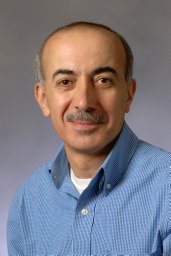 Edward F. Srour合作论文数School of Medicine, Indiana University13Joseph P. Bidwell合作论文数13
 Mohamad Safa F合作论文数Department of Human and Molecular Genetics, Virginia Commonwealth University7
 Angela Bruzzaniti合作论文数Department of Oral Biology, Indiana University School of Dentistry7j arzuaga合作论文数6David J. Olivos合作论文数5Ying-Hua Cheng合作论文数5Linlin Xu合作论文数5
 Joydeep Ghosh合作论文数Dept Med, Indiana Univ Sch Med5
 Chitteti Brahmananda R合作论文数Dept Med, Indiana Univ Sch Med5
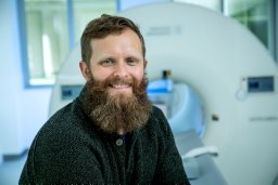 Maupin Kevin A合作论文数5
 Yongzheng He合作论文数5Evan R Himes合作论文数5Feng-Chun Yang合作论文数5Yamila Martínez合作论文数4Rita L Gerard-O'Riley合作论文数4Alexandra Aguilar-Perez合作论文数4Alex G. Robling合作论文数4
 Pavalko Fredrick M合作论文数Dept Cellular & Integrat Physiol, Indiana Univ Sch Med4
 Monique Bethel合作论文数4
 Jung Min Hong合作论文数4Keith R. Stayrook合作论文数4Ofelia Sam合作论文数3Tomás Shagarodsky合作论文数3Selene Hernandez-Buquer合作论文数3Irushi Abeysekera合作论文数3P Thunyakitpisal合作论文数3David C Scofield合作论文数3Mark Hood合作论文数3Aamir Tucker合作论文数3armando caballero合作论文数3Haley McGough合作论文数3Arbi M. Emmakah合作论文数3
 Jose Luis Rodriguez Illera合作论文数3
 idioleidys alvarez bello合作论文数3
 Maria Manuel Varela合作论文数3
 Yu Shao合作论文数3
 Tien-Min Gabriel Chu合作论文数Sch Dent, Indiana Univ3Ciovacco Wendy A合作论文数3Eleniste Pierre P合作论文数3Allen Matthew R合作论文数3Davis K M合作论文数3




