Advances in the understanding of the tumor microenvironment have led to devel-opment of immunotherapeutic strategies, such as chimeric antigen receptor T cells (CAR-Ts). However, despite success in blood malignancies, CAR-T therapies in solid tumors have been hampered by their restricted infiltration. Here, we used our understanding of early cytotoxic lymphocyte infiltration of human lymphocytes in solid tumors in vivo to investigate the receptors in normal, adjacent, and tumor tissues of primary non-small-cell lung cancer specimens. We found that CX3CL1-CX3CR1 reduction restricts cytotoxic cells from the solid-tumor bed, contributing to tumor escape. Based on this, we designed a CAR-T construct using the well-established natural killer group 2, member D (NKG2D) CAR-T expression together with overexpression of CX3CR1 to promote their infiltration. These CAR-Ts infiltrate tumors at higher rates than control-activated T cells or IL-15-overexpressing NKG2D CAR-Ts. This construct also had similar functionality in a liver-cancer model, demonstrating potential efficacy in other solid malignancies.
Myelodysplastic syndromes (MDS) are age-dependent and genetically diverse hematopoietic neoplasms associated with inflammation, cytopenias, myeloid dysplasia and risk for acute myeloid leukemia (AML) progression. Mounting evidence indicates that emergence of somatic gene mutations arising in hematopoietic stem and progenitor cells (HSPC) with age plays an important role in MDS pathogenesis. Our investigations indicate that sustained activation of innate immune response directs inflammation in the bone marrow (BM) microenvironment that contributes to hematopoietic and immune impairment through local generation of inflammatory cytokines, expansion of regulatory T- cells, and up-regulation and activation of pattern recognition receptors. Critical cellular effectors are the inflammation-associated immature myeloid cells termed myeloid-derived suppressor cells (MDSCs). Human MDSCs, distinguished phenotypically by CD33high and HLA-DR−Lin−, expand and are activated in cancers. We previously reported that MDSCs are profoundly expanded in the local BM microenvironment of MDS where they contribute to MDS hematopoietic BM failure. MDSCs suppress T-cell responses and impair CD4+/CD8+ function, suggesting that this may be a key mechanism fostering clonal escape from anti-tumor immune response. Indeed, the MDSC population size limits the benefit of immune checkpoint antagonists in the treatment of solid tumors and effective strategies to deplete MDSCs are lacking. Therefore, we hypothesized the high CD33 surface density in MDSCs can be exploited for selective immune-depletion by the T-cell engager AMV564, a novel CD33/CD3 tetravalent bispecific antibody that recognizes both CD33 and CD3, currently in clinical development for AML (NCT03144245). Depletion of MDSCs may restore immune function, hematopoiesis and improve sensitivity to checkpoint inhibitors. Primary BM mononuclear cells (BMMNC) from 15 MDS patients were treated with AMV564 or isotype IgG control for 5-7 days in vitro . AMV564 treatment of MDS BMMNC eliminated CD33+ MDSCs in a dose-dependent manner and expanded CD4+ and CD8+ T-cells compared to controls (P ≤0.001). Proliferation of CD4+ and CD8+ T-cells, measured by Brdu incorporation, more than doubled with AMV564 treatment (P ≤0.001). IFNγ, a T-cell activation biomarker, dramatically increased in AMV564 treated cells indicating T-cells are fully activated by AMV564 at concentrations as low as 0.7 ng/ml (P ≤0.01). Moreover, depletion of MDSCs and immunological restoration was accompanied by significant improvement in colony-forming capacity after AMV564 treatment (P ≤0.01). Checkpoint inhibition alone with 10 µg/ml of anti-PD1 antibody did not result in reduction of MDSCs and T-cell activation in MDS BMMNCs, demonstrating that anti-PD1 alone is insufficient to appropriately activate T-cells or modulate MDSC cell number. Interestingly, combined treatment of MDS BMMNC with anti-PD1 and AMV564 yielded more than additive T-cell activation and increased colony forming capacity (P ≤0.01), indicating the potential to augment MDS-specific immune responses. Our findings demonstrate that AMV564 treatment selectively depletes MDSCs to reactivate T lymphocytes by improving both quantity and quality of immune responses leading to the improvement of hematopoiesis. These preclinical data provide a strong rationale for clinical investigation of this innovative approach in patients with MDS.
Myelofibrosis is an indicator of poor prognosis in myeloproliferative neoplasms (MPNs), but the precise mechanism(s) contributing to extracellular matrix remodeling and collagen deposition in the bone marrow (BM) niche remains unanswered. In this study, we isolated mesenchymal stromal cells (MSCs) from mice transplanted with wild-type thrombopoietin receptor (MPLWT) and MPLW515L retroviral-transduced bone marrow. Using MSCs derived from MPLW515-transplant recipients, excessive collagen deposition was maintained in the absence of the virus and neoplastic hematopoietic cells suggested that the MSCs were reprogrammed in vivo. TGFβ production by malignant megakaryocytes plays a definitive role promoting myelofibrosis in MPNs. However, TGFβ was equally expressed by MSCs derived from MPLWT and MPLW515L expressing mice and the addition of neutralizing anti-TGFβ antibody only partially reduced collagen secretion in vitro. Interestingly, profibrotic MSCs displayed increased levels of pSmad3 and pSTAT3 suggesting that inflammatory mediators cooperating with the TGFβ-receptor signaling may maintain the aberrant phenotype ex vivo. FGFb is a known suppressor of TGFβ signaling. Reduced collagen deposition by FGFb-treated MSCs derived from MPLW515L mice suggests that the activating pathway is vulnerable to this suppressive mediator. Therefore, our findings have implications for the future investigation of therapies to reverse fibrosis in MPNs.
Recent studies suggest that aging-associated inflammation, or "inflammaging", contributes to genetic instability and MDS predisposition. Although innumerable somatic genetic events have been annotated in recent years, including many that are not unique to MDS, they are not sufficient for disease initiation. The precise underlying mechanisms conducive to the emergence of these genetic events also remain to be delineated. We reported that bone marrow (BM) myeloid derived suppressor cells (MDSC) activated by the damage associated molecular pattern (DAMP) protein S100A9, promote ineffective hematopoiesis and the development of MDS. Inflammaging associated alterations in metabolism have been implicated in predisposition to cancer development with age. Here we report that S100A9 and ROS-induced inflammaging are associated with insulin resistance and hyperglycemia in the BM microenvironment that triggers activation of adaptive oncogenic pathways and genomic instability in HSPC. Glucose concentrations were markedly elevated in MDS BM plasma vs. age-matched controls, and directly correlated with S100A9 concentration (r=0.513, P=0.003, n=41). The magnitude of BM-glucose elevation significantly exceeded that in the peripheral blood and negatively correlated with the proportion of HSPC while directly correlating with BM MDSC percentage. S100A9 transgenic (Tg) mice displayed age-dependent elevation of glucose in peripheral blood and BM when compared to wild type mice accompanied by accumulation of somatic mutations (SM) by sequencing in aged S100A9-Tg compared to younger counterparts. NGS of 38 primary MDS BM specimens showed that SM common to MDS, such as those involving ASXL1, U2AF1 and DNMT3A, we re present only in high glucose stratified MDS-BM specimens (glucose > 110 ug/ml). Furthermore, there was a strong correlation between the cellular ROS/nuclear-β-catenin to DNA damage (γH2AX+ cells) linking S100A9-induced ROS accumulation to genetic instability. Elevation in BM plasma glucose was specifically associated with upregulation of the fat mass and obesity associated (FTO) transcript and protein in both MDS BM and S100A9Tg mice. FTO is a risk factor for type 2 diabetes, and encodes an α-ketoglutarate-dependent dioxygenase that functions as an RNA demethylase specific for N 6-methyladenosine (m6A) residues, targeted by splicing factors. Both human and murine MDS specimens displayed decreased m6A mRNA methylation compared to controls. FTO knockdown with CRISPR increased mRNA m6A methylation in MDS primary specimens, whereas overexpression of FTO led to a corresponding decrease that was enhanced by S100A9 stimulation. Moreover, S100A9 treatment induced FTO and demethylation of m6A mRNA in human and murine BM cells. These effects were accompanied by disruption of spliceosomes in the nucleus, as demonstrated by delocalization of SRSF2 from nuclear speckles into the cytoplasm where they colocalize with FTO in MDS patients. Interestingly, from the S100A9Tg mouse sequencing studies, we discovered that the mutation hotspots were in locations specific for histone H3K27 acetylation which have been previously linked to genomic instability, as well as splicing regulation, and regulated by histone deacetylase 1 (HDAC1). Reduced HDAC1 levels are known to make cells hypersensitive to DNA-damaging insults, such as those inducing ROS, similar to what we observed with treatment of S100A9 in healthy human bone marrow, MDS patient samples and S100A9Tg mice. Furthermore, this correlated with increased levels of DNA damage in our patient samples and in our murine S100A9Tg model as measured by phosphorylated γH2AX histones. Our studies provide a biological rationale for the initiation of DNA-genetic damage under inflammatory senescence conditions in MDS. These findings demonstrate that S100A9-induced inflammation activates a signaling cascade that enhances development of splicing variants and somatic gene mutations critical to MDS pathogenesis. Disclosures List: Celgene Corporation: Honoraria, Research Funding.
Myelodysplastic syndrome (MDS) is one of the major types of acquired bone marrow failure characterized by impaired peripheral blood cell production (anemia and cytopenias). Although much has been learned regarding the molecular genetic events involved in the pathogenesis of the MDS hematopoietic stem/progenitor cell compartment (HSC/HPC), investigations of the environmental pressures underlying disease initiation have been limited. Prior investigations have shown that inflammation in the bone marrow microenvironment contributes to hematopoietic impairment, with inflammatory molecules providing regulatory cues driving the proliferation and apoptotic death of HSC/HPC. We have reported that, the accumulation of myeloid-derived suppressor cells (MDSCs) in the local inflammatory bone marrow microenvironment plays a major role in the direct pathogenesis of MDS. MDSCs function by producing mediators and inflammatory cytokines capable of suppressing hematopoiesis and are defined by the lack of all lineage markers and only one key surface receptor, CD33 (CD33+HLA-DR−Lin−). We recently uncovered that this receptor is greatly expressed in MDSC isolated from patients with MDS and plays an important role in MDSC-mediated hematopoietic suppressive function. We also found that this ITIM (immune-receptor tyrosine-based inhibitor motif) containing molecule could induce suppressive cytokines after its engagement with its newly identified ligand S100A9. Therefore, we tested the hypothesis that with a fully human IgG1 monoclonal antibody against CD33 (mAb33.1) we could prevent the engagement of this ligand/receptor pair as well as induce ADCC of pathogenic MDSC through the reduction in the accumulation of CD33+ MDSC, the immune suppression and the restoration of hematopoiesis in MDS bone marrow specimens. After testing primary specimens with mAb33.1 we saw a significant increase in ADCC activity via NK as evidenced by significant reduction in the proportion of MDSC in culture as compared to isotype control treated cells. This decrease correlated with an increase in CD107a granule mobilization as well as an increased cytotoxicity in a killing assay without changing the proportion of NK cells in the culture. Functionally, the concentration of secreted IL-10, TGFb and VEGF were decreased, as was the gene expression of these suppressive cytokines after treatment with mAb33.1 but was restored when the antibody was cross-linked with an anti-human IgG antibody demonstrating the blocking ability of the antibody in preventing CD33 downstream signaling. Importantly, all of these observations correlated with the restoration of hematopoiesis, as there was a significant increase in the formation of CFU-GM and BFU-E colonies on a methylcellulose assay (n=9). In addition mAb33.1 also displayed a protective effect on HSC by blocking ROS production and reducing DNA damage, as demonstrated by comet and H2AX assays. This work provides the ground for the development of a novel group of therapies directly aimed at the suppressive MDSC and blockage of their signaling, rather than directly targeting the malignant clone, with the long term goal of improving the local microenvironment. This strategy will provide the background to assess its clinical potential to serve as a therapeutic target in MDS and AML.
 Erika Adriana Eksioglu合作论文数Dept Immunol, H Lee Moffitt Canc Ctr & Res Inst4
 Sheng Wei合作论文数4Xianghong Chen (陈祥洪)合作论文数3
 Alan List合作论文数3Kathy McGraw合作论文数2
 Ashley A Basiorka合作论文数H. Lee Moffitt Cancer Center-Cancer Biology Ph.D. Program, University of South Florida2Pingyan Cheng合作论文数Chongqing Univ. of Medical Sciences, Chongqing Medical University2William A. Adams合作论文数1Elena Kostenko合作论文数1Bjoern Rueter合作论文数1Neale Lopez-Blanco合作论文数1Nhan Tu合作论文数1Tina So合作论文数1Ying Han (韩颖)合作论文数1Wendy Kandell合作论文数1Robert Dalton合作论文数1Amy F McLemore合作论文数1Alexandra Calescibetta合作论文数1Jeanmarie Guenot合作论文数1Pearlie K Epling-Burnette合作论文数Univ S Florida, Coll Med, H Lee Moffitt Canc Ctr & Res Inst, Program Immunol, Tampa, FL 33612 USA1Ling Cen合作论文数H Lee Moffitt Canc Ctr & Res Inst, Tampa, FL USA1
 Jonathan Nguyen合作论文数1
 Carlos Manuel Moran-Segura合作论文数1Lanzhu Yue合作论文数Dept Hematol, Tianjin Med Univ1
 Padron Eric合作论文数1Wright Gabriela合作论文数1
 Zonghong Shao (邵宗鸿)合作论文数Tianjin Medical University General Hospital1
 Thomas Cluzeau合作论文数1
 Ling Zhang合作论文数Department of Hematopathology and Laboratory Medicine, Moffitt Cancer Center;Department of Immunology, Moffitt Cancer Center;Department of Oncologic Sciences, University of South Florida1
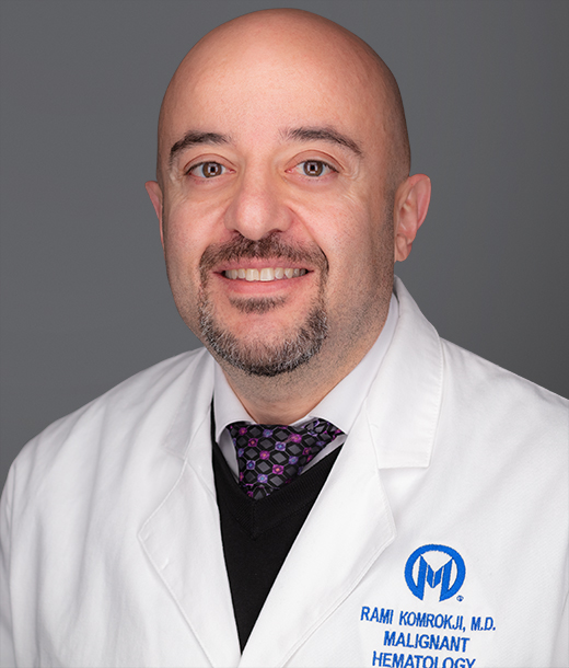 Rami Komrokji合作论文数Malignant Hematology Department, Moffitt Cancer Center;College of Medicine, University of South Florida1Danielle L. Gilvary合作论文数1
 Doug Cress合作论文数Moffitt Cancer Center1
 Kenneth L Wright合作论文数H. Lee Moffitt Cancer Center, University of South Florida1
 Xiubao Ren (任秀宝)合作论文数Tianjin Medical University Cancer Institude & Hospital1
 Jeffrey Lancet合作论文数Department of Malignant Hematology, Moffitt Cancer Center1Karl-Heinz Heider合作论文数1




