Nanobdellati (formerly DPANN) archaea are considered as primitive archaeal organisms that often live in symbiosis with archaeal hosts. In this study, we investigated the symbiotic mechanism between a Nanobdellati archaeon, Nanobdella aerobiophila strain MJ1, and its host archaeon Metallosphaera sedula strain MJ1HA, using cryo-electron tomography. In our tomographic observations, we identified a conical attachment organelle at the interface between MJ1 and MJ1HA during symbiosis. This structure consists of a concentric array of short cylindrical shells, consistent with a previous report. Subtomogram averaging, combined with AlphaFold 3 structural predictions, allowed us to identify a potential component of attachment organelles. Additionally, we inferred potential components of the S layers in MJ1 and MJ1HA based on tomographic data and subtomogram averages. Based on these analyses, we hypothesize that a MJ1 S layer component may undergo conformational changes to also serve as a component of attachment organelles, warranting further investigation.
Flowers undergo dynamic changes in morphology, color, and scent over the course of anthesis, and these changes can regulate pollinator behavior and reproductive processes. Whether similarly dynamic changes at the microscopic surface scale play a role in floral function, however, remains largely unexplored. Here, we show that trap-and-release pollination in Aristolochia subgenus Siphisia is mediated by sex-phase-dependent remodeling of the inner floral epidermis. Pollinators are attracted to and trapped within flowers during the female phase and are released several days later, after the flowers enter the male phase. Remarkably, this functional transition occurs without any externally visible change in floral morphology. Behavioral bioassays showed that pollinators could not climb the inner tube surface of female-stage flowers but readily escaped from male-stage tubes. The slipperiness in the female stage disappeared when the tubes were inverted, demonstrating that the effect was direction-dependent. Scanning electron microscopy revealed marked changes in the microstructure of the inner tube surface between the female and male stages: the downward-oriented papillate epidermal cells present in female-stage tubes collapsed during the male stage. Ultrastructural analysis by field-emission scanning electron microscopy revealed pronounced intracellular changes within these papillate cells, consistent with epidermal cell death. Field observations supported the functional importance of this microstructure: male-stage flowers with longer epidermal cells retained more pollinators than those with shorter cells. Our study provides the first evidence that plants actively reconfigure microstructures with direct functional consequences. These findings extend the known roles of cell death in flowers beyond gametophyte development and floral senescence to dynamic control of floral surface microstructures to maximize the benefit of a single pollinator visit.
Chlorophagy is an autophagy pathway that delivers chloroplast components into the vacuole for degradation, thus eliminating damaged chloroplasts. Chloroplast degradation is observed in Arabidopsis (Arabidopsis thaliana) mutants of MALTOSE-EXCESS 1 (MEX1), a maltose exporter in the chloroplast inner envelope membrane. However, whether autophagy is involved in the mex1 phenotypes is unknown. To extend our understanding of the signals that emanate from damaged chloroplasts and activate chlorophagy, we investigated how mex1 chloroplasts are degraded. Chlorotic mature leaves caused by maltose hyperaccumulation in mex1 plants contained swollen chloroplasts in the cytoplasm and degrading chloroplasts in the vacuole, together with heightened expression of autophagy-related (ATG) genes. The vacuolar degradation of mex1 chloroplasts required the core ATG proteins ATG7 and ATG10. ATG8-labeled structures accumulated on the surfaces of swollen mex1 chloroplasts. These findings indicate that maltose hyperaccumulation triggers chlorophagy via the core autophagy machinery. Notably, phenotypic analysis of mex1 atg double mutant plants suggested that excess chlorophagy aggravates the chlorosis seen in mex1 leaves. Transcriptome deep sequencing indicated that maltose-excess stress shares a similar transcriptomic response with high-light stress, which also triggers chlorophagy. Therefore, the signals inducing chlorophagy may be highly stimulated in mex1 leaves, making mex1 mutants effective tools for chlorophagy research.
Chloroplast development is primarily regulated by transcription factors, including those belonging to the GOLDEN2-LIKE (GLK) family, which alter the expression of genes encoding chloroplast proteins. However, the mechanisms by which multiple transcription factors coordinate to optimize chloroplast development remain unclear. In this study, we identified the single B-BOX (BBX) CONSTANS-LIKE (COL) proteins COL6 (also known as BBX14), COL7 (BBX16), COL8 (BBX17), and COL16 (BBX15) as novel regulators of chloroplast development in Arabidopsis thaliana. Notably, the col6 col16 (col-d) double mutant exhibited a chlorophyll-excess phenotype, which was further enhanced in the quadruple col6 col7 col8 col16 (col-q) mutant. Introduction of col-d and col-q mutations in the glk1 glk2 (glk-d) mutant partially restored the pale-green phenotype of the original glk-d mutant. COL proteins negatively and redundantly regulated a class of genes encoding chloroplast proteins, acting directly on their promoters. Moreover, COL6 and COL7 expression exhibited a diurnal cycle, with their expression patterns contrasting with those of GLK1 and GLK2, which are positive regulators of chloroplast development. Furthermore, GLK expression was markedly higher in the col-q mutants than in wild-type plants. Co-immunoprecipitation assays revealed that COL proteins interacted with the GARP DNA-binding domain in the N-terminus of GLK1, likely affecting its function. Moreover, COL proteins associate with each other to form homomeric and heteromeric complexes, suggesting functional redundancies at the protein level. In conclusion, these results suggest that the multilayered regulatory mechanisms of single BBX COL and GLK proteins optimize chloroplast development in Arabidopsis thaliana in fluctuating environments.
Cryopreservation by vitrification typically requires 30-50 v/v% of cytotoxic penetrable cryoprotective agents (CPAs) to prevent ice crystal formation during freezing and thawing, limiting its broader application. Since pressure suppresses ice crystallization, applying high pressure during vitrification may enable reducing CPA concentrations while maintaining cell viability. In this study, we used a high-pressure freezing (HPF) device, commonly used for cryofixation, to successfully cryopreserve 2D cell monolayers and 3D cell spheroids with 20-30 v/v% penetrable CPA. Compared with commonly used plunge freezing, HPF cell monolayers exhibited higher postthaw viability and better retention on the substrate, allowing for subsequent proliferation. HPF cell spheroids showed improved cell viability, metabolic activity and maintained cell-cell adhesion. Developing HPF devices specifically for cryopreservation, in combination with advanced warming techniques, holds promise for achieving vitrification with low or even no CPA.
Mitochondria are preferentially transmitted from the maternal plant in most angiosperms, including rice, and paternal mitochondria are generally eliminated during microgametogenesis and/or in zygotes. The mechanism by which paternal mitochondria are eliminated progresses during plant reproductive processes. In the present study, we examined the distribution of paternal mitochondria in rice sperm cells and zygotes produced through the in vitro fertilization (IVF) of isolated rice gametes. Male gametes of rice possess mitochondria with nucleoids, suggesting the potential transfer of paternal mitochondria and their DNA into zygotes on fertilization and subsequent selective elimination of paternal mitochondria in the zygote. To intensively monitor the fate of rice paternal mitochondria in zygotes immediately after gamete fusion, time-lapse observations were conducted in paternal mitochondria labeled with GFP from rice zygotes produced using an IVF system. The results showed that the paternal mitochondria are progressively degraded during the early developmental stage at 1 to 3 h after fusion (HAF), leaving a small number of paternal mitochondria at 6 HAF. The remaining paternal mitochondria were considered to be degraded in later developmental-stage zygotes because paternal mitochondrial DNA-derived single-nucleotide polymorphisms were not detected in the sequencing reads of genomic DNA prepared from inter-subspecific hybrid rice. In addition, treatment with autophagy inhibitors stabilized the paternal mitochondria in zygotes. This suggests that the autophagy-dependent massive and selective elimination machinery for male mitochondria functions in rice zygotes immediately after gamete fusion and supports the strict maternal inheritance of mitochondria in rice.
Grafting is an agricultural technique that joins tissues from different plants to obtain useful rootstock traits. However, cellular processes involved in joint tissue repair remain poorly understood. We analyzed Nicotiana benthamiana (Nb) and Arabidopsis thaliana (At) interfamily heterografting as a high-stress model and At homografting as a low-stress model. Transmission electron micrographs reveal the formation of autophagic structures in cells near the graft boundary over a long period in Nb/At interfamily grafts and in a short period of a few days in At homografts. Using a GFP-ATG8 marker line, the autophagosomes were observed in the cells near the graft boundary, especially on the scion side, where nutrient depletion occurred. Grafting of At autophagy-defective mutants decreases grafting success rates and post-grafting growth. NbATG5 knockdown suppresses graft establishment in Nb/At interfamily heterografts. Moreover, At autophagy-defective mutants show reduced callus formation directed to wounds under the nutrient-deficient conditions. These results suggest that autophagy is induced during grafting, promoting callus formation and contributing to tissue connectivity.
The thylakoid membrane (TM) serves as the scaffold for oxygen-evolving photosynthesis, hosting the protein complexes responsible for the light reactions and ATP synthesis. Vesicle inducing protein in plastid 1 (VIPP1), a key protein in TM remodeling, has been recognized as essential for TM homeostasis. In vitro studies of cyanobacterial VIPP1 demonstrated its ability to form large homo-oligomers (2 MDa) manifesting as ring-like or filament-like assemblies associated with membranes. Similarly, VIPP1 in Chlamydomonas reinhardtii assembles into rods that encapsulate liposomes or into stacked spiral structures. However, the nature of VIPP1 assemblies in chloroplasts, particularly in Arabidopsis, remains uncharacterized. Here, we expressed Arabidopsis thaliana VIPP1 fused to GFP (AtVIPP1-GFP) in tobacco (Nicotiana tabacum) chloroplasts and performed transmission electron microscopy (TEM). A purified AtVIPP1-GFP fraction was enriched with long filamentous tubule-like structures. Detailed TEM observations of chloroplasts in fixed resin-embedded tissues identified VIPP1 assemblies in situ that appeared to colocalize with GFP fluorescence. Electron tomography demonstrated that the AtVIPP1 oligomers consisted of bundled filaments near membranes, some of which appeared connected to the TM or inner chloroplast envelope at their contact sites. The observed bundles were never detected in wild-type Arabidopsis but were observed in Arabidopsis vipp1 mutants expressing AtVIPP1-GFP. Taken together, we propose that the bundled filaments are the dominant AtVIPP1 oligomers that represent its static state in vivo.
Triacylglycerol (TAG) has been frequently reported in cyanobacteria; however, unlike in plants and algae, the isolation of TAG as a pure substance and its subsequent chemical characterization have proven challenging. The slr2103 gene in Synechocystis sp. PCC 6803, which encodes a putative acyltransferase involved in TAG biosynthesis, has been considered evidence for the existence of TAG. However, the identification of acylplastoquinol (APQ) as the major component of the "TAG fraction" obtained through thin-layer chromatography (TLC) has raised questions about the actual presence of TAG in cyanobacteria. To address concerns regarding potential chemical and biological contamination in the detection of TAG in cyanobacteria, we developed 1D- and 2D-TLC methods to separate submicromole quantities of TAG and APQ from Synechocystis cells. Both compounds were convincingly identified using NMR and LC/MS. TAG levels depended upon culture conditions. Well-aerated cyanobacterial cultures exhibited minimal TAG levels, while TAG accumulation reached approximately 1% of total lipids in static or slowly swirled senescing cultures, where photosynthetic activity had declined substantially. Under these conditions, we observed numerous lipid globules, approximately 72 nm in diameter, located at the periphery of the cells. These findings provide critical insights into TAG and APQ accumulation in cyanobacteria, elucidating the role of lipid globules and offering perspectives on TAG biosynthesis in cyanobacteria, as well as the potential function of APQ in photosynthesis.
Cell plate formation in plants is a complex process orchestrated by the targeted delivery of Golgi-derived and endosomal vesicles containing cell plate components to the phragmoplast midzone. It has long been hypothesized that vesicles are directionally transported along phragmoplast microtubules by motor proteins. However, the mechanisms governing the accumulation and immobilization of vesicles at the phragmoplast midzone remain elusive, and the motor protein responsible has yet to be identified. Here we show that the plant-specific class II kinesin-12 (kinesin12-II) functions as a motor protein that drives vesicle transport towards the phragmoplast midzone in the moss Physcomitrium patens. In the kinesin12-II mutant, the directional movement of cell plate materials towards the midzone and their retention were abolished, resulting in delayed cell plate formation and phragmoplast disassembly. A macroscopic phenotype arising from kinesin12-II disruption was the impediment to gametophore development. We showed that this defect was attributable to the production of aneuploid and polyploid cells in the early gametophore, where chromosome missegregation and cytokinesis failure occurred. These findings suggest that plant kinesin-12 has evolved to acquire a unique and critical function that facilitates cell plate formation in the presence of phragmoplasts. Class II kinesin-12 is responsible for transporting vesicles containing cell plate materials along phragmoplast microtubules towards the midzone, facilitating efficient cell plate formation during cytokinesis and enabling sequential cell division during multicellular organ development.
Arctic (E22G) mutation in amyloid-β (Aβ enhances Aβ40 fibril accumulation in Alzheimer’s disease (AD). Unlike sporadic AD, familial AD (FAD) patients with the mutation exhibit more Aβ40 in the plaque core. However, structural details of E22G Aβ40 fibrils remain elusive, hindering therapeutic progress. Here, we determine a distinctive W-shaped parallel β-sheet structure through co-analysis by cryo-electron microscopy (cryoEM) and solid-state nuclear magnetic resonance (SSNMR) of in-vitro-prepared E22G Aβ40 fibrils. The E22G Aβ40 fibrils displays typical amyloid features in cotton-wool plaques in the FAD, such as low thioflavin-T fluorescence and a less compact unbundled morphology. Furthermore, kinetic and MD studies reveal previously unidentified in-vitro evidence that E22G Aβ40, rather than Aβ42, may trigger Aβ misfolding in the FAD, and prompt subsequent misfolding of wild-type (WT) Aβ40/Aβ42 via cross-seeding. The results provide insight into how the Arctic mutation promotes AD via Aβ40 accumulation and cross-propagation. Here, the authors use cryo-electron microscopy and solid-state NMR in concert to determine a distinctive W-shaped parallel β-sheet structure of in-vitro-prepared E22G Aβ40 fibrils.
Land plants have evolved a hydrophobic cuticle on the surface of aerial organs as an adaptation to ensure survival in terrestrial environments. Cuticle is mainly composed of lipids, namely cutin and intracuticular wax, with epicuticular wax deposited on plant surface. The composition and permeability of cuticle have a large influence on its ability to protect plants against drought stress. However, the regulatory mechanisms underlying cuticular wax biosynthesis in response to drought stress have not been fully elucidated. Here, we identified three AP2/ERF transcription factors (DREB26/ERF12, ERF13 and ERF14) involved in the regulation of water permeability of the plant surface. Transmission electron microscopy revealed thicker cuticle on the leaves of DREB26-overexpressing (DREB26OX) plants, and thinner cuticle on the leaves of transgenic plants expressing SRDX repression domain-fused DREB26 (DREB26SR). Genes involved in cuticular wax formation were upregulated in DREB26OX and downregulated in DREB26SR. The levels of very-long chain (VLC) alkanes, which are a major wax component, increased in DREB26OX leaves and decreased in DREB26SR leaves. Under dehydration stress, water loss was reduced in DREB26OX and increased in DREB26SR. The erf12/13/14 triple mutant showed delayed growth, decreased leaf water content, and reduced drought-inducible VLC alkane accumulation. Taken together, our results indicate that the DREB26/ERF12 and its closed family members, ERF13 and ERF14, play an important role in cuticular wax biosynthesis in response to drought stress. The complex transcriptional cascade involved in the regulation of cuticular wax biosynthesis under drought stress conditions is discussed.
Chloroplasts are photosynthetic organelles that evolved through the endosymbiosis between cyanobacteria-like symbionts and hosts. Many studies have attempted to isolate intact chloroplasts to analyze their morphological characteristics and photosynthetic activity. Although several studies introduced isolated chloroplasts into the cells of different species, their photosynthetic activities have not been confirmed. In this study, we isolated photosynthetically active chloroplasts from the primitive red alga Cyanidioschyzon merolae and incorporated them in cultured mammalian cells via co-cultivation. The incorporated chloroplasts retained their thylakoid structure in intracellular vesicles and were maintained in the cytoplasm, surrounded by the mitochondria near the nucleus. Moreover, the incorporated chloroplasts maintained electron transport activity of photosystem II in cultured mammalian cells for at least 2 days after the incorporation. Our top-down synthetic biology-based approach may serve as a foundation for creating artificially photosynthetic animal cells.
Serine metabolism is involved in various biological processes. Here we investigate primary functions of the phosphorylated pathway of serine biosynthesis in a non-vascular plant Marchantia polymorpha by analyzing knockout mutants of Mp PGDH encoding 3-phosphoglycerate dehydrogenase in this pathway. Growth phenotypes indicate that serine from the phosphorylated pathway in the dark is crucial for thallus growth. Sperm development requires serine from the phosphorylated pathway, while egg formation does not. Functional Mp PGDH in the maternal genome is necessary for embryo and sporophyte development. Under high CO 2 where the glycolate pathway of serine biosynthesis is inhibited, suppressed thallus growth of the mutants is not fully recovered by exogenously-supplemented serine, suggesting the importance of serine homeostasis involving the phosphorylated and glycolate pathways. Metabolomic phenotypes indicate that the phosphorylated pathway mainly influences the tricarboxylic acid cycle, the amino acid and nucleotide metabolism, and lipid metabolism. These results indicate the importance of the phosphorylated pathway of serine biosynthesis in the dark, in the development of sperm, embryo, and sporophyte, and metabolism in M. polymorpha .
Chloroplasts are usually considered spheroid organelles, but this is not the only shape of chloroplasts. The chloroplast of Chlamydomonas has been typically described as cup-shaped. However, in old studies, it was also modeled as a complex shape with “perforations” or windows. Here, we reconstructed the cellular architecture of Chlamydomonas reinhardtii and C. applanata using an array tomography system installed on a field emission scanning electron microscope. C. reinhardtii chloroplasts resembled a baseball glove or a cup without a side, featuring numerous large and small holes that may facilitate the transport of metabolites and proteins produced in the Golgi apparatus fitted in the holes. In a lipid-accumulating, high-light condition, the chloroplast volume increased by filling the side cleft with an entire wall. Many accumulated large lipid droplets were accommodated within the chloroplast holes, which could have been considered as “chloroplast lipid droplets.” Mitochondrial meshworks surrounded the chloroplast. C. applanata chloroplasts appeared like a folded starfish or a cup with many side clefts and a few holes. There was a single mitochondrion or two that branched in a complex form. Tight contacts of various organelles were also found in C. applanata. These reconstructions illustrate the complexity of chloroplast shape, which necessitates a revised understanding of the localization of lipid droplets and the evolution of chloroplasts: The prevailing image of the spheroid chloroplasts that reminds us of the similarity between chloroplasts and cyanobacteria is no longer tenable.
Tailocins are headless phage tail structures that mediate interbacterial antagonism. Although the prototypical tailocins, R- and F-pyocins, in Pseudomonas aeruginosa, and other predominantly R-type tailocins have been studied, their presence in Alphaproteobacteria remains unexplored. Here, we report the first alphaproteobacterial F-type tailocin, named rhizoviticin, as a determinant of the biocontrol activity of Allorhizobium vitis VAR03-1 against crown gall. Rhizoviticin is encoded by a chimeric prophage genome, one providing transcriptional regulators and the other contributing to tail formation and cell lysis, but lacking head formation genes. The rhizoviticin genome retains a nearly intact early phage region containing an integrase remnant and replication-related genes critical for downstream gene transcription, suggesting an ongoing transition of this locus from a prophage to a tailocin-coding region. Rhizoviticin is responsible for the most antagonistic activity in VAR03-1 culture supernatant against pathogenic A. vitis strain, and rhizoviticin deficiency resulted in a significant reduction in the antitumorigenic activity in planta. We identified the rhizoviticin-coding locus in eight additional A. vitis strains from diverse geographical locations, highlighting a unique survival strategy of certain Rhizobiales bacteria in the rhizosphere. These findings advance our understanding of the evolutionary dynamics of tailocins and provide a scientific foundation for employing rhizoviticin-producing strains in plant disease control.
Summary In Catharanthus roseus , monoterpenoid indole alkaloids (MIAs) are produced through the cooperation of four cell types, with final products accumulating in specialized cells known as idioblasts and laticifers. To explore the relationship between cellular differentiation and cell type‐specific MIA metabolism, we analyzed the expression of MIA biosynthesis in germinating seeds. Embryos from immature and mature seeds were observed via stereomicroscopy, fluorescence microscopy, and electron microscopy. Time‐series MIA and iridoid quantification, along with transcriptome analysis, were conducted to determine the initiation of MIA biosynthesis. In addition, the localization of MIAs was examined using alkaloid staining and imaging mass spectrometry (IMS). Laticifers were present in embryos before seed maturation. MIA biosynthesis commenced 12 h after germination. MIAs accumulated in laticifers of embryos following seed germination, and MIA metabolism is induced after germination in a tissue‐specific manner. These findings suggest that cellular morphological differentiation precedes metabolic differentiation. Considering the well‐known toxicity and defense role of MIAs in matured plants, MIAs may be an important defense strategy already in the delicate developmental phase of seed germination, and biosynthesis and accumulation of MIAs may require the tissue and cellular differentiation.
The molecular system regulating cellular mechanical properties remains unexplored at single-cell resolution mainly due to a limited ability to combine mechanophenotyping with unbiased transcriptional screening. Here, we describe an electroporation-based lipid-bilayer assay for cell surface tension and transcriptomics (ELASTomics), a method in which oligonucleotide-labelled macromolecules are imported into cells via nanopore electroporation to assess the mechanical state of the cell surface and are enumerated by sequencing. ELASTomics can be readily integrated with existing single-cell sequencing approaches and enables the joint study of cell surface mechanics and underlying transcriptional regulation at an unprecedented resolution. We validate ELASTomics via analysis of cancer cell lines from various malignancies and show that the method can accurately identify cell types and assess cell surface tension. ELASTomics enables exploration of the relationships between cell surface tension, surface proteins, and transcripts along cell lineages differentiating from the haematopoietic progenitor cells of mice. We study the surface mechanics of cellular senescence and demonstrate that RRAD regulates cell surface tension in senescent TIG-1 cells. ELASTomics provides a unique opportunity to profile the mechanical and molecular phenotypes of single cells and can dissect the interplay among these in a range of biological contexts. The molecular system regulating cell surface mechanics remains largely unexplored at single-cell resolution. Here, the authors report a high-throughput single-cell assay, ELASTomics, which integrates mechanical phenotyping with unbiased transcriptomics.
Sandwich freezing is a method of rapid freezing by sandwiching specimens between two copper disks, and it has been used for observing exquisite close-to-native ultrastructure of living yeast and bacteria. Recently, this method has been found to be useful for preserving cell images of glutaraldehyde-fixed cultured cells, as well as animal and human tissues. In the present study, this method was applied to observe the fine structure of living Arabidopsis plant tissues and was found to achieve excellent ultrastructural preservation of cells and tissues. This is the first report of applying the sandwich freezing method to observe plant tissues.
Properly patterned cell walls specify cellular functions in plants. Differentiating protoxylem and metaxylem vessel cells exhibit thick secondary cell walls in striped and pitted patterns, respectively. Cortical microtubules are arranged in distinct patterns to direct cell wall deposition. The scaffold protein MIDD1 promotes microtubule depletion by interacting with ROP GTPases and KINESIN-13A in metaxylem vessels. Here we show that the phase separation of MIDD1 fine-tunes cell wall spacing in protoxylem vessels in Arabidopsis thaliana . Compared with wild-type, midd1 mutants exhibited narrower gaps and smaller pits in the secondary cell walls of protoxylem and metaxylem vessel cells, respectively. Live imaging of ectopically induced protoxylem vessels revealed that MIDD1 forms condensations along the depolymerizing microtubules, which in turn caused massive catastrophe of microtubules. The MIDD1 condensates exhibited rapid turnover and were susceptible to 1,6-hexanediol. Loss of ROP abolished the condensation of MIDD1 and resulted in narrow cell wall gaps in protoxylem vessels. These results suggest that the microtubule-associated phase separation of MIDD1 facilitates microtubule arrangement to regulate the size of gaps in secondary cell walls. This study reveals a new biological role of phase separation in the fine-tuning of cell wall patterning.
 Kiminori Toyooka合作论文数RIKEN Plant Science Center86
 Mayumi Wakazaki合作论文数29
 Masami Yokota Hirai合作论文数RIKEN Center for Sustainable Resource Science, Metabolic Systems Research Team;Nagoya University7
 Kazuki Saito (斉藤和季)合作论文数RIKEN Center for Sustainable Resource Science;Chiba University7Yumi Goto合作论文数5
 Takashi Ueda合作论文数Univ Tokyo, Grad Sch Sci, Dept Biol Sci, Bunkyo Ku, Tokyo 1130033, Japan5
 Hiroo Fukuda合作论文数Department of Biological Sciences, Graduate School of Science, University of Tokyo5Takayuki Kohchi合作论文数5
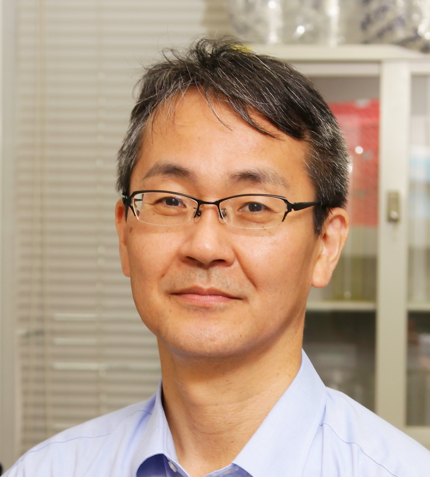 Hitoshi Sakakibara (榊原均)合作论文数Department of Applied Biosciences, Graduate School of Bioagricultural Sciences, Nagoya University5Shigeyuki Kawano合作论文数4Noriko Nagata合作论文数4Kobayashi Koichi合作论文数Graduate School of Arts and Sciences, University of Tokyo4Ken Matsuoka合作论文数Department of Bioscience and Biotechnology, Faculty of Agriculture, Kyushu University4
 Motoaki Seki合作论文数Center for Sustainable Resource Science, Institute of Physical and Chemical Research;Plant Epigenome Regulation Laboratory, Institute of Physical and Chemical Research4
 Hisayoshi Nozaki (野崎久義)合作论文数Department of Biological Sciences, Graduate School of Science, The University of Tokyo4Seiichiro Hasezawa合作论文数4
 Ryuichi Nishihama合作论文数Graduate School of Science, Nagoya University4Toshikazu Nishikawa合作论文数4Ayuko Kuwahara合作论文数4Noriko Takeda-Kamiya合作论文数4
 Kimitsune Ishizaki合作论文数Graduate School of Biostudies;Kyoto University;Graduate School of Biostudies, Kyoto University4Masataka Kajikawa合作论文数Graduate School of Biological Sciences, Nara Institute of Science and Technology4Yozo Okazaki合作论文数4
 Hiromitsu Tabeta合作论文数3
 Kien Huu Nguyen合作论文数3Kanade Tatsumi合作论文数CNRS, Univ Strasbourg3
 Naoki Minamino合作论文数Univ Tokyo, Grad Sch Sci, Dept Biol Sci, Bunkyo Ku, 7-3-1 Hongo, Tokyo 1130033, Japan3
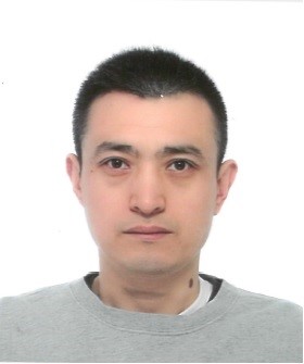 Weiqiang Li (李伟强)合作论文数Northeast Institute of Geography and Agroecology, Chinese Academy of Sciences3Yutaka Kodama合作论文数Division of Photobiology, National Institute for Basic Biology;Faculty of Sciences, Kyushu University;National Institute for Basic Biology, Kyushu University3Yasuko Watanabe合作论文数3Tanaka Maho合作论文数3Ishikawa Toshiki合作论文数3
 Ali Ferjani合作论文数3Masahiko Ikeuchi (池内榎本)合作论文数Laboratory of Plant Science, Department of Life Sciences, The University of Tokyo at Komaba3
 Kazufumi Yazaki (矢﨑一史)合作论文数Yazaki Laboratory, Research Institute for Sustainable Humanosphere, Kyoto University3Toshiyuki Takahashi合作论文数3
 Hajime Wada合作论文数Department of Biology;Faculty of Science;Kyushu University;Faculty of Science, Kyushu University3
 Lam Son Phan Tran合作论文数Institute of Genomics for Crop Abiotic Stress Tolerance, Texas Tech University;Department of Plant and Soil Science, Davis College of Agricultural Sciences & Natural Resources, Texas Tech University3
 Miyako Kusano合作论文数RIKEN Plant Science Center3Maki Kondo合作论文数3
 Tatsuru Masuda (増田建)合作论文数Graduate School of Science, The University of Tokyo3Hidehiro Fukaki合作论文数3
 Yuriko Osakabe (刑部祐里子)合作论文数School of Life Science and Technology, Tokyo Institute of Technology3
 Mikio Nishimura (西村幹夫)合作论文数Faculty of Science and Engineering, Konan University3Natsumaro Kutsuna合作论文数3Kazuo Ebine合作论文数Department of Biological Sciences;Graduate School of Science;University of Tokyo3
 Taku Demura合作论文数Plant Science Center3Keiko Sugimoto合作论文数3




