The isolation and characterization of baboon plasma factor V (FV) were performed for the development of an in vivo model for studying factor V/Va physiology in nonhuman primates. Baboon FV was purified by immunoaffinity chromatography with an antihuman FV monoclonal antibody and exhibits a specific activity of 1,940 U/mg. Baboon FV activation by thrombin proceeds through two proteolytic pathways similar to those observed with human and bovine FV. Limited amino acid sequencing of FV and its thrombin activation fragments shows 95% identity with human and 79% identity with bovine FV. 125I-Factor V and a mixture of thrombin cleaved 125I-FV activation products were infused into normal male baboons and evaluated by blood sample radioactivity measurements and by autoradiography of plasma samples following resolution by gel electrophoresis. Factor V disappeared with a half-life (t1/2) of 12.98 +/- 1.85 hours and was cleared without obvious degradation of the molecule during circulation. The radioactivity associated with the thrombin activated FV mixture, which consisted of the Mr = 220,000 activation intermediate, the Mr = 150,000 activation peptide, the heavy chain (HC) and the light chain (LC) of FVa, was cleared in a nonlinear manner. The HC and LC were removed with t1/2 < 20 minutes. The apparent molecular weight (Mr) = 220,000 and Mr = 150,000 fragments were cleared with t1/2 > 6 hours and t1/2 > 30 hours, respectively.
We have characterized the factor V protein and cDNA of a patient displaying factor V deficiency (parahemophilia) and correlated the reduced activity with a missense mutation of Ala221-to-Val. Plasma from the subject individual (C1) presented reduced factor V antigen (39% of normal) that displayed reduced activity (approximately 26% of normal). Factor V purified from this individual by standard techniques shows normal migration on sodium dodecyl sulfate gels and a normal pattern of activation by thrombin. Purified antigen from sibling C2 gives a much reduced specific activity of 263 U/mg (17% of normal). Sibling C3, the mother, and the father have antigen within the normal range (57% to 200%) that has approximately normal specific activity. The cDNA encoding the factor Va heavy and light chains of the subject individual was polymerase chain reaction-amplified and sequenced and revealed an A-to-G substitution at position 3 of codon 51 (silent mutation), a C-to-T substitution in position 2 of codon 221 (Ala221-Val), a T-to-C substitution at position 3 of codon 708 (silent mutation), and a G-to-A substitution at position 1 of codon 2185 (Thr2185-Ala). The latter mutation was also observed in control individuals and is proposed to be a possible polymorphism. Restriction analyses demonstrated the presence of one mutant and one normal allele in the father. The subject individual (C1) and sibling C2 carry only the mutant allele. The mother and sibling C3 carry only the normal allele. The inheritance pattern suggests the presence of a missing or nonexpressed allele in the mother that is passed on to all the siblings. Expression of only the mutant allele by the subject individual (C1) and sibling C2 is consistent with reduced factor V antigen and activity in these patients. We have designated this mutant as Factor VNew Brunswick.
Factor Va was incubated with 1-azidopyrene, a fluorescent lipophilic probe, in the presence of phospholipid vesicles composed of various proportions of phosphatidylcholine (PC) and phosphatidylserine (PS). The majority of the label was associated with the light chain of factor Va. The light chain was found to be labeled in the presence of phospholipid vesicles containing either 100% PC or 100%PS. After cleavage by factor Xa and incubation with PC/PS vesicles composed of 75% PC and 25% PS, label was found both on the M(r) = 30 000 fragment, derived-from the NH2-terminal portion of the bovine factor Va light chain (residues 1537-1752), and on the M(r) = 46 000/48 000 carboxyl-terminal fragment of the factor Va light chain (residues 1753-2183). The M(r) = 46 000/48 000 fragment incorporated I-azidopyrene independent of the phospholipid composition, while label incorporation into the M(r) = 30 000 fragment required phospholipid vesicles containing PC. No labeling of the M(r) = 30 000 fragment was observed with phospholipid vesicles composed of 100% PS. The label incorporation into the two portions of the molecule was found to be independent of the ionic strength in the presence of phospholipid vesicles containing 75% PC and 25% PS. In contrast, the labeling of the M(r) = 46 000/48 000 fragment with phospholipid vesicles composed of 1 00% PS was ionic strength dependent. These data suggest that two regions of factor Va light chain interact with the lipid bilayer and have different requirements for interaction: the binding site located on the M(r) = 30 000 fragment of the cofactor (A3 domain) interacts with phospholipid vesicles containing neutral phospholipid and is most likely hydrophobic in nature whereas the binding site located on the M(r) = 46 000/48 000 carboxyl-terminal fragment (C1-C2 domains ) interacts with membranes composed of anionic and neutral phospholipid and displays partly ionic binding characteristics.
The cleavage of human factor V and human factor Va by human activated protein C (APC) was analyzed in the absence and presence of phospholipid vesicles containing 75% phosphatidylcholine (PC) and 25% phosphatidylserine (PS). Membrane-bound human factor V (250 nM) is cleaved by APC (2.5 nM) to give M(r) = 200,000, 70,000, 45,000, and 30,000 fragments and an M(r) = 22/20,000 doublet. These fragments are released after four sequential cleavages of the membrane-bound procofactor at Arg306, Arg506, Arg679, and Lys994. No cofactor activity is observed following thrombin treatment of the membrane-bound APC-cleaved procofactor. In the absence of a membrane surface, no cleavage of factor V by APC is observed, and following thrombin activation factor Va retains full cofactor activity. Membrane-bound human factor Va (600 nM) loses more than 90% of its initial cofactor activity after 10 min of incubation with APC (10.9 nM), and virtually no cofactor activity is observed after 1 h of incubation. Under similar conditions but in the absence of PCPS vesicles, factor Va is cleaved but retains approximately 80% of its initial cofactor activity after 2 h of incubation with APC. In the presence of PCPS vesicles, the APC related loss of activity is correlated with cleavage of the heavy chain and appearance of fragments of M(r) = 45,000, 30,000, and of 28/26,000, and 22/20,000 doublets. These products correspond to three cleavages of the heavy chain (at Arg306, Arg506, and Arg679). Cleavage at Arg506 of factor Va precedes and appears to be required for cleavage at Arg306 and Arg679. In the absence of membrane, proteolysis at Arg506 produces an M(r) = 75,000 fragment which corresponds to the NH2-terminal portion of the human factor Va heavy chain (residues 1-506), and a carboxyl-terminal doublet of M(r) = 28/26,000 (residues 507-709) which is cleaved by APC at Arg679 to generate an M(r) = 22/20,000 doublet and an M(r) = 6,000 peptide. No cleavage of the light chain of the human cofactor is observed in the presence or absence of PCPS vesicles following 2 h of incubation with APC. Our data demonstrate that inactivation of human factor V and human factor Va only occurs in the presence of a membrane surface after cleavage at Arg306. However, while this cleavage site is exposed on membrane-bound human factor V, cleavage at Arg506 on the heavy chain of factor Va appears necessary for complete exposure of the cleavage site at Arg306.
Platelet activation leads to the incorporation of 32[PO4(2-)] into bovine coagulation factor Va and recombinant human factor VIII. In the presence of the soluble fraction from thrombin-activated platelets and (gamma-32P) adenosine triphosphate, radioactivity is incorporated exclusively into the M(r) = 94,000 heavy chain (H94) of factor Va and into the M(r) = 210,000 to 90,000 heavy chains as well into the M(r) = 80,000 light chain of factor VIII. Proteolysis of the purified phosphorylated M(r) = 94,000 factor Va heavy chain by activated protein C (APC) gave products of M(r) = 70,000, 24,000, and 20,000. Only the intermediate M(r) = 24,000 fragment contained radioactivity. Because the difference between the M(r) = 24,000 and M(r) = 20,000 fragments is located on the COOH-terminal end of the bovine heavy chain, phosphorylation of H94 must occur within the M(r) = 4,000 peptide derived from the carboxyl-terminal end of H94 (residues 663 through 713). Exposure of the radioactive factor VIII molecule to thrombin ultimately resulted in a nonradioactive light chain and an M(r) = 24,000 radioactive fragment that corresponds to the carboxyl-terminal segment of the A1 domain of factor VIII. Based on the known sequence of human factor VIII, phosphorylation of factor VIII by the platelet kinase probably occurs within the acidic regions 337 through 372 and 1649 through 1689 of the procofactor. These acidic regions are highly homologous to sequences known to be phosphorylated by casein kinase II. Results obtained using purified casein kinase II gave a maximum observed stoichiometry of 0.6 mol of 32[PO4(2-)]/mol of factor Va heavy chain and 0.35 mol of 32[PO4(2-)]/mol of factor VIII. Phosphoamino acid analysis of phosphorylated factor Va by casein kinase II or by the platelet kinase showed only the presence of phosphoserine while phosphoamino acid analysis of phosphorylated factor VIII by casein kinase II showed the presence of phosphothreonine as well as small amounts of phosphoserine. The platelet kinase responsible for the phosphorylation of the two cofactors was found to be inhibited by several synthetic protein kinase inhibitors. Finally, partially phosphorylated factor Va was found to be more sensitive to APC inactivation than its native counterpart. Our findings suggest that phosphorylation of factors Va and VIIIa by a platelet casein kinase II-like kinase may downregulate the activity of the two cofactors.
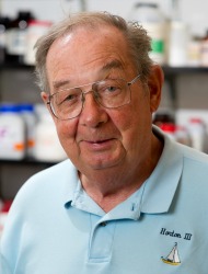 Kenneth Mann合作论文数Department of Biochemistry, Larner College of Medicine, University of Vermont;Haematologic Technologies, Inc.12
 Michael Kalafatis合作论文数Department of Chemistry;Cleveland State University;Department of Chemistry, Cleveland State University6
 Jm Murray合作论文数2Stephen R Hanson合作论文数2A Brisson合作论文数1RM Bertina合作论文数1J O Egan合作论文数1Rj Jenny合作论文数1Hugh C. Kim合作论文数1Yh Ehrlich合作论文数1Jb Lock合作论文数1C Vantveer合作论文数1Gaffney, D.P.合作论文数1
 Sriram Krishnaswamy合作论文数Perelman School of Medicine, University of Pennsylvania1S Stoylova合作论文数1




