Mitochondrial transplantation is an emerging regenerative therapy aimed at preventing ischemia-reperfusion injury by restoring oxidative metabolism in vulnerable tissues. Early clinical studies have demonstrated feasibility in pediatric myocardium and procedural safety in adult stroke patients, yet dose selection in non-cardiac, barrier-limited organs remains empiric and lacks a physiology-based framework. Establishing a quantitative approach to mitochondrial dosing is therefore a critical barrier to clinical translation. We developed a physiologically grounded computational model to estimate route-specific input dose requirements for brain, spinal cord, and kidney. The model integrates organ-level parameters including tissue mass, regional perfusion or cerebrospinal flow, permeability-surface area product, and extraction efficiency. A benchmark dose of 2 × 10⁶ mitochondria per gram of viable parenchymal tissue, derived from validated cardiac protocols, was applied across delivery scenarios. Here we show that extraction fraction is the dominant determinant of procedural feasibility, with up to twofold divergence from cardiac-derived estimates in central nervous system targets under reduced barrier permeability. Under stroke-relevant conditions with blood brain barrier permeability, required input doses converge toward myocardial benchmarks. This framework provides a quantitative foundation for route-aware mitochondrial dose planning and supports rational translational trial design in ischemia-reperfusion injury and related conditions. Mitochondria are the tiny structures inside cells that make energy. When blood flow to an organ is blocked and then restored, as happens in a stroke, a spinal cord injury or a kidney transplant, mitochondria fail and cells die. Surgeons have begun transplanting healthy mitochondria into damaged hearts and brains, but nobody knows how many are needed for organs that are protected by biological barriers that may block delivery, like parts of the brain, spinal cord, or kidney. We built a computer model that works out how many mitochondria must be injected for each organ and each injection route. We found that the fraction of injected mitochondria that actually crosses into tissue matters far more than organ size. The model gives researchers a rational starting dose for future trials. Walker et al. build a physiology-based computational model that converts a benchmark tissue mitochondrial dose into the dose that must be injected for intra-arterial and intrathecal delivery. Barrier extraction, not organ size, determines whether that dose fits within safe injection volumes.
Mitochondrial transplantation delivers viable, respiration-competent mitochondria into tissue whose own mitochondria have been damaged. It has been performed in the human heart and brain, but not in the eye, and no dose, safety profile or delivery experience existed for that compartment. Retinal ganglion cells depend heavily on oxidative phosphorylation, and in rodents mitochondria injected into the vitreous are taken up by these cells and improve survival after optic nerve injury. Here we show that fresh autologous mitochondria, isolated at the point of care, can be injected into the human vitreous without ocular or systemic toxicity. A 26-year-old woman with bilateral optic neuropathy fixed for three months after prolonged cerebral hypoperfusion received one injection into each eye, 24 hours apart, under emergency expanded access. Neither eye developed intraocular inflammation, and automated pupillary reactivity, absent across 45 readings over the preceding 71 days, returned in each eye within days of its own injection, transiently and without measurable acuity change.
Intercellular mitochondria transfer is an evolutionarily conserved process in which one cell delivers some of their mitochondria to another cell in the absence of cell division. This process has diverse functions depending on the cell types involved and physiological or disease context. Although mitochondria transfer was first shown to provide metabolic support to acceptor cells, recent studies have revealed diverse functions of mitochondria transfer, including, but not limited to, the maintenance of mitochondria quality of the donor cell and the regulation of tissue homeostasis and remodelling. Many mitochondria-transfer mechanisms have been described using a variety of names, generating confusion about mitochondria transfer biology. Furthermore, several therapeutic approaches involving mitochondria-transfer biology have emerged, including mitochondria transplantation and cellular engineering using isolated mitochondria. In this Consensus Statement, we define relevant terminology and propose a nomenclature framework to describe mitochondria transfer and transplantation as a foundation for further development by the community as this dynamic field of research continues to evolve. This Consensus Statement provides a nomenclature framework and experimental recommendations for studying mitochondrial transfer and transplantation.
Torcular dural sinus malformations (tDSMs) with high-flow fistulas pose complex management challenges due to their vascularity and the delicate neuroanatomy involved. This report presents the case of a child with tDSM and hydrocephalus, who underwent 3 staged embolization procedures but required a redo intervention due to residual malformation and venous hypertension. Utilizing the pressure cooker technique (PCT) in a redo setting allowed for high-pressure, targeted embolic delivery with minimized reflux, achieving near-complete occlusion and significant symptom relief. This case highlights PCT's potential to improve outcomes in multi-stage treatments of high-flow tDSM, reducing reflux and enhancing safety in technically demanding cases.
Mitochondrial transplantation is a promising but still experimental strategy for treating ischemic and metabolic disorders. A key barrier to its advancement is the lack of scalable, non-invasive methods for tracking transplanted extracellular mitochondria in vivo. Technetium-99m (Tc-99m) radiopharmaceuticals, widely used in SPECT imaging, may offer a clinically compatible solution. Cryopreserved mitochondria derived from HEK-293 cells were incubated with Tc-99m sestamibi, tetrofosmin, pertechnetate, or control solutions. After brief incubation and washing, mitochondrial pellets were analyzed for retained radioactivity. ATP content was measured to assess metabolic function, and electron microscopy was used to evaluate ultrastructural integrity. Tc-99m sestamibi and tetrofosmin showed labeling efficiencies of 2.74% and 2.68%, respectively. Pertechnetate demonstrated minimal uptake (0.34%). Radiolabeled mitochondria retained ATP production comparable to controls. Electron microscopy showed preserved double membranes and cristae. Controls confirmed assay specificity and viability. To our knowledge, this is the first report of radiolabeling isolated mitochondria with clinically approved Tc-99m agents. This platform supports the development of SPECT-compatible protocols for visualizing viable transplanted mitochondria in recipient tissues.
Mitochondrial transplantation is an emerging therapeutic approach for ischemia-reperfusion injury, offering the potential to restore cellular function through the engraftment of extracellular mitochondria. The successful clinical application of this strategy depends on the delivery of metabolically active mitochondria, yet the impact of circulating therapeutic agents on mitochondrial viability remains poorly understood. This study evaluates the effects of five clinically relevant agents commonly used during endovascular treatment of ischemic stroke-alteplase, cefazolin, lidocaine, phenylephrine, and heparinized saline-on extracellular mitochondria using an ex vivo model. Mitochondria were isolated from human skeletal muscle and mouse liver and exposed to these agents at clinically relevant and supra-physiological concentrations. Metabolic activity was assessed using a resazurin reduction assay as an indicator of mitochondrial viability. Even at concentrations up to 8-fold above clinical exposure, none of the agents significantly impaired mitochondrial function. These findings provide critical toxicological data demonstrating the compatibility of commonly used therapeutics with mitochondrial transplantation, supporting the development of safer and more optimized clinical protocols.
Mitochondrial transplantation is an emerging regenerative therapy aimed at preventing ischemia– reperfusion injury (IRI) by restoring oxidative metabolism in vulnerable tissues. While early clinical studies have demonstrated feasibility in pediatric myocardium and safety in adult stroke patients, dosing strategies remain undefined for barrier-limited organs where autoregulation, restricted perfusion, and anatomical constraints limit parenchymal uptake. We developed a physiologically grounded computational model to estimate route-specific input dose requirements for brain, spinal cord, and kidney. The model integrates organ-level parameters including tissue mass, regional perfusion or cerebrospinal flow, permeability–surface area product, and extraction efficiency. A benchmark dose of 2 × 10⁶ mitochondria per gram, derived from validated cardiac protocols, was applied across delivery scenarios. Sensitivity analysis explored the effects of extraction fraction and tissue mass. Route-adjusted input requirements diverged quantitatively from cardiac-derived estimates, with up to 2-fold increases in central nervous system (CNS) targets and approximately 20% higher dosing required for the kidney. This framework provides a quantitative foundation for translational dose planning in IRI and related conditions. ### Competing Interest Statement MRL: Unrestricted educational grants from Medtronic and Stryker; consulting agreement with Aeaean Advisers, Metis Innovative, Genomadix, AIDoc, Phenox and Arsenal Medical; equity interest in Proprio, Stroke Diagnostics, Apertur, Stereotaxis, Fluid Biomed, Synchron and Hyperion Surgical; editorial board of Journal of NeuroInterventional Surgery; Data safety monitoring board of Arsenal Medical. Other authors have no disclosures.
Background: Post stroke depression (PSD) is a common and debilitating complication of acute ischemic stroke (AIS), yet its biological basis remains unclear. AIS induces immune dysregulation and blood brain barrier (BBB) disruption, which may allow reactivation of latent viruses such as JC virus (JCV), a neurotropic polyomavirus that uses the serotonin receptor 5HT2A to enter neurons involved in mood regulation. Methods: We evaluated peripheral expression of host genes required for JCV entry, replication, and trafficking in three AIS transcriptomic datasets and assessed endothelial compromise using a complementary panel of BBB associated genes. We then examined the anatomical relevance of viral entry receptors using publicly available brain expression datasets. Results: AIS samples showed upregulation of host chaperone and trafficking genes, with concurrent downregulation of 5HT2A. BBB related profiles revealed reduced expression of structural junction proteins and increased expression of endothelial activation markers. Brain mapping localized high 5HT2A expression to regions implicated in mood regulation. Conclusions: These findings support a biologically plausible model in which AIS transiently enables JCV reactivation and CNS entry, particularly in serotonin rich brain regions that may contribute to PSD pathogenesis. ### Competing Interest Statement M.R.L.: Unrestricted educational grants from Medtronic and Stryker; consulting agreement with Aeaean Advisers, Metis Innovative, Genomadix, AIDoc, Phenox and Arsenal Medical; equity interest in Proprio, Stroke Diagnostics, Apertur, Stereotaxis, Fluid Biomed, Synchron and Hyperion Surgical; editorial board of Journal of NeuroInterventional Surgery; Data safety monitoring board of Arsenal Medical. D.M.K.: Scientific Advisory Board of Curevo. Grant support from Sanofi Pasteur. Royalty payments for institutionally-owned patents related to vaccines. J.R.Z. and M.W. declare that they have no conflict of interest. ### Funding Statement This study did not receive any funding ### Author Declarations I confirm all relevant ethical guidelines have been followed, and any necessary IRB and/or ethics committee approvals have been obtained. Yes The details of the IRB/oversight body that provided approval or exemption for the research described are given below: Gene expression datasets were obtained from the National Center for Biotechnology Information's Gene Expression Omnibus (GEO). Three datasets were selected based on relevance to acute ischemic stroke, availability of whole blood or PBMC profiles, and adequate sample size for group comparisons. These included GSE58294 (https://www.ncbi.nlm.nih.gov/geo/query/acc.cgi?acc=GSE58294), GSE16561 (https://www.ncbi.nlm.nih.gov/geo/query/acc.cgi?acc=GSE16561.), and GSE37587 (https://www.ncbi.nlm.nih.gov/geo/query/acc.cgi?acc=GSE37587) GEO series matrix files and corresponding platform annotation files (GPL570, GPL6883, GPL10558) were downloaded and parsed using open-source Python libraries. I confirm that all necessary patient/participant consent has been obtained and the appropriate institutional forms have been archived, and that any patient/participant/sample identifiers included were not known to anyone (e.g., hospital staff, patients or participants themselves) outside the research group so cannot be used to identify individuals. Yes I understand that all clinical trials and any other prospective interventional studies must be registered with an ICMJE-approved registry, such as ClinicalTrials.gov. I confirm that any such study reported in the manuscript has been registered and the trial registration ID is provided (note: if posting a prospective study registered retrospectively, please provide a statement in the trial ID field explaining why the study was not registered in advance). Yes I have followed all appropriate research reporting guidelines, such as any relevant EQUATOR Network research reporting checklist(s) and other pertinent material, if applicable. Yes All data produced in the present work are contained in the manuscript.
Amyloidogenic proteins play a central role in a range of pathological conditions, yet their presence in thrombi has only recently been recognized. Whether computational prediction tools can identify amyloid- forming potential in thrombus proteomes remains unclear. AmyloGram is a computational tool that estimates amyloid-forming potential based on n-gram sequence encoding and random forest classification. Using AmyloGram, we analyzed 204 proteins in UniProt that were tagged by humans as amyloidogenic. We then applied the same approach to proteins identified in thrombi retrieved using mechanical thrombectomy from patients with cardioembolic and atherothrombotic stroke. In addition, we used AmyloGram to analyze the amyloidogenicity of 83,567 canonical human protein sequences. Among the UniProt-annotated 'amyloid' set, nearly all proteins received AmyloGram scores above 0.7, including 23 of the 24 human proteins. Even the lowest-scoring human protein, lysozyme (scoring 0.675), is known to form amyloid under certain conditions. In thrombi from both stroke subtypes in four different studies, all detected proteins (with a single exception) had AmyloGram scores above 0.7, suggesting a high likelihood of amyloid content. A majority of unannotated proteins also achieve AmyloGram scores exceeding 0.7. AmyloGram reliably identifies known amyloid-forming proteins and reveals that stroke thrombi are enriched for proteins with high amyloidogenic potential. These findings support the hypothesis that thrombus formation in stroke involves amyloid-related mechanisms and warrant further investigation using histological and functional validation.
Aneurysms on variant arteries, especially involving the persistent trigeminal artery (PTA), azygos artery, and lenticulostriate arteries (LSAs), are rare. This report presents a case and subsequent management of a patient with multiple unruptured aneurysms arising from variant anatomical structures. A 48-year-old patient with a history of worsening headaches presented with three unruptured aneurysms at the PTA, azygos artery, and left LSA, confirmed via catheter angiography with three-dimensional reconstruction. Given the potential effects on conventional cerebrovascular structures, managing multiple aneurysms in rare and variant locations requires a tailored strategy. This case was successfully treated with an endovascular approach and a period of observation, emphasizing the need for individualized treatment planning.
Introduction: Anesthesia options for ischemic stroke patients undergoing endovascular thrombectomy (MT) can include local anesthesia, conscious sedation, or general anesthesia. Recent meta-analyses show that patients requiring emergency conversion (EC) to general anesthesia (GET) have higher mRS post-discharge than non-converters. The aim of this study was to assess the immediate outcomes in EC patients and identify factors associated with EC risk. Methods: This retrospective study included 264 sequential patients undergoing MT for large vessel occlusion at a comprehensive stroke center. EC was defined as intra-procedural induction of GET during MT. Results: Twelve patients (4.5%) required EC to GET. Median times from puncture to first pass (49 vs. 27 mins) and puncture to reperfusion (88 vs. 47 mins) were nearly doubled in EC patients compared to non-converters. Though the median TICI scores were similar, EC patients required double the number of passes and experienced significantly more symptomatic hemorrhagic complications (OR 5.82; 95% CI 1.10-30.688; p=0.018). An increased trend towards in-hospital death was noted (OR 1.46; 95% CI 0.382-5.602; p=0.289). Tobacco use was the only correlated clinical factor reaching statistical significance (RR 2.05; 95% CI 0.878-4.783; p=0.048). Conclusion: Although EC patients have similar reperfusion scores based on radiographic criteria, more complications were observed, and nearly double the time was required for reperfusion. While there is no consensus on anesthesia type during MT, EC introduces unnecessary risk and should be avoided. Given the potential harm from EC, multidisciplinary communication prior to puncture is essential.
This article assesses the association between anterior circulation morphometry and the presence of intracranial aneurysm using three-dimensional rotational angiography (3DRA). A retrospective analysis at a Peruvian academic medical center between December 2018 and February 2020 identified 206 patients with unruptured intracranial aneurysms and matched controls who underwent 3DRA. Angiographic images were obtained per standard of care, and measurements of the vasculature were performed using 3DRA vascular automated software. A total of 163 aneurysms and 43 control angiograms were evaluated. Women represented 82.5% of the cases and the mean age was 55.9 years (standard deviation 114.2). In multivariate analysis, five specific features were found to be statistically significant predictors for presence of an anterior circulation aneurysm: female sex (odds ratio [OR] =2.71; p = 0.048), C-shape of the middle cerebral artery (MCA) (OR =2.73; p = 0.018), distal internal carotid artery (ICA) diameter (OR = 3.42; p = 0.012), ICA bifurcation angle (OR = 1.02; p = 0.036), and length of the carotid siphon (OR = 1.08; p = 0.047). Features detected on 3DRA suggest morphological characteristics of the ICA and MCA may be predictive for intracranial aneurysm. Our findings build from prior reports by demonstrating five specific patient and imaging features associated with anterior circulation aneurysms. While 3DRA is the standard of care in many settings, medical centers with resource limitations may not have access to this technique. The demographic and morphological features identified in our study may have correlates that if detected on contrast computed tomography or magnetic resonance imaging studies, may be used to help screen for a higher level of care in select patients.
Introduction: Cerebral vasospasm is a major cause of morbidity and mortality after aneurysmal subarachnoid hemorrhage (aSAH). Though used routinely in clinical practice, objective metrics to evaluate effectiveness of intra-arterial vasodilatory treatment (IAT) have not been identified. To quantify effects of IAT on flow dynamics in cerebral vasospasm, we retrospectively compared Lindegaard ratios (LR) of aSAH patients pre-and post-IAT. Methods: Subjects included 24 aSAH patients undergoing IAT in the bilateral middle cerebral arteries (MCA) for cerebral vasospasm, diagnosed by standard clinical and radiographic criteria. Subjects were excluded if balloon angioplasty was performed prior to/within 2 days of the index procedure. All subjects received 10 mg IA milrinone, a non-catecholamine phosphodiesterase inhibitor drug, per MCA. Transcranial doppler (TCD) ultrasound was measured pre-IAT (D0) and at D+1 and D+2 post-IAT. LR was calculated as the ratio of mean flow velocities in the middle cerebral artery (MCA) to the ipsilateral extracranial internal carotid artery. Standard threshold for vasospasm is defined as LR>3. Results: Right and left LR were aggregated, total N=48. Average pre-IAT LR was 2.96 (R 2.67/L 3.25±1.29). Post-IA LR were 2.72±1.12 and 3.15±1.55 at D+1 and D+2, respectively. Subjects with pre-IA LR>3 showed an average LR decrease of 21% post-IAT D+1 (p=0.01) and an 8% decrease at D+2 (p=0.16) [Fig.1A]. Subjects with pre-IAT<3 experienced an average LR increase of 28% by D+2 (p=0.02) [Fig 1B]. Conclusion: Subjects with TCD confirmed vasospasm (LR>3) at D0 showed a significant improvement in cerebral blood flow one day after therapy. Subjects who did not meet TCD threshold for vasospasm (LR<3) showed little change one day post-IAT but worsening cerebral blood flow by day two post-IAT. Further study is needed to determine whether LR can be used to predict benefit or objectively assess effectiveness of IAT in aSAH patients with cerebral vasospasm.
Cerebral vasospasm (CV) is a critical determinant of outcomes in patients with aneurysmal subarachnoid hemorrhage (aSAH). Despite advances in neurocritical care, modifiable risk factors for CV remain poorly understood, and identifying them could significantly enhance patient management and treatment strategies. The present study explored the potential link between the reactivation of herpes simplex virus type 1 (HSV-1), a common resident virus in cranial nerves, and CV severity. It was hypothesized that higher HSV-1 viral load in saliva may be associated with increased CV severity. Saliva samples were collected on days 4, 7, 10 and 14 post-aSAH, and HSV-1 DNA levels were measured using quantitative PCR. CV severity was assessed using the Lindegaard ratio (LR), with an LR >3 considered the diagnostic threshold for CV. A total of 36 patients were enrolled, and 139 saliva samples were collected. HSV-1 DNA was detected in 19.4% of samples (27/139), and 44% of patients (16/36) developed CV. HSV-1 seropositive patients made up 88.9% (32/36) of the cohort, with 50% exhibiting viral shedding during the study period. None of the HSV-1 seronegative patients (11.1%, 4/36) exhibited viral shedding or developed CV. Regression analysis showed a positive association between HSV-1 viral load and CV severity, with viral load explaining 27.8% of the variability (P=0.005). Age was also significant, with older patients experiencing less severe CV (P<0.001). Supervised machine learning identified viral load thresholds that aligned with standard LR values for moderate and severe CV. While the small sample size and observational design limit the generalizability of the results, these findings suggested that earlier detection and intervention for CV could be informed by assessing HSV-1 serostatus and monitoring viral activity through saliva samples or other non-invasive methods, highlighting the need for larger, controlled studies to validate these results.
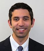 Michael Levitt合作论文数University of Washington34
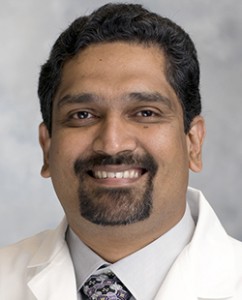 Basavaraj Ghodke合作论文数Department of Radiology, University of Washington15
 Emma Federico合作论文数11
 Eytan Raz合作论文数Division of Interventional Neuroradiology, Department of Radiology, New York University7
 Cory Kelly合作论文数6Samuel Levy合作论文数6
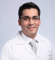 Giancarlo Saal-Zapata合作论文数Hosp Nacl Guillermo Almenara Irigoyen EsSalud, Serv Neurocirugia Endovasc6
 Thanh Nguyen合作论文数Department of Radiology, Weill Cornell Medicine, Cornell University6
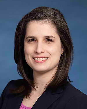 Anna Luisa Kuhn合作论文数University of Massachusetts Memorial Medical Center;University of Massachusetts Chan Medical School6
 Ajit Singh Puri合作论文数Neurological Surgery, School of Medicine, University of Massachusetts Medical School6Renieri Leonardo合作论文数6
 Chris L Dupont合作论文数J Craig Venter Inst6
 Ossama Mansour合作论文数5
 Ameer E Hassan合作论文数5
 Mohamad Abdalkader合作论文数Sch Med, Boston Univ5
 Mario Martinez-Galdamez合作论文数Department of Interventional Neuroradiology, Radiology, Fundación Jiménez Díaz5
 Siegler James E合作论文数5
 Michel Piotin合作论文数5
 Raul Nogueira合作论文数Department of Emergency Medicine, Emory University School of Medicine;Department of Neurology, Emory University School of Medicine5Juan F. Arenillas合作论文数5Malveeka Sharma合作论文数4Alexandra Gonzalez合作论文数4David I Bass合作论文数4
 Marios Psychogios合作论文数Clin Radiol & Nucl Med, Univ Hosp Basel4
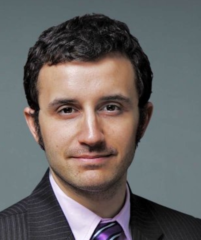 Maksim Shapiro合作论文数Department of Neurology, Department of Medicine, New York University4
 Italo Linfante合作论文数4Khot Sandeep P合作论文数4
 Khandelwal Priyank合作论文数4
 Fawaz Almufti合作论文数4
 Nabeel A. Herial合作论文数4
 Natalia Perez de la Ossa Herrero合作论文数4
 Michael Hill合作论文数Department of Clinical Neuroscience, University of Calgary;Hotchkiss Brain Institute, University of Calgary4
 Pascal M. Jabbour合作论文数Department of Neurological Surgery, The Sidney Kimmel Medical College, Thomas Jefferson University4
 Louis J. Kim合作论文数University of Washington4
 Pablo M Lavados合作论文数3Patrik Gabikian合作论文数3Viet Quy Nguyen合作论文数3Alejandro Rodriguez Vasquez合作论文数3Jane G Morris合作论文数3Wazim Izzath合作论文数3Emma Griffin合作论文数3Hugh Stephen Winters合作论文数3
 Artem Kaliaev合作论文数3Brijesh P. Mehta合作论文数3Huynh Vu Le合作论文数3
 Raghid Nabih Kikano合作论文数3Héctor H García合作论文数3Luz Toribio合作论文数3
 Gianfranco Arroyo合作论文数3




