Rigaku ran a series of practical schools to address the loss of crystallographic training opportunities resulting from the cancellation of conventional schools around the world due to the COVID-19 pandemic. With the return to normalcy, we stopped holding the schools. However, in the last year a number of requests were made to reprise the lectures. This year, we ran one school but took a different approach. Rather than performing the lectures live, we prerecorded them with the intention of using them for a future on-demand school. We held live Q&A sessions after the lectures to capture this very useful dialog with students for inclusion in the course material. As before, we limited the school to ten lectures of 60-90 minutes in length covering practical aspects of small molecule crystallography, including data collection and data processing in CrysAlisPro and structure solution in Olex2. In the school, we covered some advanced topics that students commonly see in their work: absolute structure determination, twinning, and disorder. With this edition of the school, we added a number of microED topics. So that students might practice independently, we used freely available data reduction and structure solution software and data sets with which to practice. To give students credit for course completion, we provided an online exam and an electronic certificate of completion. In this presentation, we will discuss the setup and execution of the school and provide some results for this last cohort of students.
Microcrystal electron diffraction (MicroED/3D ED), which solves atomic structures from crystals in the sub-micron size range using electron radiation, has evolved from an exploratory technique into a cutting-edge analytical method practiced in laboratories worldwide. However, viewing MicroED solely as an extension of single-crystal diffraction, traditionally performed with X-rays on smaller crystals, underestimates its capabilities. With thousands of crystallites available simultaneously on a sample grid and measurements completed within minutes, we can obtain a more comprehensive view of our specimen by collecting crystallographic data from dozens or even hundreds of crystals. This approach allows for the analysis of powders containing mixtures of compounds, different polymorphs, or even small traces of contaminants, including ab-initio solutions, even in cases too complex for powder diffraction or where only minute sample amounts are available. In this contribution, we will discuss how high-throughput workflows in the Rigaku XtaLAB Synergy-ED electron diffractometer enable various beyond-single-crystal investigations with a high degree of automation and ease of use.
3D electron diffraction (3D ED)/Micro electron diffraction (MicroED) has extended the limits of crystallography by enabling the determination of three dimensional molecular structures from sub-μm microcrystals. However, 3D ED/microED measurements using current state-of-the-art electron microscopes require experts in both electron microscopy and crystallography making the method rather difficult for researchers who simply need structures. Here, we present a diffractometer specifically designed for 3D ED/microED and show how it works for determining crystal structures. The newly developed electron diffractometer will provide many researchers with an easy path to structure determination of crystals that are less than 1 μm in size.
Since our launch of the XtaLAB Synergy-ED in 2021, users have produced hundreds of new and re-visited structures via MicroED, including over 500 unique structures from Rigaku application labs alone. Most of the early studies were conducted at ambient temperature. We commonly perform low- or variable-temperature experiments, allowing more in-depth study of crystalline materials that are sensitive to vacuum and/or electron-beam damage, and that undergo temperature-accessible phase transitions. Since the Synergy-ED goniometer is compatible with many of the cryo-holders already available for TEM instruments, structural scientists may now use MicroED at non-ambient temperatures, including variable-temperature studies. Cryo-transfer specimen holders such as the Gatan ELSA provide protection of samples before introduction to the vacuum, allowing the study of solvates and other vacuum-sensitive species. The programmable temperature control also allows exploration of phase-transition behavior. We will discuss results from samples for which cryo-transfer proved essential. The Hummingbird Scientific MEMS biasing/heating holder offers the possibility to increase temperature, allowing for exploration of the phase behavior of samples such as porous materials. Our recent results using single-crystal data from electron diffraction on a MOF system, Cu(ta)2 (Hta = 1H-1,2,3-triazole), at room temperature and at 200 °C, were compared to a previous study of the same material from 2012 done using SC-XRD and PXRD.
Recognizing the potential of Single-Crystal Electron Diffraction (SC-ED), also known as 3DED and MicroED, Rigaku and JEOL announced a collaboration in 2020 to develop an instrument designed in a fashion that will make it easy for any crystallographer to use.The resulting instrument, the XtaLAB Synergy-ED [1], a new and fully integrated electron diffractometer, was released about one year later, in May 2021.Many materials only form nanosized crystals or are challenging to produce in large quantities.Before the development of the SC-ED technique, synthetic chemists were forced to rely on other methods, such as NMR, often in combination with each other, to postulate 3D structure.Unfortunately, for complicated molecules such as natural products, the NMR results can be challenging to interpret.SC-ED has become a revolutionary technique for the advancement of structural science.The XtaLAB Synergy-ED is an electron diffractometer operated via CrysAlisPro for a seamless workflow from data collection to structure determination of three-dimensional molecular structures from nanocrystals.Since its launch, our demo lab XtaLAB Synergy-ED has generated over 160 structures of various samples, from organics to MOFs, with a range of compositions and cell dimensions as shown in the pie chart in Figure 1 and the histogram in Figure 2, respectively.In this presentation, we will provide an overview of the capabilities of the Synergy-ED and review some of more interesting structures that have been determined since May of 2021.
The XtaLAB Synergy Flow turns any Synergy cabinet diffractometer into an automated, high-throughput machine by incorporating a 6-axis UR3 Universal Robot and a 3-puck dewar. The Flow system can automatically screen and collect 48 crystal samples with minimal human intervention. CrysAlisPro has been upgraded with tools to control all aspects of robotics and sample queuing. A unique X-ray safe dewar-drawer system allows loading and unloading of pucks without opening the X-ray enclosure or disturbing data collection. Ultimately, the XtaLAB Synergy Flow system is the perfect solution to allow full-time use of your diffractometer during a time when human interaction and contamination must be minimized.
Development of a diffractometer specialized for electron diffraction experiments.
Conclusion An automated procedure has been developed that involves robust data processing and modeling to directly extract intermolecular packing functions from Total Diffraction data collected on organic glassy / amorphous materials. The amorphous intermolecular packing function can be regarded as a material fingerprint for the specific amorphous / glassy material being studied. Due to the metastable nature of amorphous and glassy materials, this fingerprint is likely to be sensitive to the thermal history of the sample (manufacturing and storage) and could potentially be indicative of physical properties such as stability. With suitable and robust data reduction procedures, glassy / amorphous packing function analysis can be performed for organic systems using either Mo or Cu X-rays on either a general-purpose powder diffractometer or a single crystal diffractometer.
There is no question that atomic pair distribution function analysis has had a profound impact on the analysis of crystalline and amorphous materials [1].As a complement to the use of synchrotron sources for collecting PDF data, we have explored the use of home laboratory-based single crystal diffractometers to analyze both crystalline and amorphous materials.In order to generate the most useful reduced radial distribution functions, G(r), we have found it necessary to modify existing code in CrysAlis Pro[2] and develop new code to generate G(r) data for refinement in PDFgui [3].In this presentation we will explore the collection and analysis of total scattering data on both crystalline and amorphous materials with wavelengths readily available to home laboratory systems.
MicroED has extended the limits of crystallography by enabling the determination of 3D-molecular structures from sub-μm microcrystals. However, measurements using microED require experts in both electron microscopy and crystallography, and its use is not easy for researchers who need structures. Here, we present a diffractometer specifically designed for microED and show how it works for determining crystal structures. The newly developed electron diffractometer will provide many researchers with an easy path to microcrystal structure determination.
In order to address the loss of crystallographic training opportunities resulting from the cancellation of conventional schools around the world due to the COVID-19 pandemic we have started an online crystallography school with live lectures and live Q&A using Zoom Webinar.In 2020 we ran three versions of the school: two 10 one-hour classes on basic topics in crystallography and five 1.5hour classes on advanced topics.In June 2021 we plan to run a fourth school consisting of 10 1.5 hour classes on advanced topics.We have reported on the execution and results of the two basic schools held in 2020 previously (1).For the June 2021 school, we have scheduled ten 1.5 hour lectures on advanced topics including: electron diffraction, refinement, twinning, powder and PDF analysis, solution scattering and macromolecular crystallography, non-spherical atom refinement and charge density analysis, and data mining.This presentation will review the execution and outcomes of the
In order to address the loss of crystallographic training opportunities resulting from the cancelation of conventional schools around the world due to the COVID-19 pandemic, we have started an online crystallography school with live lectures and live Q&A using Zoom Webinar. Since we were trying to reach a large audience in a relatively short period, we have limited the school to ten 1 h lectures covering practical aspects of small molecule crystallography including data collection, data processing, and structure solution. In the school, we also covered some advanced topics that students commonly see in their work: absolute structure determination, twinning, and disorder. To round out the education, we provided lectures on macromolecular crystallography and powder diffraction. For students to practice on their own, we used freely available data reduction and structure solution software, as well as datasets with which to practice. To give students credit for course completion, we provided an online exam and an electronic certificate of completion. In this editorial, we will provide some insight into the issues of holding lectures with up to 750 students of very diverse backgrounds and review the efficacy of the school in teaching crystallography for the two cohorts of students.
There is a misconception in the literature (Gruene, [i]et.al.[/i], 2018) that one needs crystals as large as 50 µm on a side to perform single X-ray diffraction studies.It has also been suggested that MicroED measurements are needed to probe smaller samples.In this presentation, we will demonstrate that this is simply not true.Furthermore, we will show the lower limit of what is possible with a home X-ray source is somewhere just above the upper range of what is accessible to MicroED, that is, about 1-3 µm.We will also compare some X-ray and ED structural quality and make some suggestions regarding classification of different types of structures based on quality of the results.
The structure of organic compounds, including their absolute configuration, is extremely critical to the activity of medicines, aroma chemicals, and agrochemicals. Nearly all of these structures have been determined by single-crystal X-ray diffraction (SC-XRD) analysis. However, it is widely believed that SC-XRD analysis is not versatile since it requires considerable crystal growth and expertise. Recently, crystal growth is becoming less critical by utilizing the microcrystal electron diffraction method (MicroED). However, there is still a gap between the two methods because MicroED has limitations with respect to crystal size and quality of the results. Moreover, modern instruments for SC-XRD analysis have evolved to analyze smaller crystals and are able to deliver a structure easier. Here, we propose a new approach to fill the above gap with the tool named "What is this? (WIT)". WIT is a fully automated process from evaluating the crystal to providing the 'structure'. This method assumes the situation where a researcher happens to obtain a shiny grain during a course of chemical synthesis and wants to know the structure of the molecule making up the grain. Therefore, WIT assumes no chemical information and collects a mere 60% of the full dataset to enable faster characterization of the molecule with enough quality to fulfill the requirement. To assess the utility of WIT, we compared the results obtained by MicroED on a crystal of the same compound and similar in size. In summarizing the results, we propose a possible functional classification of the analyzed structures. We have developed a new tool, “What is this?” (WIT) for the fully automated structure determination of small molecules concurrent with single crystal data collection and processing. WIT assumes the case where a researcher happens to obtain a shiny grain of unknown composition during a chemical synthesis and opts to try to determine the three-dimensional structure of the molecule making up the grain.
 Fraser J. White合作论文数School of Chemistry, The University of Edinburgh22Joseph D. Ferrara合作论文数18Damian Kucharczyk合作论文数10Pierre Le Magueres合作论文数10
 Akihito Yamano合作论文数7
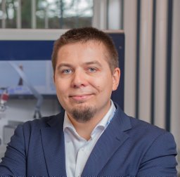 Jakub Wojciechowski合作论文数Institute of General and Ecological Chemistry, Technical University (Politechnika),7Mark Del Campo合作论文数7Yoshitaka Aoyama合作论文数6Przemyslaw Stec合作论文数6Michał Jasnowski合作论文数5Zoltan Gal合作论文数5Adrian Jones合作论文数5Sho Ito合作论文数5Simon Bates合作论文数4Griffin Alexandra M E合作论文数4Eiji Okunishi合作论文数4Tadeusz Skarzynski合作论文数4
 Oliver Presly合作论文数3Sato Hiroyasu合作论文数3
 Dyanne L. Cruickshank合作论文数Department of Chemistry, University of Cape Town3paul n swepston合作论文数2Alexandra Stanley合作论文数2Christian Schuermann合作论文数2Daniel Baker合作论文数2A. Yamano合作论文数2Keisuke Saito合作论文数2
 Robert Bucker合作论文数2R. Buecker合作论文数1Christian R. Goeb合作论文数1Takashi Sato合作论文数1
 Florian Kleemiß合作论文数Fakultät für Chemie und Pharmazie, Universität Regensburg1Christian Göb合作论文数Rigaku Europe SE1Horst Puschmann合作论文数1
 Tomislav Stolar合作论文数Div Phys Chem, Rudjer Boskovic Inst1Takashi Matsumoto合作论文数1
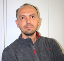 Užarević Krunoslav合作论文数Rudjer Boskovic Inst1
 Khai-Nghi Truong合作论文数1Criswell Angela R合作论文数1Y Aoyama合作论文数1
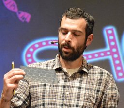 Luca Grisanti合作论文数1Eric W. Reinheimer合作论文数1
 Peter A. Wood合作论文数Department of Geography, University College1
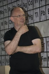 Ernest Meštrović合作论文数PLIVA - Research and Development Ltd.1




