Rigaku ran a series of practical schools to address the loss of crystallographic training opportunities resulting from the cancellation of conventional schools around the world due to the COVID-19 pandemic. With the return to normalcy, we stopped holding the schools. However, in the last year a number of requests were made to reprise the lectures. This year, we ran one school but took a different approach. Rather than performing the lectures live, we prerecorded them with the intention of using them for a future on-demand school. We held live Q&A sessions after the lectures to capture this very useful dialog with students for inclusion in the course material. As before, we limited the school to ten lectures of 60-90 minutes in length covering practical aspects of small molecule crystallography, including data collection and data processing in CrysAlisPro and structure solution in Olex2. In the school, we covered some advanced topics that students commonly see in their work: absolute structure determination, twinning, and disorder. With this edition of the school, we added a number of microED topics. So that students might practice independently, we used freely available data reduction and structure solution software and data sets with which to practice. To give students credit for course completion, we provided an online exam and an electronic certificate of completion. In this presentation, we will discuss the setup and execution of the school and provide some results for this last cohort of students.
3D electron diffraction (3D ED)/Micro electron diffraction (MicroED) has extended the limits of crystallography by enabling the determination of three dimensional molecular structures from sub-μm microcrystals. However, 3D ED/microED measurements using current state-of-the-art electron microscopes require experts in both electron microscopy and crystallography making the method rather difficult for researchers who simply need structures. Here, we present a diffractometer specifically designed for 3D ED/microED and show how it works for determining crystal structures. The newly developed electron diffractometer will provide many researchers with an easy path to structure determination of crystals that are less than 1 μm in size.
Along with the adoption of three-dimensional electron diffraction (3D ED/MicroED) as a mainstream tool for structure determination from sub-micron single crystals, questions about best practices regarding each step along the workflow, from data collection to structure solutions, arise. In this paper, we discuss three particular aspects of a 3D ED/MicroED experiment which, after hundreds of structures solved in Rigaku’s laboratories, we have found to be important to consider carefully. First, for a representative model system of a hydrated compound (trehalose dihydrate), we show that cryo-transfer of the sample into the diffractometer is an effective means to prevent dehydration, while cooling of the sample without cryo-transfer yields a marginal improvement only. Next, we demonstrate for a small (tyrosine) and a large (clarithromycin) organic compound, how a simplified and fast workflow for dynamical diffraction calculations can determine absolute crystal structures with high confidence. Finally, we discuss considerations and trade-offs for choosing an optimal effective crystal-to-detector distance; while a long distance is mandatory for a protein (thaumatin) example, even a small molecule with difficult diffraction behavior (cystine) yields superior results at longer distances than the one used by default.
Conditions have been identified in which phenolic aldoximes and ketoximes of the types used in commercial solvent extraction processes can be doubly deprotonated and generate polynuclear Cu complexes with lower extractant:Cu molar ratios than those found in commercial operations. Electrospray mass spectrometry has provided an insight into the solution speciation in extraction experiments and has identified conditions to allow isolation and characterization of polynuclear Cu-complexes. Elevation of pH is effective in enhancing the formation of trinuclear complexes containing planar {Cu3-μ3-O}4+ or {Cu3-μ3-OH}5+ units. DFT calculations suggest that such trinuclear complexes are more stable than other polynuclear species. Solid structures of complexes formed by a salicylaldoxime with a piperidino substituent ortho to the phenolic OH group (L9H2) contain two trinuclear units in a supramolecular assembly, {[Cu3OH(L9H)3(ClO4)](ClO4)} 2, formed by H-bonding between the central {Cu3-μ3-OH}5+ units and oxygen atoms in the ligands of an adjacent complex. Whilst the lower ligand:Cu molar ratios provide more efficient Cu-loading in solvent extraction processes, the requirement to raise the pH of the aqueous phase to achieve this will make it impractical in most commercial operations because extraction will be accompanied by the precipitation (as oxyhydroxides) of Fe(III) which is present in significant quantities in feed solutions generated by acid leaching of most Cu ores.
Recognizing the potential of Single-Crystal Electron Diffraction (SC-ED), also known as 3DED and MicroED, Rigaku and JEOL announced a collaboration in 2020 to develop an instrument designed in a fashion that will make it easy for any crystallographer to use.The resulting instrument, the XtaLAB Synergy-ED [1], a new and fully integrated electron diffractometer, was released about one year later, in May 2021.Many materials only form nanosized crystals or are challenging to produce in large quantities.Before the development of the SC-ED technique, synthetic chemists were forced to rely on other methods, such as NMR, often in combination with each other, to postulate 3D structure.Unfortunately, for complicated molecules such as natural products, the NMR results can be challenging to interpret.SC-ED has become a revolutionary technique for the advancement of structural science.The XtaLAB Synergy-ED is an electron diffractometer operated via CrysAlisPro for a seamless workflow from data collection to structure determination of three-dimensional molecular structures from nanocrystals.Since its launch, our demo lab XtaLAB Synergy-ED has generated over 160 structures of various samples, from organics to MOFs, with a range of compositions and cell dimensions as shown in the pie chart in Figure 1 and the histogram in Figure 2, respectively.In this presentation, we will provide an overview of the capabilities of the Synergy-ED and review some of more interesting structures that have been determined since May of 2021.
The study of the structure of single crystals has typically been achieved with X-ray diffraction while many decades of progress and research have led to hardware improvements which have pushed the limits of X-ray diffraction.The current generation of home lab instruments allow the study of crystals down to about 1 micron in size with sources such as the FR-X, a high-power rotating anode 1 .In the quest to study even smaller samples than this, microED has become increasingly popular in recent years 2 .As electrons interact more strongly with a crystalline sample than X-rays do, the study of samples smaller than 1 micron becomes possible and, in fact, necessary.We would like to introduce our solution for microED, the Synergy ED, along with results we have obtained using it, and efforts we have made to improve the quality of results.
Development of a diffractometer specialized for electron diffraction experiments.
MicroED has extended the limits of crystallography by enabling the determination of 3D-molecular structures from sub-μm microcrystals. However, measurements using microED require experts in both electron microscopy and crystallography, and its use is not easy for researchers who need structures. Here, we present a diffractometer specifically designed for microED and show how it works for determining crystal structures. The newly developed electron diffractometer will provide many researchers with an easy path to microcrystal structure determination.
In order to address the loss of crystallographic training opportunities resulting from the cancellation of conventional schools around the world due to the COVID-19 pandemic we have started an online crystallography school with live lectures and live Q&A using Zoom Webinar.In 2020 we ran three versions of the school: two 10 one-hour classes on basic topics in crystallography and five 1.5hour classes on advanced topics.In June 2021 we plan to run a fourth school consisting of 10 1.5 hour classes on advanced topics.We have reported on the execution and results of the two basic schools held in 2020 previously (1).For the June 2021 school, we have scheduled ten 1.5 hour lectures on advanced topics including: electron diffraction, refinement, twinning, powder and PDF analysis, solution scattering and macromolecular crystallography, non-spherical atom refinement and charge density analysis, and data mining.This presentation will review the execution and outcomes of the
In order to address the loss of crystallographic training opportunities resulting from the cancelation of conventional schools around the world due to the COVID-19 pandemic, we have started an online crystallography school with live lectures and live Q&A using Zoom Webinar. Since we were trying to reach a large audience in a relatively short period, we have limited the school to ten 1 h lectures covering practical aspects of small molecule crystallography including data collection, data processing, and structure solution. In the school, we also covered some advanced topics that students commonly see in their work: absolute structure determination, twinning, and disorder. To round out the education, we provided lectures on macromolecular crystallography and powder diffraction. For students to practice on their own, we used freely available data reduction and structure solution software, as well as datasets with which to practice. To give students credit for course completion, we provided an online exam and an electronic certificate of completion. In this editorial, we will provide some insight into the issues of holding lectures with up to 750 students of very diverse backgrounds and review the efficacy of the school in teaching crystallography for the two cohorts of students.
There is a misconception in the literature (Gruene, [i]et.al.[/i], 2018) that one needs crystals as large as 50 µm on a side to perform single X-ray diffraction studies.It has also been suggested that MicroED measurements are needed to probe smaller samples.In this presentation, we will demonstrate that this is simply not true.Furthermore, we will show the lower limit of what is possible with a home X-ray source is somewhere just above the upper range of what is accessible to MicroED, that is, about 1-3 µm.We will also compare some X-ray and ED structural quality and make some suggestions regarding classification of different types of structures based on quality of the results.
The structure of organic compounds, including their absolute configuration, is extremely critical to the activity of medicines, aroma chemicals, and agrochemicals. Nearly all of these structures have been determined by single-crystal X-ray diffraction (SC-XRD) analysis. However, it is widely believed that SC-XRD analysis is not versatile since it requires considerable crystal growth and expertise. Recently, crystal growth is becoming less critical by utilizing the microcrystal electron diffraction method (MicroED). However, there is still a gap between the two methods because MicroED has limitations with respect to crystal size and quality of the results. Moreover, modern instruments for SC-XRD analysis have evolved to analyze smaller crystals and are able to deliver a structure easier. Here, we propose a new approach to fill the above gap with the tool named "What is this? (WIT)". WIT is a fully automated process from evaluating the crystal to providing the 'structure'. This method assumes the situation where a researcher happens to obtain a shiny grain during a course of chemical synthesis and wants to know the structure of the molecule making up the grain. Therefore, WIT assumes no chemical information and collects a mere 60% of the full dataset to enable faster characterization of the molecule with enough quality to fulfill the requirement. To assess the utility of WIT, we compared the results obtained by MicroED on a crystal of the same compound and similar in size. In summarizing the results, we propose a possible functional classification of the analyzed structures. We have developed a new tool, “What is this?” (WIT) for the fully automated structure determination of small molecules concurrent with single crystal data collection and processing. WIT assumes the case where a researcher happens to obtain a shiny grain of unknown composition during a chemical synthesis and opts to try to determine the three-dimensional structure of the molecule making up the grain.
The relationship between the structure and the properties of a drug or material is a key concept of chemistry. Knowledge of the three-dimensional structure is considered to be of such importance that almost every report of a new chemical compound is accompanied by an X-ray crystal structure - at least since the 1970s when diffraction equipment became widely available. Crystallographic software of that time was restricted to very limited computing power, and therefore drastic simplifications had to be made. It is these simplifications that make the determination of the correct structure, especially when it comes to hydrogen atoms, virtually impossible. We have devised a robust and fast system where modern chemical structure models replace the old assumptions, leading to correct structures from the model refinement against standard in-house diffraction data using no more than widely available software and desktop computing power. We call this system NoSpherA2 (Non-Spherical Atoms in Olex2). We explain the theoretical background of this technique and demonstrate the far-reaching effects that the improved structure quality that is now routinely available can have on the interpretation of chemical problems exemplified by five selected examples.
The HyPix Arc 150° detector takes the latest, direct X-ray detection, hybrid photon counting technology and forms it into a curved detector for unparalleled data collection efficientcy and quality.
A tetrameric pentacene, PT, has been used to explore the effects of exciton delocalization on singlet fission (SF). For the first time, triplet decorrelation through intramolecular triplet diffusion was observed following SF. Transient absorption spectroscopy was used to examine different decorrelation mechanisms (triplet diffusion versus structural changes) for PT and its dimeric equivalent PD on the basis of the rate and activation barrier of the decorrelation step. Charge-separation experiments using tetracyano-p-quinodimethane (TCNQ) to quench triplet excitons formed through SF demonstrate that enhanced intersystem crossing, that is, spin catalysis, is a widely underestimated obstacle to quantitative harvesting of the SF products. The importance of spatial separation of the decorrelated triplet states is emphasized, and independent proof that the decorrelated triplet pair state consists of two (T-1) states per molecule is provided.
 Simon Parsons合作论文数School of Chemistry, College of Science and Engineering, The University of Edinburgh24
 Mathias Meyer合作论文数22Peter A. Tasker合作论文数18
 Euan Brechin合作论文数School of Chemistry, University of Edinburgh15Joseph D. Ferrara合作论文数13
 Akihito Yamano合作论文数9
 Oliver Presly合作论文数8Yoshitaka Aoyama合作论文数8
 Ross Forgan合作论文数University of Glasgow8Zoltan Gal合作论文数7
 Peter Sadler合作论文数Department of Chemistry, University of Warwick7David K. Henderson合作论文数From the National Institutes of Health7Damian Kucharczyk合作论文数6Sho Ito合作论文数6
 Jason Love合作论文数School of Chemistry, The University of Edinburgh6Griffin Alexandra M E合作论文数6Anna Collins合作论文数6Patricia Richardson合作论文数5
 Bailey Philip J合作论文数School of Chemistry, The University of Edinburgh, The King's Buildings, West Mains Road, Edinburgh, EH9 3JJ, UK. Philip.Bailey@ed.ac.uk5
 Md. Abu Affan合作论文数5Eiji Okunishi合作论文数5
 Jakub Wojciechowski合作论文数Institute of General and Ecological Chemistry, Technical University (Politechnika),5Mark Del Campo合作论文数5
 Fasihuddin B. Ahmad合作论文数Centre For Water Soluble Polymers, The North East Wales Institute5
 Giannis S. Papaefstathiou合作论文数4Michael Melchart合作论文数4Michał Jasnowski合作论文数4
 Ross Inglis合作论文数4Roach Benjamin D合作论文数4
 Neil Robertson合作论文数School of Chemistry, The University of Edinburgh4
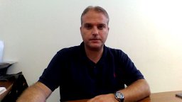 Constantinos J. Milios合作论文数School of Chemistry, The University of Edinburgh4Enrique Colacio Rodríguez合作论文数Departamento de Química Inorgánica, Facultad de Ciencias, Universidad de Granada4Laura Budd合作论文数3Horst Puschmann合作论文数3
 Jack Clegg合作论文数School of Chemistry and Molecular Biosciences, Faculty of Science, The University of Queensland3
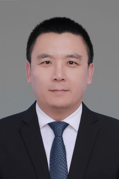 Gang Zhang (张纲)合作论文数College of Chemical Engineering, Nanjing Forestry University3
 Leonard F. Lindoy合作论文数School of Chemistry, University of Sydney3
 Robert Bucker合作论文数3Leeland James W合作论文数3
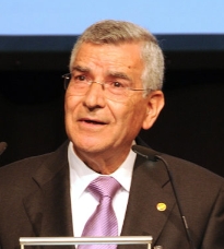 Giovanni Natile合作论文数Dipartimento di Farmacia, Università degli Studi di Bari Aldo Moro3Ruth J. McQuitty合作论文数3Sato Hiroyasu合作论文数3John Campbell合作论文数3
 Nazzareno Re合作论文数Università Degli Studi"GdAnnunzio"Chieti-Pescara3Iris M. Oppel合作论文数3
 Alessandro Marrone合作论文数Facoltà di Farmacia, Università degli Studi “G. D’Annunzio” Chieti-Pescara3Norrihan Sam合作论文数3Carmen R. Barone合作论文数3
 Antonio J. Mota合作论文数Faculty of Pharmacy, University of Granada3




