BACKGROUND:Acute pancreatitis is a common acute surgical condition worldwide as well as in Fiji. The incidence, epidemiology, management, and outcomes have not been previously documented. The aims of this study were to determine the causes, treatment and outcomes of acute pancreatitis at Colonial War Memorial Hospital (CWMH) in Fiji. METHOD:This was a retrospective, single center, descriptive study conducted over a 5-year period for all adult patients diagnosed with acute pancreatitis at CWMH, a tertiary hospital and regional referral center. RESULTS:There were 156 patients; 50% were female; 53% were of I-Taukei origin; and a median age of 44.5 years. The most common cause identified was idiopathic/unknown (38%) followed by gallstones (36%), alcohol (13%), hyperlipidaemia (7%) and others (6%). The incidence was 49 per 100 000 with majority mild (89%) in severity, 10% moderately severe and 1% severe. Inappropriate prophylactic antibiotics were given to 30% of the patients. For gallstone pancreatitis, only 17% had cholecystectomy on index admission while 20% underwent cholecystectomy within 6 weeks. Major morbidity occurred in 8%. There were no inpatient mortality recorded. No patients received minimally invasive treatment for complicated pancreatitis. CONCLUSION:The management of acute pancreatitis in Fiji is largely in line with international standards, but in some aspects severely limited by lack of resources, a common issue in developing countries. Establishment of pathways for antibiotic use and index admission cholecystectomy is recommended. Further resourcing for infrastructure and training is needed to improve diagnosis for etiology, as well as radiological and endoscopic treatment of complications.
We report a selection strategy for linking a drug-pharmacophore to a degenerate DNA library for use in cell- and whole-animal systematic evolution of ligands by exponential enrichment (SELEX) to direct the selection of aptamers to a specific target. This approach enables the discovery of aptamers with both high affinity and high tissue specificity, guided by a tethered small molecule and enhanced by an aptamer. We applied this approach to a critical application in prostate cancer (PCa) by conjugating a fluorescent analogue of the FDA-approved drug Pluvicto to a degenerate N40-DNA library to direct the selection of aptamers against the prostate-specific membrane antigen (PSMA). Seeking antibody-like functionality, we introduce two modified dNTPs─phenolic-dT and naphthyl-dC─to enhance the affinity and serum stability of selected aptamers. After 31 rounds of cell SELEX, followed by one round in a mouse bearing an LNCaP xenograft, next-generation sequencing informed the selection of several aptamers, of which an exemplar shows very high affinity for PSMA (Kd ∼ 0.8 nM). Its affinity depends on both the small-molecule drug and the modified nucleosides. Appreciating the outstanding challenge of identifying agents that differentiate PSMA on tumors from salivary glands, we identify aptamers that selectively bind PSMA-expressing tumors while sparing salivary glands. Use of a PSMA-targeting pharmacophore as a molecular bait represents the first example of SELEX against specific targets expressed on tumors while avoiding binding to the same target expressed on normal tissues. The resulting aptamers represent hybrid biologics that enhance the affinity and tumor specificity of the small-molecule drug-pharmacophore.
Prostate cancer frequently metastasizes to bone, leading to a spectrum of osteosclerotic and osteolytic lesions that cause debilitating symptoms. Accurate differentiation of bone tissue or lesion types can provide opportunity for local pathologic investigation, which is critical for understanding the bone metastasis and remains challenging. Current imaging methods lack the ability to directly differentiate tissues based on collagen organization and may induce invasive effect on bone tissues. We introduce Polarization-Sensitive Optical Coherence Tomography (PS-OCT) to investigate normal, osteosclerotic, and osteolytic bone tissues. High-resolution PS-OCT imaging reveals collagen fiber arrangement, enabling nuanced distinction of degree of collagen alignment among different bone tissue types or regions. We present a novel feature named degree of ordered organization (DOO), derived from the multiple contrasts of PS-OCT that can quantitatively evaluate bone samples from different pathologic groups, including control,osteoblastic and osteolytic tissues. The capacity of PS-OCT to differentiate trabecular/lamellar and irregular (woven bone) regions within the same specimen is tested and validated on ex-vivo samples extracted from 13 subjects. Our study is the first time that PS-OCT is applied to metastatic bone disease with the aim of enhancing the understanding of bone-related pathologies, and potentially impacting clinical practice. This work demonstrates that PS-OCT can provide useful insight into bone microstructures, and thus it has potential applications across diverse bone disorders.### Competing Interest StatementThe authors have declared no competing interest.
INTRODUCTION:Sarcopenia is a known risk factor for adverse outcomes across multiple disease states, including severe trauma. Factors such as age, hyperinflammation, prolonged immobilization, and critical illness may not only exacerbate progression of this disease but may also contribute to the development of induced sarcopenia, or sarcopenia secondary to hospitalization. This study seeks to (1) determine the effects of severe traumatic injury on changes in skeletal muscle mass in older adults; (2) test whether changes in skeletal muscle mass are associated with clinical frailty, physical performance, and health-related quality of life; and (3) examine trauma-induced frailty and temporal changes in myokine and chemokine profiles. METHODS:A prospective, longitudinal cohort study of 47 critically ill, older (≥45 years) adults presenting after severe blunt trauma was conducted. Repeated measures of computed tomography-based skeletal muscle index, frailty, and quality of life were obtained in addition to selected plasma biomarkers over 6 months. RESULTS:Severe trauma was associated with significant losses in skeletal muscle mass and increased incidence of sarcopenia from 36% at baseline to 60% at 6 months. Severe trauma also was associated with a transient worsening of induced frailty and reduced quality of life irrespective of sarcopenia status, which returned to baseline by 6 months after injury. Admission biomarker levels were not associated with skeletal muscle index at the time points studied but demonstrated distinct temporal changes across our entire cohort. CONCLUSIONS:Severe blunt trauma in older adults is associated with increased incidence of induced sarcopenia and reversible induced frailty. Despite muscle wasting, functional decline is transient, with a return to baseline by 6 months, suggesting a need for holistic definitions of sarcopenia and further investigation into long-term functional outcomes in this population.
Neuroendocrine prostate cancer (NEPC) is a rare and aggressive subtype of prostate cancer (PCa), emerging from advanced treatments and characterized by loss of androgen receptor (AR) signaling and neuroendocrine features, leading to rapid progression and treatment resistance. The third symposium on treatment-induced NEPC, held from 21 to 23 June 2024, at Harrison Hot Springs Resort, BC, Canada, united leading global researchers and clinicians. Sponsored by the Vancouver Prostate Centre (VPC), Canadian Institute of Health Research, Prostate Cancer Foundation Canada and Pharma Planter Inc, the event focused on the latest NEPC research and innovative treatment strategies. Co-chaired by Drs. Yuzhuo Wang and Martin Gleave, the symposium featured sessions on NEPC's historical context, molecular pathways, epigenetic regulation and the role of the tumor microenvironment and metabolism in its progression. Keynotes from experts like Dr. Himisha Beltran and Dr. Martin Gleave highlighted the complexity of NEPC. The Emerging Talent session showcased new research, pointing to the future of NEPC treatment. The symposium concluded with a consensus on the need for early detection, targeted therapies and personalized medicine to effectively combat NEPC, emphasizing the importance of global collaboration in advancing NEPC understanding and treatment.
Bone metastases are the most severe and prevalent consequences of prostate cancer (PC), affecting more than 80% of patients with advanced PC. PCBMs generate pain, pathological fractures, and paralysis. As modern therapies increase survival, more patients are suffering from these catastrophic consequences. Radiographically, PCBMs are predominantly osteosclerotic, but the mechanisms of abnormal bone formation and how this pathological increase in bone density is related to fractures are unclear. In this study, we conducted a comprehensive analysis on a cohort of 76 cadaveric PCBM specimens and 12 cancer-free specimens as controls. We used micro-computed tomography to determine 3D organization and quantify bone characteristics, quantitative backscattering electron microscopy to characterize mineral content and details in bone structure, nanoindentation to determine mechanical properties, and histological and immunohistochemical analysis of bone structure and composition. We define 4 PCBM phenotypes: osteolytic, mixed lytic-sclerotic, and 2 subgroups of osteosclerotic lesions-those with residual trabeculae, and others without residual trabeculae. The osteosclerotic lesions are characterized by the presence of abnormal bone accumulated on trabeculae surfaces and within intertrabecular spaces. This abnormal bone is characterized by higher lacunae density, abnormal lacunae morphology, and irregular lacunae orientation. However, mineral content, hardness, and elastic modulus at micron-scale were indistinguishable between this irregular bone and residual trabeculae. The collagen matrix of this abnormal bone presents with irregular organization and a prominent collagen III composition. These characteristics suggest that osteosclerotic PCBMs initiate new bone deposition as woven bone; however, the lack of subsequent bone remodeling, absence of lamellar bone deposition on its surface, and presence of collagen III distinguish this pathologic matrix from conventional woven bone. Although the mineralized matrix retains normal bone hardness and stiffness properties, the lack of fibril anisotropy presents a compromised trabecular structure, which may have clinical implications.
Aberrant expression of the E26 transformation -specific (ETS) transcription factors characterizes numerous human malignancies. Many of these proteins, including EWS:FLI1 and EWS:ERG fusions in Ewing sarcoma (EwS) and TMPRSS2:ERG in prostate cancer (PCa), drive oncogenic programs via binding to GGAA repeats. We report here that both EWS:FLI1 and ERG bind and transcriptionally activate GGAA-rich pericentromeric heterochromatin. The respective pathogen -like HSAT2 and HSAT3 RNAs, together with LINE , SINE , ERV, and other repeat transcripts, are expressed in EwS and PCa tumors, secreted in extracellular vesicles (EVs), and are highly elevated in plasma of patients with EwS with metastatic disease. High human satellite 2 and 3 ( HSAT2,3 ) levels in EWS:FLI1- or ERG -expressing cells and tumors were associated with induction of G2/M checkpoint, mitotic spindle, and DNA damage programs. These programs were also activated in EwS EV-treated fibroblasts, coincident with accumulation of HSAT2,3 RNAs, proinflammatory responses, mitotic defects, and senescence. Mechanistically, HSAT2,3 -enriched cancer EVs induced cGAS-TBK1 innate immune signaling and formation of cytosolic granules positive for double -strand RNAs, RNA -DNA, and cGAS. Hence, aberrantly expressed ETS proteins derepress pericentromeric heterochromatin, yielding pathogenic RNAs that transmit genotoxic stress and inflammation to local and distant sites. Monitoring HSAT2,3 plasma levels and preventing their dissemination may thus improve therapeutic strategies and blood -based diagnostics.
In cancer cells, inhibition of integrin-linked kinase (ILK) increases centrosome declustering causing mitotic arrest and cell death. Yet, not all cancer cells are susceptible to anti-ILK treatment alone. We investigate a combination drug strategy targeting ILK and another oncogenic kinase, Abelson kinase (ABL). Drug-concentration viability assays (i.e., MTT assays) indicate that ILK and ABL inhibitors in combination decreased the viability of glioblastoma cells over the ILK drug QLT-0267 alone. Combination strategies also increased aberrant mitoses and cell death over QLT-0267 alone. This was evident from an increase in mitotic arrest, apoptosis and a sub-G1 peak following FAC analysis. In vitro, ILK and ABL localized to the centrosome and the putative ILK kinase domain was important for this localization. Increased levels of cytosolic ABL are associated with its transformative abilities. ILK inhibitor effects on survival correlated with its ability to decrease cytosolic ABL levels and inhibit ABL's localization to mitotic centrosomes in glioblastoma cells. ILK inhibitor effects on ABL's centrosomal localization were reversed by the proteasomal inhibitor MG132 (a drug that inhibits ABL degradation). These results indicate that ILK regulates ABL at mitotic centrosomes and that combination treatments targeting ILK and ABL are more effective then QLT-0267 alone at decreasing the survival of dividing glioblastoma cells.
Supplementary Tables 5-8 from Characterization of ERG, AR and PTEN Gene Status in Circulating Tumor Cells from Patients with Castration-Resistant Prostate Cancer
Supplementary information describing methods for cell culture, HDL assay,qPCR and western blotting analysis, steroid analysis and BLT-1 pharmacokinetic analysis. Supplemental statistical analysis of IHC samples and results of in vivo profiling of BLT-1
Supplementary Figure 3 from Oncolysis of Prostate Cancers Induced by Vesicular Stomatitis Virus in PTEN Knockout Mice
To explore the pathways and mechanisms driving inflammation and fibrosis in stented ureters. In total, six healthy female pigs underwent cystoscopic unilateral ureteral stent insertion (6 Fr). After 14 days indwelling time, ureteral tissue was harvested in three pigs, while the remaining three pigs had their stents removed, and were recovered for 7 days. Three separate pigs served as controls. Tissue from stented and contralateral ureters was analysed histologically to evaluate tissue remodelling and classify the degree of inflammation and fibrosis, while genome, proteome and immunohistochemistry analysis was performed to assess changes at the transcriptional and translational levels. Finally, immunofluorescence was used to characterize the cell composition of the immune response and pathways involved in inflammation and fibrosis. Statistical analysis was performed using GraphPad Prism and RStudio for Welch ANOVA, Kruskal–Wallis and Dunnett’s T3 multiple comparison test. Stents cause significant inflammation and fibrosis of ureters. Gene set enrichment analysis confirmed fibrotic changes and tissue proliferation and suggests that epithelial–mesenchymal transition is a driver of fibrosis. Moreover, IL-6/JAK/STAT and TNFα via NF-κB signalling might contribute to chronic inflammation promoting a profibrotic environment. Immunostaining confirmed epithelial–mesenchymal transition in the urothelium and NF-κB expression in ureters stented for 14 days. Tissue alterations do not fully recover after 7 days. Histological evaluation showed that contralateral, unstented ureters are affected by mild inflammation. Our study showed that stenting has a significant impact on the ureter. Chronic inflammation and epithelial–mesenchymal transition are drivers of fibrosis, potentially impairing ureteral functionality in the long term. Furthermore, we observed mild inflammation in contralateral, unstented ureters.
Supplementary Video 1 from Phosphorylated Caveolin-1 Regulates Rho/ROCK-Dependent Focal Adhesion Dynamics and Tumor Cell Migration and Invasion
Supplementary Tables 1-4 from Characterization of ERG, AR and PTEN Gene Status in Circulating Tumor Cells from Patients with Castration-Resistant Prostate Cancer
Supplementary Figure 1 from Phosphorylated Caveolin-1 Regulates Rho/ROCK-Dependent Focal Adhesion Dynamics and Tumor Cell Migration and Invasion
Prostate-specific membrane antigen (PSMA) is expressed in epithelial cells of the prostate gland and is strongly upregulated in prostatic adenocarcinoma, with elevated expression correlating with metastasis, progression, and androgen independence. Because of its specificity, PSMA is a major target of prostate cancer therapy; however, detectable levels of PSMA are also found in other tissues, especially in salivary glands and kidney, generating bystander damage of these tissues. Antibody target therapy has been used with relative success in reducing tumor growth and prostate specific antigen (PSA) levels. However, since antibodies are highly stable in plasma, they have prolonged time in circulation and accumulate in organs with an affinity for antibodies such as bone marrow. For that reason, a second generation of PSMA targeted therapeutic agents has been developed. Small molecules and minibodies have had promising clinical trial results, but concerns about their specificity had arisen with side effects due to accumulation in salivary glands and kidneys. Herein we study the specificity of small molecules and minibodies that are currently being clinically tested. We observed a high affinity of these molecules for PSMA in prostate, kidney and salivary gland, suggesting that their effect is not prostate specific. The search for specific prostate target agents must continue so as to optimally treat patients with prostate cancer, while minimizing deleterious effects in other PSMA expressing tissues.
Supplementary Figures 1-3 from Characterization of ERG, AR and PTEN Gene Status in Circulating Tumor Cells from Patients with Castration-Resistant Prostate Cancer
<p>XLSX file - 10K, Supplementary Table 1: Growth inhibition of transplantable tumor lines after Bicalutamide treatment.</p>
Supplementary Video 3 from Phosphorylated Caveolin-1 Regulates Rho/ROCK-Dependent Focal Adhesion Dynamics and Tumor Cell Migration and Invasion
 Guy Eslick合作论文数Department of Social Services53
 Martin Gleave合作论文数Vancouver Prostate Centre, Faculty of Medicine, The University of British Columbia;Department of Urologic Sciences, The University of British Columbia31
 Brakenridge Scott合作论文数27
 Philip A. Efron合作论文数27
 Ladan Fazli合作论文数Vancouver Prostate Centre27
 Lyle L. Moldawer合作论文数Department of Surgery, College of Medicine, University of Florida25
 Frederick A Moore合作论文数Department of Surgery, College of Medicine, University of Florida;University of Florida Health25
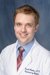 Russell B. Hawkins合作论文数Department of Surgery, University of Florida College of Medicine24
 Alicia Mohr合作论文数Department of Surgery, College of Medicine, University of Florida Health23
 Eltit Felipe合作论文数Faculty of Dentistry, Finis Terrae University16
 Julie Stortz合作论文数St. John’s Mercy Medical Center, St Louis University16
 Dijoia Darden合作论文数15
 Mazyar Ghaffari合作论文数Pulmonary Research Group, University of Alberta15
 Paul S Rennie合作论文数14
 Zhongkai Wang合作论文数Dept Biostat, Univ Florida14
 Christiaan Leeuwenburgh合作论文数Department of Physiology and Aging, College of Medicine, University of Florida;Center for Translational Research in Neurodegenerative Disease, College of Medicine, University of Florida14
 Sarah Parsons合作论文数14
 Gabriela Ghita合作论文数Dept Biostat, Univ Florida13
 Jaimar C Rincon合作论文数Facultad de Medicina, Universidad del Zulia Instituto de Investigaciones Clinicas “Dr. Americo Negrette”13
 Christopher Ong合作论文数12
 Marvin Dirain合作论文数12
 Azra Bihorac合作论文数Division of Nephrology, Hypertension & Renal Transplantation, Department of Medicine, College of Medicine, University of Florida Health12Ricardo Ungaro合作论文数12
 Robert H Bell合作论文数12Elaine Vickers合作论文数11Manju Sharma合作论文数11Yubin Guo合作论文数11
 Henry V Baker合作论文数Department of Molecular Genetics and Microbiology, University of Florida College of Medicine11Dina C. Nacionales合作论文数11
 Brumback Babette A合作论文数Dept Biostat, Univ Florida10
 Timothy V Beischlag合作论文数10James Toouli合作论文数10Naokazu Ibuki合作论文数10Antonio Hurtado-Coll合作论文数10McKenzie Hollen合作论文数9Anne Haegert合作论文数9
 Colin Collins合作论文数Department of Urologic Sciences, Faculty of Medicine, The University of British Columbia9
 Yuzhuo Wang (王玉琢)合作论文数BC Cancer;Vancouver Prostate Centre;Department of Urologic Sciences, University of British Columbia;Living Tumor Laboratory9
 Samuel Leung合作论文数8
 Jodie Palmer合作论文数Exercise & Sport Sci, Univ Western Australia8
 Sam Wiseman合作论文数Department of Surgery, Faculty of Medicine, The University of British Columbia8
 Ivan Robert Nabi合作论文数Department of Cellular & Physiological Sciences, Faculty of Medicine, University of British Columbia;Life Sciences Institute Cell and Developmental Biology, University of British Columbia8
 Liliana Kojic合作论文数8Mariacecilia Lopez合作论文数8
 Steven Jones合作论文数Department of Medical Genetics, Faculty of Medicine, University of British Columbia;Genome Sciences Centre, BC Cancer8
 Bharat H. Joshi合作论文数Center for Biologics Evaluation and Research, Food and Drug Administration8
 Colleen Nelson合作论文数Centre for Genomics and Personalised Health, Queensland University of Technology;Vancouver Prostate Centre, University of British Columbia8Howard Tearle合作论文数8Mitali Pandey合作论文数8




