PrP lowering is effective against prion disease in animal models and is being tested clinically. Therapies in the current pipeline lower PrP production, leaving pre-existing PrP to be cleared according to its own half-life. We hypothesized that PrP's half-life may be a rate-limiting factor for the time to effect of PrP-lowering drugs, and one reason why late treatment of prion-infected mice is not as effective as early treatment. Using isotopically labeled diet with targeted mass spectrometry, as well as antisense oligonucleotide treatment followed by timed PrP measurement, we estimate a half-life of 5-6 days for PrP in the brain. PrP turnover is not affected by over- or under-expression. Mouse PrP and human PrP have similar turnover rates measured in wild-type or humanized knock-in mice. CSF PrP appears to mirror brain PrP in real time in rats. PrP in the colon is readily quantifiable and has a half-life just slightly shorter than in brain. An under-expressed pathogenic mutant PrP, corresponding to D178N in humans, exhibits an accelerated turnover rate. Our data may inform the design of both preclinical and clinical studies of PrP-lowering drugs.
Prion protein (PrP) lowering is effective in animal models of prion disease and is being tested clinically in prion disease patients, but there remains a need for more potent PrP-lowering drug candidates. Inspired by the reported potency and duration of action of divalent short interfering RNA (siRNA), a new oligonucleotide drug modality for the central nervous system, we sought to discover and develop a new PrP-lowering drug candidate. Herein we identify a mouse Prnp-targeting divalent siRNA molecule, 1682-s4, that lowers PrP to 49% residual brain expression in wild-type mice, and, in the context of intracerebral infection with Rocky Mountain Laboratory (RML) prions, achieves a 2.7-fold increase in survival time with pre-symptomatic chronic treatment and 64% increase in survival time with a single dose after symptom onset. We describe the generation of two transgenic mouse lines, Tg25109 and Tg26372, expressing the full human PRNP gene and its non-coding sequence, and demonstrate their utility for in vivo discovery of potent human PRNP-targeting oligonucleotides. We discover siRNA sequence 2439 against human PRNP and compare its potency in different divalent siRNA chemical scaffolds. We determine that both the fixed UU tail and extended nucleic acid linkages of scaffold s4 contribute to superior potency compared to other scaffolds tested, offering 9.4 and 15.9 percentage points respectively of additional PrP knockdown. A single dose of 348 μg of 2439-s4 lowered whole brain hemisphere human PrP in transgenic mice to 17% residual after 30 days, while 52 μg lowered PrP to 49% residual. 1-2% of the dose of 2439-s4 delivered into cerebrospinal fluid is retained in the brain, and the median effective tissue concentration is estimated at 1.2 micrograms per gram of tissue. Good Laboratory Practices toxicology studies identified no significant liabilities, and the U.S. FDA has cleared an Investigational New Drug application to bring 2439-s4 into clinical trials.
Prion disease is a fatal neurodegenerative disease caused by the misfolding of prion protein (PrP) encoded by the PRNP gene. While there is currently no cure for the disease, depleting PrP in the brain is an established strategy to prevent or stall templated misfolding of PrP. Here we developed in vivo cytosine and adenine base strategies delivered by adeno-associated viruses to permanently modify the PRNP locus to achieve PrP knockdown in the mouse brain. Systemic injection of dual-adeno-associated virus PHP.eB encoding BE3.9max and single guide RNA installing PRNP R37X resulted in 37% average installation of the desired edit, 50% reduction of PrP in the mouse brain and 52% extension of lifespan in transgenic human PRNP mice inoculated with pathogenic human prion isolates representing the most common sporadic and genetic subtypes of prion disease. We further engineered base editing systems to achieve improved in vivo potency and reduced base editor expression in nontargeting tissues, resulting in 63% average PrP reduction in the mouse brain from a 6.7-fold lower viral dose, with no detected off-target editing of anticipated clinical significance observed in either human cells or mouse tissues. These findings support the potential of in vivo base editing as one-time treatment for prion disease.
Prion disease is caused by misfolding of the prion protein (PrP) into pathogenic self-propagating conformations, leading to rapid-onset dementia and death. However, elimination of endogenous PrP halts prion disease progression. In this study, we describe Coupled Histone tail for Autoinhibition Release of Methyltransferase (CHARM), a compact, enzyme-free epigenetic editor capable of silencing transcription through programmable DNA methylation. Using a histone H3 tail-Dnmt3l fusion, CHARM recruits and activates endogenous DNA methyltransferases, thereby reducing transgene size and cytotoxicity. When delivered to the mouse brain by systemic injection of adeno-associated virus (AAV), Prnp -targeted CHARM ablates PrP expression across the brain. Furthermore, we have temporally limited editor expression by implementing a kinetically tuned self-silencing approach. CHARM potentially represents a broadly applicable strategy to suppress pathogenic proteins, including those implicated in other neurodegenerative diseases.
During development, inflammation or tissue injury, macrophages may successively engulf and process multiple apoptotic corpses via efferocytosis to achieve tissue homeostasis1. How macrophages may rapidly adapt their transcription to achieve continuous corpse uptake is incompletely understood. Transcriptional pause/release is an evolutionarily conserved mechanism, in which RNA polymerase (Pol) II initiates transcription for 20-60 nucleotides, is paused for minutes to hours and is then released to make full-length mRNA2. Here we show that macrophages, within minutes of corpse encounter, use transcriptional pause/release to unleash a rapid transcriptional response. For human and mouse macrophages, the Pol II pause/release was required for continuous efferocytosis in vitro and in vivo. Interestingly, blocking Pol II pause/release did not impede Fc receptor-mediated phagocytosis, yeast uptake or bacterial phagocytosis. Integration of data from three genomic approaches-precision nuclear run-on sequencing, RNA sequencing, and assay for transposase-accessible chromatin using sequencing (ATAC-seq)-on efferocytic macrophages at different time points revealed that Pol II pause/release controls expression of select transcription factors and downstream target genes. Mechanistic studies on transcription factor EGR3, prominently regulated by pause/release, uncovered EGR3-related reprogramming of other macrophage genes involved in cytoskeleton and corpse processing. Using lysosomal probes and a new genetic fluorescent reporter, we identify a role for pause/release in phagosome acidification during efferocytosis. Furthermore, microglia from egr3-deficient zebrafish embryos displayed reduced phagocytosis of apoptotic neurons and fewer maturing phagosomes, supporting defective corpse processing. Collectively, these data indicate that macrophages use Pol II pause/release as a mechanism to rapidly alter their transcriptional programs for efficient processing of the ingested apoptotic corpses and for successive efferocytosis.
IC50 Values of CNL-Induced Decreases in Cell Viability at 48 hours in Seven HNSCC Cell Lines and Non-Transformed PGF Cells.
Abstract Limb regeneration in the Mexican axolotl requires the dedifferentiation of mature limb cells into progenitor cells known as blastema cells. The blastema cells become competent to the signals that inform the blueprint, or pattern, of the regenerating cells to organize into the complex limb structures. Signaling downstream of the limb nerves plays a role in the induction of patterning competency; however, the underpinnings of this process are poorly understood. Studying patterning competency in amputated axolotl limbs is challenging because the complex network of signals and tissue interactions that occur during regeneration are activated. Here, we address this issue by developing an assay that we call the competency accessory limb model (CALM) assay, which is based on a simplified limb injury model where various signaling inputs can be systemically added and their individual contributions to patterning competency can be evaluated independently. We used the CALM as a platform to generate foundational knowledge by characterizing the specific timing of competency induction and maintenance and identifying nerve-dependent signals sufficient for induction and epigenetic modifications induced in wounded cells by these signals.
Methylpyruvate Partially Rescues from CNL+CQ-driven Autophagic/Mitophagic Response in UNC-10 Cells
Inducing or Inhibiting Early Stage Autophagy Does Not Alter CNL-Induced Cell Death in HNSCC
Methylpyruvate Rescues from CNL+CQ-driven Oxidative Stress and Mitochondrial Function in UNC-10 Cells
Introduction: Little is known about how the newly regenerated limb tissues in the Mexican axolotl seamlessly integrate with the remaining stump tissues to form a functional structure, and why this doesn't occur in some regenerative scenarios. In this study, we evaluate the phenomenological and transcriptional characteristics associated with integration failure in ectopic limb structures generated by treating anterior-located ectopic blastemas with Retinoic Acid (RA) and focusing on the "bulbus mass" tissue that forms between the ectopic limb and the host site. We additionally test the hypothesis that the posterior portion of the limb base contains anterior positional identities. Methods: The positional identity of the bulbus mass was evaluated by assaying regenerative competency, the ability to induce new pattern in the Accessory Limb Model (ALM) assay, and by using qRTPCR to quantify the relative expression of patterning genes as the bulbus mass deintegrates from the host site. We additionally use the ALM and qRTPCR to analyze the distribution of anterior and posterior positional identities along the proximal/distal limb axis of uninjured and regenerating limbs. Results: The bulbus mass regenerates limb structures with decreased complexity when amputated and is able to induce complex ectopic limb structure only when grafted into posterior-located ALMs. Expressional analysis shows significant differences in FGF8, BMP2, TBX5, Chrdl1, HoxA9, and HoxA11 expression between the bulbus mass and the host site when deintegration is occuring. Grafts of posterior skin from the distal limb regions into posterior ALMs at the base of the limb induce ectopic limb structures. Proximally-located blastemas express significantly less HoxA13 and Ptch1, and significantly more Alx4 and Grem1 than distally located blastemas. Discussion: These findings show that the bulbus mass has an anterior-limb identity and that the expression of limb patterning genes is mismatched between the bulbus mass and the host limb. Our findings additionally show that anterior positional information is more abundant at the limb base, and that anterior patterning genes are more abundantly expressed in proximally located blastemas compared to blastemas in the more distal regions of the limb. These experiments provide valuable insight into the underlying causes of integration failure and further map the distribution of positional identities in the mature limb.
The retina is highly metabolically active, relying on glucose uptake and aerobic glycolysis. Situated in close contact to photoreceptors, a key function of cells in the retinal pigment epithelium (RPE) is phagocytosis of damaged photoreceptor outer segments (POS). Here we identify RPE as a local source of insulin in the eye that is stimulated by POS phagocytosis. We show that Ins2 messenger RNA and insulin protein are produced by RPE cells and that this production correlates with RPE phagocytosis of POS. Genetic deletion of phagocytic receptors (‘loss of function’) reduces Ins2 , whereas increasing the levels of the phagocytic receptor MerTK (‘gain of function’) increases Ins2 production in male mice. Contrary to pancreas-derived systemic insulin, RPE-derived local insulin is stimulated during starvation, which also increases RPE phagocytosis. Global or RPE-specific Ins2 gene deletion decreases retinal glucose uptake in starved male mice, dysregulates retinal physiology, causes defects in phototransduction and exacerbates photoreceptor loss in a mouse model of retinitis pigmentosa. Collectively, these data identify RPE cells as a phagocytosis-induced local source of insulin in the retina, with the potential to influence retinal physiology and disease.
Apoptosis of cells and their subsequent removal through efferocytosis occurs in nearly all tissues during development, homeostasis, and disease. However, it has been difficult to track cell death and subsequent corpse removal in vivo. We developed a genetically encoded fluorescent reporter, CharON (Caspase and pH Activated Reporter, Fluorescence ON), that could track emerging apoptotic cells and their efferocytic clearance by phagocytes. Using Drosophila expressing CharON, we uncovered multiple qualitative and quantitative features of coordinated clearance of apoptotic corpses during embryonic development. When confronted with high rates of emerging apoptotic corpses, the macrophages displayed heterogeneity in engulfment behaviors, leading to some efferocytic macrophages carrying high corpse burden. Overburdened macrophages were compromised in clearing wound debris. These findings reveal known and unexpected features of apoptosis and macrophage efferocytosis in vivo.
When the Accessory Limb Model (ALM) regenerative assay was first published by Endo, Bryant, and Gardiner in 2004, it provided a robust system for testing the cellular and molecular contributions during each of the basic steps of regeneration: the formation of the wound epithelium, neural induction of the apical epithelial cap, and the formation of a positional disparity between blastema cells. The basic ALM procedure was developed in the axolotl and involves deviating a limb nerve into a lateral wound and grafting skin from the opposing side of the limb axis into the site of injury. In this chapter, we will review the studies that lead to the conception of the ALM, as well as the studies that have followed the development of this assay. We will additionally describe in detail the standard ALM surgery and how to perform this surgery on different limb positions.
Epithelial tissues such as lung and skin are exposed to the environment and therefore particularly vulnerable to damage during injury or infection. Rapid repair is therefore essential to restore function and organ homeostasis. Dysregulated epithelial tissue repair occurs in several human disease states, yet how individual cell types communicate and interact to coordinate tissue regeneration is incompletely understood. Here, we show that pannexin 1 (Panx1), a cell membrane channel activated by caspases in dying cells, drives efficient epithelial regeneration after tissue injury by regulating injury-induced epithelial proliferation. Lung airway epithelial injury promotes the Panx1-dependent release of factors including ATP, from dying epithelial cells, which regulates macrophage phenotype after injury. This process, in turn, induces a reparative response in tissue macrophages that includes the induction of the soluble mitogen amphiregulin, which promotes injury-induced epithelial proliferation. Analysis of regenerating lung epithelium identified Panx1-dependent induction of Nras and Bcas2 , both of which positively promoted epithelial proliferation and tissue regeneration in vivo. We also established that this role of Panx1 in boosting epithelial repair after injury is conserved between mouse lung and zebrafish tailfin. These data identify a Panx1-mediated communication circuit between epithelial cells and macrophages as a key step in promoting epithelial regeneration after injury.
Neurodevelopment and efferocytosis have fascinated scientists for decades. How an organism builds a nervous system that is precisely tuned for efficient behaviors and survival and how it simultaneously manages constant somatic cell turnover are complex questions that have resulted in distinct fields of study. Although neurodevelopment requires the overproduction of cells that are subsequently pruned back, very few studies marry these fields to elucidate the cellular and molecular mechanisms that drive nervous system development through the lens of cell clearance. In this review, we discuss these fields to highlight exciting areas of future synergy. We first review neurodevelopment from the perspective of overproduction and subsequent refinement and then discuss who clears this developmental debris and the mechanisms that control these events. We then end with how a more deliberate merger ofneurodevelopment and efferocytosis could reframe our understanding of homeostasis and disease and discuss areas of future study.
Synthetic gene circuits that precisely control human cell function could expand the capabilities of gene- and cell-based therapies. However, platforms for developing circuits in primary human cells that drive robust functional changes in vivo and have compositions suitable for clinical use are lacking. Here, we developed synthetic zinc finger transcription regulators (synZiFTRs), which are compact and based largely on human-derived proteins. As a proof of principle, we engineered gene switches and circuits that allow precise, user-defined control over therapeutically relevant genes in primary T cells using orthogonal, US Food and Drug Administration-approved small-molecule inducers. Our circuits can instruct T cells to sequentially activate multiple cellular programs such as proliferation and antitumor activity to drive synergistic therapeutic responses. This platform should accelerate the development and clinical translation of synthetic gene circuits in diverse human cell types and contexts.
 Kodi Ravichandran合作论文数Division of Immunobiology, Department of Pathology & Immunology, School of Medicine, Washington University in St. Louis10
 Costa-Pinheiro Pedro合作论文数Research Center, Portuguese Oncology Institute8
 Todd E. Fox合作论文数Department of Pharmacology, Penn State College of Medicine8Mark Kester合作论文数8Jeremy J P Shaw合作论文数7
 Justin Perry合作论文数Graduate School of Medical Sciences, Weill Cornell Medicine7Alexandra Hickman合作论文数6Emily K. Venner合作论文数6T L Boyer合作论文数6
 Sonia Vallabh合作论文数Broad Institute;Prion Alliance6
 Eric V. Minikel合作论文数Broad Institute of MIT and Harvard, Cambridge, MA, USA.;Analytical and Translational Genetics Unit, Massachusetts General Hospital;Broad Institute, MIT and Harvard6
 Mark Jameson合作论文数6
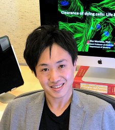 Sho Morioka合作论文数Department of Biological Sciences, Environmental and Molecular Toxicology, North Carolina State University5Suna Onengut-Gumuscu合作论文数5J. I. Etchegaray合作论文数4
 Chris Bernardo Medina合作论文数Departments of Chemistry and Biochemistry and., the4
 Lucas Christopher D合作论文数1] MRC Centre for Inflammation Research, The Queen's Medical Research Institute, University of Edinburgh, Edinburgh EH16 4TJ, UK. [2].4Pamela Brauer合作论文数3Meredith A Mortberg合作论文数3Barron Brady合作论文数Interdisciplinary Plant Group and the Missouri Maize Center, University of Missouri3
 Ben Deverman合作论文数Stanley Center for Psychiatric Research, Broad Institute of MIT and Harvard3
 Catherine D Mccusker合作论文数Department of Veterinary and Animal Sciences;University of Massachusetts;Department of Veterinary and Animal Sciences, University of Massachusetts3
 Ahmad 'Mo' Khalil合作论文数Department of Biomedical Engineering, College of Engineering, Boston University;K2 Biotechnologies, Inc.;Fynch Bio, Inc.2
 Vlad Serbulea合作论文数2Abigail Heher合作论文数2Jessie R Davis合作论文数2Meirui An合作论文数2Shannon Kelley合作论文数2Jonathan M Levy合作论文数2
 Turan Tufan合作论文数Sch Med, Univ Virginia2
 Jeffrey Rathmell合作论文数Ben May Department of Cancer Research, University of Chicago2
 Norbert Leitinger合作论文数Department of Pharmacology, School of Medicine, University of Virginia2Jividen Kasey合作论文数2
 Kristen Penberthy合作论文数1 Carter Immunology Center.2
 Gregory Newby合作论文数McKusick-Nathans Department of Genetic Medicine, School of Medicine, Johns Hopkins University;Institute for Nanobiotechnology, Johns Hopkins University;Department of Biomedical Engineering, Johns Hopkins University2
 Vieira Warren合作论文数2
 David R. Liu合作论文数Department of Chemistry and Chemical Biology, Faculty of Arts and Sciences, Harvard University2
 Marta Stremska合作论文数Institute of Mother and Child2
 Jiri Safar合作论文数Institute for Neurodegenerative;Departments of 2 Neurology;Departments of 2 Neurology, Institute for Neurodegenerative2
 Sarah Kucenas合作论文数School of Medicine, Saint Louis University2Andrew J. Davidson合作论文数1Keith A. Gagnon合作论文数1Seth R Goldman合作论文数1Kendra E Liu合作论文数1
 Magdalena E.M. Oremek合作论文数1Kristina Kelley合作论文数1Deborah E Cabin合作论文数1Vikram Seshadri合作论文数1Tyler Caron合作论文数1




