Purpose To evaluate the immediate and long-term safety as well as thrombus-capturing efficacy for 5 weeks after implantation of an absorbable inferior vena cava (IVC) filter in a swine model. Materials and Methods This study was approved by the institutional animal care and use committee. Eleven absorbable IVC filters made from polydioxanone suture were deployed via a catheter in the IVC of 11 swine. Filters remained in situ for 2 weeks (n = 2), 5 weeks (n = 2), 12 weeks (n = 2), 24 weeks (n = 2), and 32 weeks (n = 3). Autologous thrombus was administered from below the filter in seven swine from 0 to 35 days after filter placement. Fluoroscopy and computed tomography follow-up was performed after filter deployment from weeks 1-6 (weekly), weeks 7-20 (biweekly), and weeks 21-32 (monthly). The infrarenal IVC, lungs, heart, liver, kidneys, and spleen were harvested at necropsy. Continuous variables were evaluated with a Student t test. Results There was no evidence of IVC thrombosis, device migration, caval penetration, or pulmonary embolism. Gross pathologic analysis showed gradual device resorption until 32 weeks after deployment. Histologic assessment demonstrated neointimal hyperplasia around the IVC filter within 2 weeks after IVC filter deployment with residual microscopic fragments of polydioxanone suture within the caval wall at 32 weeks. Each iatrogenic-administered thrombus was successfully captured by the filter until resorbed (range, 1-4 weeks). Conclusion An absorbable IVC filter can be safely deployed in swine and resorbs gradually over the 32-week testing period. The device is effective for the prevention of pulmonary embolism for at least 5 weeks after placement in swine. © RSNA, 2017.
BACKGROUND:The purpose of this study was to evaluate percutaneous transhepatic portal vein stenting (PVS) for palliation of refractory ascites and/or variceal bleeding caused by extrahepatic portomesenteric venous stenosis in patients with pancreaticobiliary cancer.MATERIALS AND METHODS:A single-institution, retrospective review of patients who underwent PVS between January 2007 and July 2015 was performed. A total of 38 patients were identified, of whom 28 met the inclusion criterion of PVS performed primarily for refractory ascites or variceal bleeding. In addition to technical success and overall survival, clinical success was measured by fraction of remaining life palliated. The palliative effect of PVS was also quantified by measuring changes in liver and ascites volumes after the procedure.RESULTS:Technical success was 93% (26/28). Stent deployment involved more than one portomesenteric vessel in most patients (20/26). The cumulative probability of symptom recurrence at 6, 12, 18, and 24 months was 12%, 16%, 26%, and 40%, respectively. There was a significant difference (p < .001) in the probability of symptom recurrence, recurrence of abdominal ascites, and increase in liver volume between patients whose stents remained patent and those whose stents demonstrated partial or complete occlusion. The mean fraction of remaining life palliated was 87%. All but two patients were found to have improvement in clinical symptoms for the majority of their lives after the procedure. There were no major or minor complications.CONCLUSION:As a low-risk procedure with a high clinical success rate, PVS can play a substantial role in improving quality of life in patients with portomesenteric stenoses.IMPLICATIONS FOR PRACTICE:Portomesenteric venous stenosis is a challenging complication of pancreaticobiliary malignancy. Portomesenteric stenoses can lead to esophageal, gastric, and mesenteric variceal bleeding, as well as abdominal ascites. The purpose of this study was to evaluate the safety and efficacy of portal vein stenting (PVS) in patients with cancer who have symptomatic portal hypertension caused by portomesenteric venous compression. As a low-risk procedure with a high clinical success rate, PVS can play a substantial role in improving quality of life in patients with portomesenteric stenoses.
To describe the technique and results of MRI-guided transgluteal prostate biopsy performed in a closed MRI on a select group of patients, and to demonstrate the feasibility, safety, and clinical utility of this approach. 47 men (average age of 61 years, range 41-77) were referred for prostate MRI and subsequently transgluteal MR-guided prostate biopsy. The referrals fell into three general categories: (A) patients with prior transrectal ultrasound (TRUS) biopsies showing prostate cancer referred for re-evaluation due to discordant PSA levels (PSA out of proportion relative to Gleason) (n = 25 of 47); (B) patients with one or more (range 1 to 4) prior negative TRUS biopsies and discordant PSA levels (n = 15 of 47); and (C) patients with a history of total prostatectomy ± colectomy, i.e. not amenable to TRUS, and suspicion of disease recurrence based on PSA. All patients received pre-procedure multiparametric diagnostic MRI which included dynamic contrast enhanced imaging, T2 sequencing, and diffusion weighted imaging. Only patients with suspicious focal findings on MRI were referred for MRI-guided biopsy. A safe transgluteal route to the area of interest was determined. Only one to two lesions were targeted per patient. All biopsies were technically successful, as defined by satisfactory image confirmation of targeting, and all were without immediate complication. 23 (49%) of the biopsies returned a diagnosis of prostate cancer. 15 (32%) had upgrading of previously biopsy proven prostate cancer. 7(15%) patients had prior negative prostate biopsies and were diagnosed with clinically significant (Gleason 4 + 3 or higher) prostate cancer. 22(47%) of patients were transitioned from watchful waiting to definitive therapy (Surgery, Radiation and/or Medicinal) by the referring urologist as a result of positive findings on MRI-guided biopsy. The clinical utility of the MR-guided transgluteal biopsy is manifest in its ability to diagnose new cancers and upgrade previously diagnosed cancers of the prostate, thereby allowing a significant number of patients to transition from watchful waiting and active surveillance to more definitive therapy.
PURPOSE:To evaluate the use of a self-expanding tract sealant device (BioSentry™) on the rates of pneumothorax and chest tube insertion after percutaneous lung biopsy. MATERIALS AND METHODS:In this retrospective study, we compared 318 patients who received BioSentry™ during percutaneous lung biopsy (treated group) with 1956 patients who did not (control group). Patient-, lesion-, and procedure-specific variables, and pneumothorax and chest tube insertion rates were recorded. To adjust for potential selection bias, patients in the treated group were matched 1:1 to patients in the control group using propensity score matching based on the above-mentioned variables. Patients were considered a match if the absolute difference in their propensity scores was ≤equal to 0.02. RESULTS:Before matching, the pneumothorax and chest tube rates were 24.5 and 13.1% in the control group, and 21.1 and 8.5% in the treated group, respectively. Using propensity scores, a match was found for 317 patients in the treatment group. Chi-square contingency matched pair analysis showed the treated group had significantly lower pneumothorax (20.8 vs. 32.8%; p = 0.001) and chest tube (8.2 vs. 20.8%; p < 0.0001) rates compared to the control group. Sub-analysis including only faculty who had >30 cases of both treatment and control cases demonstrated similar findings: the treated group had significantly lower pneumothorax (17.6 vs. 30.2%; p = 0.002) and chest tube (7.2 vs. 18%; p = 0.001) rates. CONCLUSIONS:The self-expanding tract sealant device significantly reduced the pneumothorax rate, and more importantly, the chest tube placement rate after percutaneous lung biopsy.
CONTEXT- Optical imaging techniques are currently available for imaging tissues without the need for any type of extensive tissue preparation. There are several applications for their potential use in surgical pathology practice.OBJECTIVE- To evaluate the feasibility of using a confocal fluorescence microscopy (CFM) platform for ex vivo examination of tissues obtained from surgical resections of breast, lung, kidney, and liver.DESIGN- Tissue fragments (0.5-1.0 cm) were immersed in 0.6 mM acridine orange for 6 seconds and imaged using a CFM platform at a 488-nm wavelength. The imaged tissues were subsequently fixed in formalin and processed routinely to generate hematoxylin-eosin-stained tissue sections. Mosaics of the grayscale CFM images were studied at different magnifications for recognition of the tissue and were compared with conventional histopathologic examination of hematoxylin-eosin tissue sections.RESULTS- We imaged 55 tissue fragments obtained from 16 breast (29%), 18 lung (33%), 14 kidney (25%), and 7 liver (13%) surgical excision specimens. Acridine orange labeled the nuclei, creating the contrast between nucleus and cytoplasm and thereby recapitulating the tissue architecture. We could obtain CFM images of good quality within 5 to 10 minutes that allowed recognition of the cytomorphologic details for categorization of the imaged tissue and were similar to histologic examination of hematoxylin-eosin tissue sections.CONCLUSIONS- The ease and speed of acquisition of CFM images together with the resolution and resemblance of the CFM images to hematoxylin-eosin sections suggest that the CFM platform has excellent potential for use in surgical pathology practice.
The best friend of the HPB surgeon is the interventional radiologist. Advanced laparoscopic HPB surgery can be done safely only in an environment where skillful interventional radiology exists. Therefore excellent communication between HPB surgeon and interventional radiologist is critical for accurate diagnostic and challenging therapeutic interventions. This chapter reviews the important role that interventional radiology plays in the management of HPB patients from diagnosis to management of surgical complications. In addition, options for unresectable patients (i.e. chemoembolization, radioembolization, and thermal ablation) are presented.
Overview In the past decades, there has been a substantial expansion in the use of image‐guided procedures for diagnosing and treating various types of cancer. Percutaneous biopsy is usually the first step in obtaining a definitive diagnosis and aids in the development of the treatment plan. Patients with primary or metastatic disease involving the liver are candidates for a wide range of image‐guided interventions such as hepatic arterial embolization procedures, liver tumor ablation, and portal vein embolization. Cancer patients also benefit from a variety of palliative image‐guided interventions, such as vena cava filter placement, biliary drainage and stent placement, and renal artery embolization, to name a few. Interventional radiology offers a multitude of minimally invasive procedures that are a key component in the management of cancer patients.
Determine preliminary safety and efficacy of an absorbable vena cava filter for the prevention of pulmonary embolism in a porcine model. Study was approved by the IACUC. 11 Absorbable Inferior Vena Cava (IVC) filters were woven from polydioxanone suture size 00 (Riverpoint Medical, OR) and catheter deployed in 11 porcine. Autologous thrombus measuring 5mm (diameter) x 18 mm (length) to 15 x 55 mm were iatrogenically administered inferior to the filter in 7 animals from 0 – 35 days. Thrombus, pulmonary embolism, and filter migration was assessed by fluoroscopy and CT weekly for weeks 1-6, semi-monthly for weeks 7-20, and monthly for weeks 21-32. Necropsies were performed to determine absorption characteristics: (i) 2 animals at 2 weeks to quantify neointimal hyperplasia, (ii) 2 animals at 5 weeks to reveal filter basket integrity at planned indication terminus, (iii) 2 animals at 12 weeks, expected time for initial basket disintegration, (iv) 2 animals at 24 weeks, expected resorption of filter, and (v) 3 animals at 32 weeks to reveal chronic IVC change. All injected thrombi were trapped by the absorbable filters and held through resorption, requiring 1-4 weeks for autologous thrombolysis, resulting in 100% capture efficiency. There were no instances of pulmonary embolism, filter migration, caval penetration, or tilt greater than 14 degrees. Severe infrarenal caval narrowing occurred in one subject following filter mal-deployment that subsequently resolved. Necropsy results revealed (i) neointimal hyperplasia encased the stent portion of the filter at 2 weeks, (ii) filter baskets were intact at 12 weeks, (iii) filter suture not humanly visible at 24 weeks, and (iv) polydioxanone remnants measuring less than 0.1mm were observed with 10x magnification at 32 weeks. Blood count, gases, chemistry, coagulation and liver panels remained normal throughout the study. Preliminary large animal results suggest that an absorbable polydioxanone vena cava filter can be both safe and effective in trapping iatrogenically administered autologous thrombus through resorption with minimal presence of the filter at 32 weeks.
Background: Risk of disappearing colorectal liver metastases (CLM) has increased with the advances in preoperative chemotherapy. Less than 50% of disappearing CLM have complete pathological response, leading to high recurrence rates after curative surgery when disappearing CLM are not resected. Objective: To evaluate the clinical relevance of fiducial placement with curative strategy for CLM at risk of disappearance. Methods: All consecutive patients that received a platinum microcoil fiducial placement for CLM surgery tracking with curative intent at a tertiary center were reviewed. Under computed-tomography, magnetic resonance imaging, or ultrasound guidance, coils were placed percutaneously at the deep margin of the CLM at risk for disappearing (≤20mm and >1cm deep). Results: Among 1377 patients undergoing CLM resection between 2005 and 2015, 35 patients underwent fiducial placement. Three patients were excluded due to disease progression before surgery, and 32 patients underwent surgery with curative intent for 104 CLM, including 41 marked CLM. Among marked CLM, 11 (27%) represented a solitary metastasis, 36 (88%) were in the right liver, 34 (83%) were located > 1cm deep in the liver parenchyma, and median size was 1.15 cm (0.6–2cm). No complication occurred after fiducial placement. After chemotherapy, 18 (44%) of the 41 metastases disappeared radiographically. All tracked CLM were treated ‒ 31 were resected, and 10 treated with thermal ablation. After a median follow-up of 13.9 months (range, 0–64.3 months), no local recurrences were observed. Conclusion: Fiducial placement represents a safe procedure that facilitates accurate tracking of small CLM at risk of disappearance after chemotherapy.
To describe the incidence of multiple renal artery pseudoaneurysms (PSA) in patients referred for renal artery embolization following partial nephrectomy and to study its relationship to RENAL nephrometry scores.
The risk of colorectal liver metastases (CLM) disappearing on cross-sectional imaging has increased with advances in preoperative chemotherapy, but <50 % of disappearing CLM demonstrate complete pathological response.
PURPOSE:The safety and efficacy of the combined use of sorafenib and yttrium-90 resin microspheres (Y90 RMS) to treat advanced hepatocellular carcinoma (HCC) is not well established. We determined the incidence of adverse events with this combination therapy in patients with advanced HCC at our institution and analyzed the treatment and survival outcomes.MATERIALS AND METHODS:We reviewed the records of 19 patients with Barcelona Clinic Liver Cancer class B or C HCC who underwent treatment with Y90 RMS (for 21 sessions) while receiving full or reduced doses of sorafenib between January 2008 and May 2010. Therapy response was evaluated using Response Evaluation Criteria in Solid Tumors. We evaluated median overall survival (OS) and progression-free survival (PFS) as well as hepatic and extrahepatic disease PFS and incidence of adverse events.RESULTS:The median patient age was 67 years, and portal or hepatic venous invasion was present in eight patients (42%). Ten patients received reduced doses of sorafenib. The median Y90 radiation activity delivered was 41.2 mCi. The partial response of Response Evaluation Criteria in Solid Tumors was observed in four patients (19%). The median hepatic disease PFS was 7.82 months, extrahepatic disease PFS was 8.94 months, OS was 19.52 months, and PFS was 6.63 months. Ninety days after treatment with Y90 RMS, five patients (26%) had grade II adverse events and four patients (21%) had grade III adverse events.CONCLUSION:OS and PFS outcomes were superior to those observed in prior studies evaluating sorafenib alone in patients with a similar disease status, warranting further study of this treatment combination.
Background Liver metastases are associated with a poor prognosis. We investigated the use of hepatic arterial infusion (HAI) of irinotecan combination therapy in patients with liver metastases. Patients and methods Patients with histologically confirmed advanced cancer with liver metastases that was refractory to standard therapy were eligible. A standard “3 + 3” phase I study design was used to determine the dose-limiting toxicity (DLT) and the maximum tolerated dose (MTD). Three cohorts were evaluated: HAI of irinotecan with systemic intravenous (IV) (a) bevacizumab, (b) oxaliplatin and bevacizumab, or (c) bevacizumab and cetuximab. Results From October 2009 through December 2013, 98 patients with various tumor types were enrolled (median age, 62 years, range, 34-85; and median number of prior therapies, 4, range, 1–11). In cohorts A and C, dose escalation continued until the highest dose level—considered the MTD—was reached. In cohort B, dose escalation continued until dose level 3, and dose level 2 was considered the MTD. Rates of grade 3/4 adverse events were as follows: diarrhea, 8 %; fatigue, 4 %; neutropenia, 4 %; thrombocytopenia, 2 %; and skin rash, 2 %. Seventy-seven patients were evaluable for response. Partial response was noted in 5 (6.5 %) patients (neuroendocrine cancer, n = 2; CRC, n = 2; NSCLC, n = 1); and stable disease ≥ 6 months in 17 (22.1 %) patients (CRC, n = 13; breast, n = 1; neuroendocrine, n = 1; NSCLC, n = 1; pancreatic, n = 1). Conclusions HAI irinotecan in combination with bevacizumab; oxaliplatin plus bevacizumab; or cetuximab plus bevacizumab was safe and may be a treatment option for selected patients with advanced cancer and liver involvement.
To determine the feasibility of infusing resorbable inferior vena cava (IVC) filter with iodine-based contrast agents to produce a radiopaque, computed tomography (CT)-visible IVC filter. Infused poly(p-dioxanone) (PPDO) was obtained by incubating PPDO in different concentrations of 4-iodobenzoyl chloride (IBC) and 2,3,5-triiodobenzoic acid (TIBA). Characterizations of infused and nascent PPDO were done using elemental analysis, micro-CT, tensile strength analysis, scanning electron microscopy, and differential scanning calorimetry. Elemental analysis showed percentage loading of 1.07 ± 0.08 for IBC and 0.73 ± 0.01 for TIBA. The iodine loading remained the same within 2 weeks for TIBA but decreased to about 80 % with IBC when subjected to physiological conditions. Micro-CT images showed increased attenuation of the infused PPDO compared with the nascent PPDO. The Hounsfield unit values for infused and nascent sutures were 110 ± 40 and 153 ± 53 for PPDO infused with 2 mg/mL IBC and TIBA, respectively, but only 11.35 ± 2 for nascent PPDO. In contrast the HU for bone was 116 ± 37. Tensile strength analysis showed maximum loads of 1.01 ± 0.43 kg and 10.02 ± 0.54 kg for IBC and TIBA, respectively, and 10.10 ± 0.64 kg for nascent PPDO. Scanning electron microscopy showed that the morphology of the PPDO surface did not change after coating and preliminary cytotoxicity assay showed no killing effect on Hela cells. PPDO infused with a contrast agent is significantly more radiopaque than nascent PPDO on micro-CT imaging. This radiopacity could allow the position and integrity of infused resorbable IVC filter to be monitored while it is in place, thus increasing its safety and efficacy as a medical device.
Objective: The objectives of this study were to evaluate tensile strength retention of polydioxanone as a function of time in a swine venous system and to assess the feasibility of an absorbable inferior vena cava (IVC) filter made from polydioxanone in a pilot swine study.Methods: Twenty strands (60 cm each) of size 1 polydioxanone absorbable suture (Ethicon, Somerville, NJ) were placed in the central venous system of domestic swine. Strands were harvested at weekly intervals during 10 weeks for tensile strength testing.Results were compared with control samples obtained from an in vitro engineered circulation system containing sodium phosphate buffer solution. Three IVC filters braided from polydioxanone suture were also catheter deployed in three swine to assess absorbable IVC filter feasibility. Results: Polydioxanone retained 82% tensile strength in vitro vs 79% in vivo at 35 days (P > .22), the desired prophylactic duration. For IVC filters made from polydioxanone, technical success of placement was achieved in all three filters deployed (100%). Autologous thrombus deployed inferior to the filter remained trapped in the filter until thrombus resorption, with no evidence of pulmonary emboli on follow-up computed tomography. There were no instances of caval penetration, filter-induced IVC thrombosis, filter migration, or tilt >15 degrees with imaging and clinical follow-up carried out to 32 weeks.Conclusions: Strength retention of polydioxanone suture placed in the venous system of swine is similar to earlier in vitro studies out to 10 weeks (P > .06 for all weeks) and is more than sufficient (8.20 0.37 kg mean load at break for size 1) to trap thrombus. Pilot animal study suggests that an absorbable polydioxanone IVC filter can be catheter deployed to capture and to hold iatrogenically administered autologous thrombus through resorption.
To determine the feasibility of coating resorbable filter device made up of polydioxanone (PDDO) with gold nanoparticles as a radiopaque, computed tomography (CT)-visible inferior vena cava (IVC) filter device. Coated PPDO was obtained by incubating PPDO in different concentrations of gold nanoparticles having 2 and 4 nm mean diameter using dichloromethane as a solvent at room temperature. After 24 h, PPDO was dried, washed with ethyl acetate, and dried under vacuum. Characterization was done using micro-CT, X-ray, scanning electron microscopy (SEM), and tensile strength. Micro-CT and X-ray images of the coated PPDO showed significant signal enhancement compared to uncoated PDDO. Elemental analysis showed loading of 50 ± 1 ppm for 2 nm versus 30 ± 10 ppm for 4 nm. Figure 1 shows the signal attenuation of AuNP infusion into the PDDO in micro-CT and x-ray imaging. Tensile strength showed 9.89 ± 0.05 and 10.09 ± 0.14 kg load at break for 2nm and 4 nm, respectively, as compared with the control of 10.46 ± 0.23 kg. SEM analysis depicted that the morphology of the PPDO surface does not change after coating. The significant increase in signal intensity with coated PDDO compared to bare PDDO using x-ray and CT imaging indicates its major advantage as a radiopaque resorbable IVC filter. The radiopacity allows for monitoring the position and integrity of the filter while in place, therefore increasing its safety and efficacy as a medical device.
 Kamran Ahrar合作论文数59
 David C. Madoff合作论文数Department of Radiology & Biomedical Imaging, Yale University School of Medicine33
 Ravi Murthy合作论文数32
 Joe Ensor合作论文数Dept Biostat, Houston Methodist Res Inst28
 Alda L. Tam合作论文数Department of Interventional Radiology, Division of Diagnostic Imaging, The University of Texas MD Anderson Cancer Center;MD Anderson UT Health Houston Graduate School of Biomedical Sciences28
 John F. Cardella合作论文数26
 Sanjay Gupta合作论文数The James and Eillen Dicke Urology Research Laboratory, Department of Urology, School of Medicine, Case Western Reserve University21Sanjoy Kundu合作论文数20
 Marshall E. Hicks合作论文数Division of Diagnostic Imaging, Department of Interventional Radiology, The University of Texas MD Anderson Cancer Center19Avritscher Rony合作论文数17
 Bruno C. Odisio合作论文数Department of Interventional Radiology, Division of Diagnostic Imaging, The University of Texas MD Anderson Cancer Center17Fa Morello合作论文数16Steven Yevich合作论文数15Abdelsalam Mohamed合作论文数15ME Hicks合作论文数14Kenneth C. Wright合作论文数14
 Armeen Mahvash合作论文数13
 Sharjeel H Sabir合作论文数12
 Jean-Nicolas Vauthey合作论文数Department of Surgical Oncology, The University of Texas MD Anderson Cancer Center;Division of Surgery, The University of Texas MD Anderson Cancer Center11
 Steven Curley合作论文数Division of Surgical Oncology Baylor College of Medicine;Christus Trinity Mother Frances Health System11
 Thomas A. Aloia合作论文数Ascension10
 Savitri Krishnamurthy合作论文数Department of Pathology, Division of Pathology and Laboratory Medicine, The University of Texas MD Anderson Cancer Center10Rony Avritscher合作论文数10
 Steven Rose合作论文数University of California San Diego10Marites P. Melancon合作论文数9
 Donald L Miller合作论文数Center for Devices and Radiological Health, U.S. Food and Drug Administration, U.S. Department of Health and Human Services9Richard B. Towbin合作论文数9
 Surena F. Matin合作论文数Department of Urology, Division of Surgery, The University of Texas MD Anderson Cancer Center8Sanaz Javadi合作论文数8
 Katherine Perrin Dixon合作论文数8Wojak Joan C合作论文数7
 David Sacks合作论文数Radiology, Reading Hospital7
 Sattva Neelapu合作论文数Graduate School of Biomedical Sciences, University of Texas MD Anderson Cancer Center;Department of Lymphoma/Myeloma, Division of Cancer Medicine, The University of Texas MD Anderson Cancer Center7Clement J. Grassi合作论文数7Stephen E. McRae合作论文数7Stokes Leann S合作论文数6
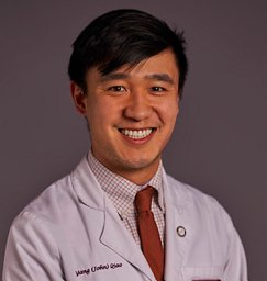 Qiao Yang合作论文数6Li Chun合作论文数6Burapol Singhana合作论文数6Jody Vykoukal合作论文数The University of Texas MD Anderson Cancer Center6Arshad Ahmed Khan合作论文数6Judy Ahrar合作论文数6E. Joseph合作论文数5
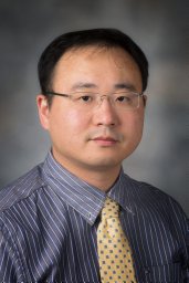 Jun Zhao合作论文数Purdue School of Engineering and Technology, Indiana University-Purdue University at Indianapolis5
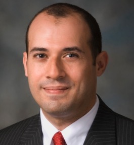 Ahmed O Kaseb合作论文数Department of Gastrointestinal Medical Oncology, Division of Cancer Medicine, The University of Texas MD Anderson Cancer Center5Timothy L. Swan合作论文数5Roland L. Bassett合作论文数The University of Texas MD Anderson Cancer Center5
 Junichi Shindoh合作论文数Faculty of Medicine, University of Tokyo;Fax +81-3-5684-3989 e-mail: narusek-sur@h.u-tokyo.ac.jp JP;Faculty of Medicine, University of Tokyo5




