Frontal sinus fractures (FSF) can lead to detrimental intracranial infections, yet there is variability in the literature regarding the efficacy of antibiotic prophylaxis. This study aims to provide guidance on the role of prophylactic antibiotics in mitigating infection following FSF. A retrospective cohort study was conducted from January 2018 to December 2022 comparing the incidence of infection between patients managed with versus without antibiotics. Patients with FSF diagnosed by CT scan and at least 18 years old were included. Our primary outcome was the incidence of infection following FSF. Of 201 patients, 26 (12.9%) had a dural tear or cerebrospinal fluid leak (CSF). Patients managed with antibiotics were significantly more likely to have a dural tear or CSF leak [25 (15.4%), 1 (2.6%), P=0.032], and none of these patients developed an infection. A total of 6 (3%) patients developed infection, including 4 meningitis cases and 2 sinusitis cases. Infections occurred in 5 of 162 patients (3.1%) who received antibiotics, compared with 1 infection (2.6%) in the 39 patients without antibiotic prophylaxis, with no statistically significant difference (P=1.0). The most common prophylactic antibiotic among patients who developed infection was IV cefazolin. The majority of fractures (149, 74%) were managed conservatively, including all patients who developed infections. Although further randomized controlled trials are needed to better define the role of antibiotic prophylaxis in preventing infections, antibiotics may not be necessary for all FSF patients. In the subset of patients with CSF leak, the absence of infection might indicate the protective effects of antibiotics.
OBJECTIVE:This study describes the management and outcomes of temporal bone fractures resulting from falls. METHODS:We retrospectively reviewed patients with traumatic temporal bone fractures from 2018 through 2022. RESULTS:We analysed 171 patients with temporal bone fractures, 62 (36.3 per cent) of which occurred secondary to falls. Fall patients were significantly older than non-fall patients (mean age 46 vs. 38 years; p = 0.0079) and had higher Modified Frailty Index-5 scores (0.63 vs. 0.20; p = 0.0003). Fall patients had shorter hospital stays (10.1 vs. 15.8 days; p = 0.015), were more frequently discharged home (66.1 vs. 44.0 per cent; p = 0.007) and were less likely to experience non-resolving facial nerve weakness (6.5 vs. 21.1 per cent; p = 0.030). CONCLUSION:Patients with temporal bone fractures from falls are older and frailer than non-fall patients and have unique preventative and rehabilitation needs.
Background: For decades, there has been an ongoing debate about the ideal timing of orbital fracture repair (OFR) in adults. Methods: The authors conducted a retrospective review of patients who underwent OFR at 2 centers (2015 to 2019). Excluded were patients younger than 18 years and those with follow-up less than 2 weeks. The study’s primary outcome was the incidence/persistence of postoperative enophthalmos/diplopia at least 2 weeks following OFR. The association between surgical timing and postoperative ocular complications was assessed in patients with extraocular muscle (EOM) entrapment, enophthalmos and/or diplopia, and different fracture sizes. Results: Of 253 patients, 13 (5.1%) had preoperative EOM entrapment. Of these, patients who had OFR within 2 days of injury were less likely to develop postoperative diplopia compared with patients who had OFR within 8 to 14 days (1 of 8 patients [12.5%] versus 3 of 3 patients [100%]; P = 0.018). Patients who had OFR for nearly total defects within 1 week of injury were significantly less likely to have postoperative enophthalmos (0 patients [0.0%]) compared with those who had surgery after 2 weeks (2 patients [33.3%] after 15 to 28 days versus 8 patients [34.8%] after 28 days from injury; P < 0.001). Patients who had delayed OFR for large fractures smaller than nearly total defects, preoperative persistent diplopia, or enophthalmos were not at significantly greater likelihood of postoperative ocular complications compared with those who had early OFR. Conclusions: The authors recommend OFR within 2 days of injury for EOM entrapment and 1 week for nearly total defects. Surgical delay up to at least 4 weeks is possible in case of less severe fractures, preoperative persistent diplopia, or enophthalmos. CLINICAL QUESTION/LEVEL OF EVIDENCE: Risk, IV.
PURPOSE: Soft tissue reconstruction of the hind-foot presents a reconstructive challenge, often requiring free tissue transfer. The ideal free flap has to provide thin, pliable tissue, achieve optimal contour and also withstand weight bearing. METHODS: Free tissue transfers performed by a single surgeon for hind-foot reconstruction at a trauma center over three years were reviewed. The mechanism of injury, surgical and functional outcomes were recorded. RESULTS: A total of 20 free tissue transfers were performed. Motor vehicle accidents were the most common injury mechanism (5/10), followed by falls (5/10) and ballistic wounds (4/10). The median interval from injury to reconstruction was 10 days. Twelve defects were covered with an anterolateral thigh flap; the remaining with muscle flaps (5 latissimus dorsi, 3 vastus lateralis). All arterial anastomoses were performed end-to-end and two recipient veins were used in each case. No total flap failures were encountered. Major complications included hematoma (1/20), soft tissue infection (3/20) and partial flap necrosis (1/20). Minor complications included delayed healing (1/20). Follow-up ranged from 2-28 months. Two patients required below knee amputation at 4 and 11 months after reconstruction, respectively; the remaining had a stable reconstruction at the completion of follow up. CONCLUSION: Stable and durable hind-foot reconstruction is feasible with both fasciocutaneous and muscle flaps. Flap selection takes into consideration the defect location, underlying hardware and need for weight bearing. The complication rate remains high and a below knee amputation might be needed despite successful soft tissue reconstruction.
Background: Postoperative diplopia is reported in up to 52% of orbital bone fracture (OBF) repair. Evidence on these risk factors is based on low-quality data, single-institution studies, and small sample sizes. Our study is the largest and first multicenter study to determine the predictors of postoperative diplopia following OBF repair. Methods: The authors conducted a retrospective review of patients who underwent OBF repair at 2 centers from 2015 to 2019. The authors’ primary outcome was the incidence or persistence of postoperative diplopia at least 2 weeks after OBF repair. Descriptive statistics were calculated. Multivariable logistic regression was performed to determine significant predictors of postoperative diplopia. Results: Of 254 patients, the median age was 36.1 years (interquartile range, 27.8 to 50.7 years), and the median follow-up was 79.5 days (interquartile range, 40.3 to 157.3 days). The most common postoperative ocular symptom was diplopia (51 of 254 [20.1%]). Patients who had preoperative limited ocular motility or enophthalmos had greater odds of developing postoperative diplopia, compared with patients who did not have these preoperative symptoms (adjusted ORs, 2.33 [95% CI, 1.03 to 5.24] and 2.35 [95% CI, 1.06 to 5.24], respectively). Patients who had combined orbital floor and medial wall and moderate OBF (>2-cm2 defect or >3-mm displacement) on preoperative computed tomographic scan had greater odds (adjusted ORs, 2.16 [95% CI, 1.04 to 4.46] and 3.77 [95% CI, 1.44 to 9.83], respectively) of developing postoperative diplopia, compared with patients without these preoperative computed tomographic findings. Conclusion: During primary assessment of the patient with OBF, preoperative ocular signs and symptoms, fracture severity, and location of OBF are key predictors of postoperative diplopia. CLINICAL QUESTION/LEVEL OF EVIDENCE: Risk, III.
Purpose: The decision for open reduction and internal fixation (ORIF) of orbital fractures is usually based on clinical severity and soft tissue and bony findings. This study aimed to identify prognostic factors for a successful surgical outcome. Materials and Methods: We included all orbital fractures treated by ORIF referred to the Ophthalmology clinic for assessment over a 12-year period. A successful outcome was defined as (i) a single operation, (ii) improved diplopia and globe position at 6 months, (iii) no surgical complications, and (iv) patient satisfaction. Data was collected on presenting symptoms, orthoptic measurements, time interval from injury to surgery, fracture geometry and involvement of internal, and external bony landmarks. Univariate and multivariate regression was used to identify predictive factors for success. Results: There were 143 cases with median age 35.4 years and 81.8% (117/143) male. 51% (73/143) were complex fractures involving multiple orbital walls. 63.6% (91/143) achieved significant improvement in both enophthalmos and diplopia at 6 months. 15.3% (22/143) had significant preoperative soft tissue or neurogenic injury. 11.8% (17/143) required orbital plate repositioning or removal. 1.4% (2/143) developed orbital haematoma and 4.2% (6/143) had cicatricial entropion. Pre-operative nerve or muscle damage (OR 0.05, p = 0.01) and infraorbital fissure fracture (OR 0.38, p = 0.04) were associated with poor outcomes, whereas an intact posterior ledge was associated with successful outcomes (OR 3.03, p = 0.02). Conclusion: Careful ocular motility evaluation to ascertain neurogenic injury and muscle compartment syndrome, and radiological analysis of the integrity of the posterior ledge and the inferior orbital fissure can facilitate management and expectations of ORIF surgery.
Introduction: Orbital fractures often involve the orbital floor. They can be isolated or involve multiple orbital walls, also in conjunction with other facial fractures. Common symptoms include diplopia, enophthalmos, hypoglobus and cheek numbness. Repair of orbital trauma is believed by many specialists to be time sensitive, where outcomes can be significantly affected by timing and delays. We wanted to evaluate our outcomes in relation to timing.
Ventral hernia repair with concurrent panniculectomy (VHR-PAN) is associated with high wound complication rates despite reported increased patient satisfaction. Some surgeons believe negative-pressure therapy after primary closure of the surgical incision (ciNPT) may lower wound complications in high-risk abdominal wounds. This study aims to evaluate if ciNPT improves outcomes in patients undergoing VHR-PAN.
Thyroid-associated orbitopathy is the most common cause of unilateral or bilateral proptosis in adults. A mainstay of surgical treatment is orbital decompression utilizing osteotomies to increase the size of the affected bony orbit to accommodate the larger soft tissue volume. Over the past several decades, numerous approaches have been described for orbital decompression. However, given the intricate osseous and soft tissue anatomy within the orbit, orbital decompression is a potentially hazardous intervention. With advances in three-dimensional imaging and virtual planning, extensive orbital decompressions can be performed safely and efficiently. In this report, we describe two cases of three-wall orbital decompressions using three-dimensional planning.
Dr. Carraway: The types of canthoplasty and canthopexy that we will be considering are tarsal strip procedure, lateral canthal tightening, transposition of the lateral canthal tendon, the dermal-orbicular pennant canthoplasty, and the inferior lateral retinacular canthopexy. Dr. Patipa, what is your indication for use of lateral canthal tightening? James H. Carraway, MD Dr. Patipa When I examine a patient's lower eyelid and midface, looking for laxity of the lateral canthal tendon, I primarily assess mobility of the lateral canthal angle. I will then tighten the lateral canthal tendon as part of the surgical procedure to achieve the desired result. Boaz J. Lissauer, MD Dr. Carraway: Dr. Lissauer, how would you use the tarsal strip procedure? What kind of patients would be appropriate? Dr. Lissauer: I use lateral tarsal strip procedures in patients in whom there is significant lengthening of the tarsoligamentous sling with horizontal lid laxity that clinically presents as frank eyelid malposition or cosmetically presents as a rounding or bowing of the temporal aspect of the lower eyelids. Michael P. Grant, MD, PhD Dr. Carraway: Let us now discuss the dermal-orbicular pennant canthoplasty as described by Marsh Edgerton.1 Dr. Grant, do you use this procedure and, if so, under what circumstances? Dr. Grant: This procedure falls in between lateral canthal tightening procedures, as Dr. Patipa described, and a frank lateral tarsal strip. I would use this procedure where there is laxity at the lateral canthal angle and, perhaps, some lateral lid retraction, but not frank horizontal lid laxity with the more pronounced potential for displacement at the lateral canthal angle. Thus I see this as a compromise procedure, similar to other types of suture suspension procedures, but less aggressive than a lateral canthal tightening procedure. Michael Patipa, MD Dr. Patipa: I would like to clarify that in the …
 Merbs Shannath L合作论文数11Nicholas T. Iliff合作论文数11Daniel C. Garibaldi合作论文数6
 John S. Leigh合作论文数Department of Radiology, Perelman School of Medicine, University of Pennsylvania5Scott Seidman合作论文数4Pn Manson合作论文数3James H. Carraway合作论文数3
 Paul Manson合作论文数Beth Israel Deaconess Medical Center, Beth Israel Lahey Health3JP Hanna合作论文数2Lf Dellosso合作论文数2Eric Resnick合作论文数2Seray Er合作论文数2Bashar Hassan合作论文数2Yara Al-Omari合作论文数2
 Fan Liang合作论文数R Adams Cowley Shock Trauma Center, University of Maryland School of Medicine2
 Robin Yang合作论文数Plastic and Reconstructive Surgery, School of Medicine, Johns Hopkins University2Sc Landis合作论文数2
 Robert L. Tomsak合作论文数Department of Neurology, Case Western Reserve University School of Medicine2Story C. Landis合作论文数2Robin Yang, MD合作论文数1Amir H Dorafshar合作论文数1Rj Tusa合作论文数1Bartlomiej Kachniarz合作论文数1Adam M. Kaufman合作论文数1AJ Nam合作论文数1Stephanie Figueroa合作论文数1Tina Tolson合作论文数1Dg Keenum合作论文数1
 Cynthia Yusuf合作论文数1Paul N. Manson合作论文数1Joshua S Yoon合作论文数1Sydney R Coleman合作论文数1Jennifer Hartt Elisseeff合作论文数1Elizabeth Yang合作论文数1Alan McDougall合作论文数Dept Neurol & Neurophysiol, Liverpool Hosp1Vickie Lee合作论文数1Oliver Schein合作论文数Epidemiology, Bloomberg School of Public Health, Johns Hopkins University1
 Shameema Sikder合作论文数Department of Otolaryngology-Head and Neck Surgery, School of Medicine, Johns Hopkins University1
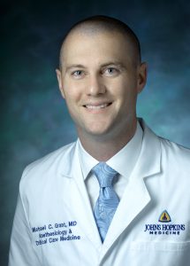 Michael Grant合作论文数Division of Cardiac Anesthesia and Surgical Intensive Care, Department of Anesthesiology and Critical Care Medicine, School of Medicine, Johns Hopkins University1
 Ledibabari M Ngaage合作论文数Division of Plastic & Reconstructive Surgery, University of Maryland School of Medicine1
 Farhan Merali合作论文数1
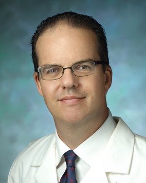 Charles G. Eberhart合作论文数Johns Hopkins University School of Medicine1Siegel R E合作论文数1
 Holly B Hindman合作论文数1Deitz Luke合作论文数1
 Aziz Ahmad Nazif合作论文数Faculty of Science and Technology, Universiti Malaysia Terengganu (UMT),1Sawyer R N合作论文数1Diaconu Silviu C合作论文数School of Medicine &, George Washington University;Health Sciences1




