We present our technique for isolated arthroscopic rotator interval closure in the treatment of mild symptomatic glenohumeral instability in the absence of a labral tear. A careful history, physical examination, and imaging help to identify a select subset of atraumatic instability patients who may benefit from this procedure, and diagnostic arthroscopy can provide further evidence. By use of a posterior viewing portal with anterior and anterolateral working portals, the anterior capsuloligamentous complex is mobilized, and the rotator interval is closed with carefully placed sutures to advance the capsule superiorly and provide tension through decreased capsular volume.
Vitamin D acts to maintain calcium and phosphate homeostasis within the body. It is now estimated that 1 billion people worldwide are vitamin D deficient. This problem is particularly important to athletes of all ages, as vitamin D plays a significant role in bone health, immune function, and physical performance. In the deficient state, the athlete may be at an increased risk for potential problems such as stress fractures, respiratory infections, and muscle injuries. The purpose of this article is to examine vitamin D deficiency and review its relationship to the athlete.
We describe 3 cases of posterior hip instability associated with femoroacetabular impingement. In each case, we obtained a detailed medical history, performed a physical examination, evaluated imaging, recorded intraoperative findings, and clinically followed the patient for 1 year. Two of the 3 patients sustained a traumatic posterior hip subluxation caused by noncontact injuries. All patients had decreased internal rotation on physical examination, radiographic evidence of acetabular retroversion, a cam lesion, an elevated α angle, and a posterior acetabular rim fracture with associated labral injury. All patients underwent hip arthroscopy and direct repair of the bony acetabular fragment using 3 to 5 suture anchors. One-year follow-up in all cases demonstrated good to excellent results and full return to activities without restriction. Patients with femoroacetabular impingement may be predisposed to traumatic posterior dislocation or subluxation and a concomitant posterior acetabular rim fracture with labral injury. We propose that FAI predisposed these athletes to posterior hip instability.
In competitive athletes, stress fractures of the tibia, foot, and ankle are common and lead to considerable delay in return to play. Factors such as bone vascularity, training regimen, and equipment can increase the risk of stress fracture. Management is based on the fracture site. In some athletes, metabolic workup and medication are warranted. High-risk fractures, including those of the anterior tibial diaphysis, navicular, proximal fifth metatarsal, and medial malleolus, present management challenges and may require surgery, especially in high-level athletes who need to return to play quickly. Noninvasive treatment modalities such as pulsed ultrasound and extracorporeal shock wave therapy may have some benefit but require additional research.
Background: Despite improvements in arthroscopic rotator cuff repair technique and technology, a significant rate of failed tendon healing persists. Improving the biology of rotator cuff repairs may be an important focus to decrease this failure rate. The objective of this study was to determine the mRNA biomarkers and histological characteristics of repaired rotator cuffs that healed or developed persistent defects as determined by postoperative ultrasound. Hypothesis: Increased synovial inflammation and tendon degeneration at the time of surgery are correlated with the failed healing of rotator cuff tendons. Study Design: Case-control study; Level of evidence, 3. Methods: Biopsy specimens from the subscapularis tendon, supraspinatus tendon, glenohumeral synovium, and subacromial bursa of 35 patients undergoing arthroscopic rotator cuff repair were taken at the time of surgery. Expression of proinflammatory cytokines, tissue remodeling genes, and angiogenesis factors was evaluated by quantitative real-time polymerase chain reaction. Histological characteristics of the affected tissue were also assessed. Postoperative (>6 months) ultrasound was used to evaluate the healing of the rotator cuff. General linear modeling with selected mRNA biomarkers was used to predict rotator cuff healing. Results: Thirty patients completed all analyses, of which 7 patients (23%) had failed healing of the rotator cuff. No differences in demographic data were found between the defect and healed groups. American Shoulder and Elbow Surgeons shoulder scores collected at baseline and follow-up showed improvement in both groups, but there was no significant difference between groups. Increased expression of matrix metalloproteinase 1 (MMP-1) and MMP-9 was found in the supraspinatus tendon in the defect group versus the healed group (P = .006 and .02, respectively). Similar upregulation of MMP-9 was also found in the subscapularis tendon of the defect group (P = .001), which was consistent with the loss of collagen organization as determined by histological examination. From a general linear model, the upregulation of MMP-1 and MMP-9 was highly correlated with failed healing of the rotator cuff (R2 = .656). Conclusion: The upregulation of tissue remodeling genes in the torn rotator cuff at the time of surgery provides a snapshot of the biological environment surrounding the torn rotator cuff that is closely related to the healing of repaired rotator cuffs.
The purpose of the present study is to describe the technique of margin convergence for U-shaped rotator cuff tears and report the clinical outcomes and ultrasonography with a minimum of 2 years follow-up. Three hundred eleven patients with a rotator cuff tear were prospectively enrolled in a registry at one institution. Inclusion criteria included any patient undergoing arthroscopic margin convergence for a rotator cuff tear. Exclusion criteria included open or mini-open rotator cuff repairs or suture anchor fixation to the cuff insertion without margin convergence. The outcome measurements included physical examination, manual muscle testing, the American Shoulder and Elbow Surgeons (ASES) score, and ultrasonography. Nineteen patients met the study criteria and 13 were available for 2-year follow-up (68.4%). The mean age of this cohort was 62.2 ± 7.5 years with a mean pre-operative rotator cuff tear size of 4.0 ± 1.6 cm. The ASES score increased significantly from 50.0 ± 17.7 before surgery to 83.3 ± 19.5 at 2 years ( P = 0.01). The active forward elevation also improved from 156.2 ± 11.9° before surgery to 168.0 ± 12.1 at 2 years ( P = 0.03). The active external rotation 54.4 ± 14.5 at baseline and improved to 57.1 ± 19.1 at 2 years ( P = 0.04). The strength also increased significantly from 6.7 ± 6.4 to 10.6 ± 4.9 lb at 1 year ( P = 0.048). The post-operative ultrasound demonstrated that 46.2% of rotator cuff tears were healed at 2 years. In conclusion, margin convergence is a useful technique for U-shaped tears that are difficult to mobilize.
Labral tears typically occur anterosuperiorly in association with femoroacetabular impingement or dysplasia. Less commonly, labral pathology may occur in an atypical direct anterior location adjacent to the iliopsoas tendon in the absence of bony abnormalities. We hypothesize that this pattern of injury is related to compression or traction on the anterior capsulo-labral complex by the iliopsoas tendon where it crosses the acetabular rim. In a retrospective review of prospectively collected data, we identified 25 patients that underwent isolated, primary, unilateral iliopsoas release and presented for at least 1 year follow-up (mean 21 months). Pre-operative demographics, clinical presentation, intra-operative findings, and outcome questionnaires were analyzed. The injury was treated with a tenotomy of the iliopsoas tendon at the level of the joint line and either labral debridement or repair. Mean post-operative outcome scores were 87.17, 92.46, and 78.8 for the modified Harris Hip Score, activities of daily living Hip Outcome Score, and sports-related score, respectively. The atypical labral injury identified in this study appears to represent a distinct pathological entity, psoas impingement, with an etiology which has not been previously described.
Background: The objective of this study was to determine whether the tear size of a supraspinatus tendon correlated with synovial inflammation and tendon degeneration in patients who underwent shoulder arthroscopy for rotator cuff repair. We hypothesized that increased synovial inflammation would correlate with greater tear size of the supraspinatus tendon at the time of surgery.Materials and methods: Tissue from the synovium, bursa, torn supraspinatus tendon, and subscapularis tendon was obtained from patients during shoulder arthroscopy to evaluate the messenger RNA expression of proinflammatory cytokines, tissue remodeling, and angiogenesis factors in the tendon, bursa, and synovium. Additional tissue was fixed to determine histologic changes including inflammation, vascular ingrowth, and collagen organization.Results: Increased expression of interleukin 1 beta, interleukin 6, cyclooxygenase 2, matrix metalloproteinase (MMP) 9, and vascular endothelial growth factor was found in the synovium of patients with full-thickness tears versus partial-thickness tears (P < .05). In the supraspinatus tendon, increased expression of MMP-1, MMP-9, MMP-13, and vascular endothelial growth factor was found in the full-thickness group. The upregulation of these genes in the full-thickness group was consistent with enhanced synovial inflammation, greater vascular ingrowth, and the loss of collagen organization in both supraspinatus and subscapularis tendons as determined by histology.Conclusion: Increased synovial inflammation and tissue degeneration correlate with the tear size of the supraspinatus tendon. A better understanding of the relationship between synovial inflammation and the progression of tendon degeneration can help in the design of novel and effective treatments to limit the advancement of rotator cuff disease and to improve their clinical outcomes.Level of evidence: Basic Science, Molecular and Cell Biology Study. (C) 2011 Journal of Shoulder and Elbow Surgery Board of Trustees.
Gulotta, Lawrence V MD; Voos, James E MD; Shindle, Michael K MD; Weiss, Leigh ATC, DPT; Barnes, Ronnie ATC; Rodeo, Scott A MD; Warren, Russell F MD Author Information
With increased use of magnetic resonance imaging (MRI) and arthroscopy, intra-articular ganglion cysts of the posterior cruciate ligament (PCL) have received more recognition as a possible cause of knee discomfort. Reported treatment options have ranged from ultrasound-guided cyst aspiration to arthroscopic cyst resection. In this report, we present the case of a patient who, on MRI, was diagnosed with a symptomatic intrasubstance PCL ganglion that later, during surgery, demonstrated mucinous degeneration of the entire ligament. Treatment was complete resection of the PCL. Five years after surgery, the patient demonstrated excellent, asymptomatic knee function. Although the best treatment for an intrasubstance PCL ganglion cyst that has caused degeneration of the entire ligament is unknown, PCL resection without reconstruction should be considered a viable option. It can result in a large increase in range of motion and function.
Within the discipline of sports medicine, articular cartilage injuries in the hip have received considerably less attention than other joints, largely due to the difficulty that practitioners have had with accurate assessment. Non-arthritic cartilage injuries in the hip refer to focal chondral defects on either the femoral or acetabular side of the joint. Focal chondral defects on the femoral side are relatively uncommon, however, and may result from axial loading or shear injury of the head within the socket. Subluxation events of the femoral head seen in high-energy contact sports may result in these types of focal chondral injuries. Cartilage injuries on the acetabular side are more common and typically present as localized cartilage delamination in the anterior-superior weight-bearing zone of the acetabular rim. The most common underlying condition resulting in these types of cartilage defects is femoroacetabular impingement. This chapter discusses current surgical indications and techniques appropriate for management of these injuries as well as clinical and radiographic methods to detect focal cartilage lesions in the hip joint.
PURPOSE: Vitamin D deficiency has become an epidemic for all age groups in the United States and Europe. Vitamin D has been recently discovered to have numerous effects throughout many tissues, including skeletal muscle. We hypothesize that the prevalence of Vitamin D insufficiency is high in an American professional football team. METHODS:Eighty-nine professional football players from a single NFL team underwent laboratory testing of Vitamin D levels in the spring of 2010 as part of their routine pre-season evaluation. The mean age of the players was 25.0 years old (s.d. 2.6, range 21-32). Data was obtained from the team to determine the number of players with time lost due to muscle injuries. RESULTS:Forty-five (50.6%) players had levels consistent with vitamin D insufficiency (20-31.9 ng/mL). Seventeen players (19.1%) had values within normal limits (>32 ng/mL). There were 31 white players and 58 black players. The mean vitamin D level in white players was 30.3 (range 17-46 ng/ml) and 20.4 in black players (range 8-40 ng/mL) (p < 0.001). Seventy-three players had no muscle injury with a mean vitamin D level of 24.7 (range 9-46 ng/mL) and 16 players suffered a muscle injury with a mean vitamin D level of 19.9 (8-33 ng/mL) (p < 0.04).Twenty-seven (30.3%) players had deficient total vitamin D 25(OH) levels (<20 ng/mL). CONCLUSIONS: There is a high prevalence of vitamin D insufficiency (80%) identified in a professional American football team. Black players and players who suffered muscle injuries had significantly lower mean vitamin D levels. Due to these findings, we recommend screening and treatment of vitamin D insufficiency in professional athletics.
The sternoclavicular joint (SCJ) is the only true articulation between the axial and appendicular skeleton. It is a saddle-type, diarthrodial joint where less than half of the clavicle articulates with the sternum 26 Rockwood Jr., C.A. Wirth M. Disorders of the sternoclavicular joint. in: Rockwood Jr., C.A. Matsen F. The shoulder. Saunders, Philadelphia1998: 555-610 Google Scholar ; sprains, fractures, or dislocations, especially from indirect forces, are often attributed to this relatively bony incongruity. 20 Medvecky M.J. Zuckerman J.D. Sternoclavicular joint injuries and disorders. Instr Course Lect. 2000; 49: 397-406 PubMed Google Scholar Its location close to the center of the body and its strong ligamentous restraints, however, limit the frequency of injury to this joint. 9 Cave E.F. Shoulder girdle injuries. in: Cave E.F. Fractures and other injuries. Yearbook Medical Publishers, Chicago1958: 258-259 Google Scholar , 25 Rockwood Jr., C.A. Williams G.R. Young D.C. Disorders of the acromioclavicular joint. in: Rockwood Jr., C.A. Matsen F. The shoulder. Saunders, Philadelphia1998: 483-553 Google Scholar Biomechanically, the SCJ is characterized by approximately 30° to 35° of forward elevation and 35° of flexion/extension during normal shoulder range of motion (ROM), with 4° of sternoclavicular motion for every 10° of shoulder elevation. 25 Rockwood Jr., C.A. Williams G.R. Young D.C. Disorders of the acromioclavicular joint. in: Rockwood Jr., C.A. Matsen F. The shoulder. Saunders, Philadelphia1998: 483-553 Google Scholar
Several all-arthroscopic techniques have been described to address patellar instability. Most arthroscopic procedures focus on soft tissue plication or "tightening" of the medial retinacular structures to correct lateral patellar instability. We found these techniques to be ineffective when the medial stabilizers were avulsed from the patella. As a result, we have developed an arthroscopic technique to repair medial patellofemoral ligament avulsions to the patella using suture anchors.
Purpose of reviewIce hockey is a fast-paced sport played by an increasing number of children and teens across the nation.Recent findingsThe risk of injury in youth hockey is high due to contact from body checking. Youth hockey programs need to educate players, coaches, and parents about the importance of knowing and following the rules as well as the dangers of body checking another player from behind.SummaryIn this article, we will present an overview of the types and rates of injuries that occur in ice hockey and then present a detailed review of hip/groin injuries that are commonly diagnosed in these athletes.
With the improvements in flexible instrumentation, hip arthroscopy is being increasingly used to treat a variety of hip pathology, including labral tears. However, up to this point, there has not been a case report of an anterior and a posterior labral tear successfully repaired arthroscopically. We present a case report of a 27-year-old male firefighter who presented to our institution with an anterior and posterior labral tear, as well as a cam lesion and loose body, following a traumatic hip dislocation. The purpose of this case report is to illustrate that both anterior and posterior labral tears can be repaired using hip arthroscopy. Anterior and posterior labral tears can be caused by a traumatic hip dislocation, and both can be successfully repaired using arthroscopic techniques.
The increased risk of symptomatic progression towards osteoarthritis after chondral damage has led to the development of multiple treatment options for cartilage repair. These procedures have evolved from arthroscopic lavage and debridement, to marrow stimulation techniques, and more recently, to osteochondral autograft and allograft transplants, and autogenous chondrocyte implantation. The success of mosaicplasty procedures in the knee has led to its application to other surfaces, including the talus, tibial plateau, patella, and humeral capitellum. In this report, we present two cases of a chondral defect to the femoral head after a traumatic hip dislocation, treated with an osteochondral autograft (OATS) from the ipsilateral knee, and the inferior femoral head, respectively, combined with a surgical dislocation of the hip. At greater than 1 year and greater than 5 years of follow-up, MRI studies have demonstrated good autograft incorporation with maintenance of articular surface conformity, and both patients clinically continue to have no pain and full active range of motion of their respective hips. In our opinion, treatment of osteochondral defects in the femoral head surface using a surgical dislocation combined with an OATS procedure is a promising approach, as full exposure of the femoral head can be obtained while preserving its vasculature, thus enabling adequate restoration of the articular cartilage surface.
Background: Tears of the gluteus medius tendon at the greater trochanter have been termed "rotator cuff tears of the hip." Previous reports have described the open repair of these lesions.Hypothesis: Endoscopic repair of gluteus medius tears results in successful clinical outcomes in the short term.Study Design: Case series; Level of evidence, 4.Methods: Of 482 consecutive hip arthroscopies performed by the senior author, 10 patients with gluteus medius tears repaired endoscopically were evaluated prospectively. Perioperative data were analyzed on this cohort of patients. There were 8 women and 2 men, with an average age of 50.4 years (range, 33-66 years). Patients had persistent lateral hip pain and abductor weakness despite extensive conservative measures. Diagnosis was made by physical examination and magnetic resonance imaging and was confirmed at the time of endoscopy in all cases. At the most recent follow-up, patients completed the Modified Harris Hip Score and Hip Outcomes Score surveys.Results: At an average follow-up of 25 months (range, 19-38 months), all 10 patients had complete resolution of pain; 10 of 10 regained 5 of 5 motor strength in the hip abductors. Modified Harris Hip Scores at 1 year averaged 94 points (range, 84-100), and Hip Outcomes Scores averaged 93 points (range, 85-100). There were no adverse complications after abductor repairs. Seven of 10 patients said their hip was normal, and 3 said their hip was nearly normal.Conclusion: With short-term follow-up, endoscopic repair of gluteus medius tendon tears of the hip appears to provide pain relief and return of strength in select patients who have failed conservative measures. Further long-term follow-up is warranted to confirm the clinical effectiveness of this procedure.
 Bryan T. Kelly合作论文数18
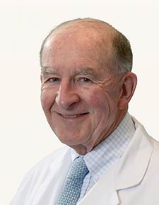 Russell F Warren合作论文数Department of Orthopaedic Surgery, Weill Cornell Medical College;Hospital for Special Surgery18James E. Voos合作论文数11
 Shane J. Nho合作论文数Schulthess Clinic Hip Service Zurich9
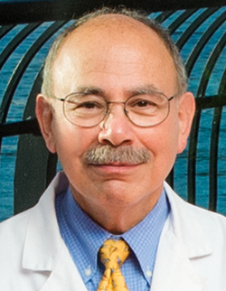 Joseph M. Lane合作论文数Metabolic Bone Disease Service, Hospital for Special Surgery8
 John D Macgillivray合作论文数7
 A. Jay Khanna合作论文数Johns Hopkins Orthopaedic Surgery, Good Samaritan Hospital7
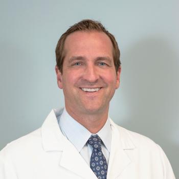 Michael J. Gardner合作论文数Department of Orthopaedic Surgery, School of Medicine, Stanford University6Paul D Sponseller合作论文数5
 Scott A. Rodeo合作论文数Department of Orthopaedic Surgery, Hospital for Special Surgery;Weill Cornell Medical College5Tony Wanich合作论文数4
 Robert Buly合作论文数Weill Medical College, Cornell University4Benton E. Heyworth合作论文数4Matthew H Griffith合作论文数3
 Benjamin G Domb合作论文数3Wasserman Bruce A合作论文数3Yadav Hemang合作论文数Mayo School of Graduate Medical Education, Mayo Clinic3Marx Robert G合作论文数3
 Andrew J Weiland合作论文数3Dean G. Lorich合作论文数3Denis Nam合作论文数3
 Michael B Cross合作论文数3
 Frank A. Cordasco合作论文数Sports Medicine and Shoulder Service, Hospital for Special Surgery, 535 East 70th Street, New York, NY 10021, USA3Douglas N. Mintz合作论文数3Lisa Shindle合作论文数3Jonas R. Rudzki合作论文数2Luis E. Moya合作论文数2Folorunsho Edobor-Osula合作论文数2David M. Green合作论文数2Leigh Weiss合作论文数2
 Ronald S. Adler合作论文数Division of Musculoskeletal Imaging, Department of Radiology, New York University2
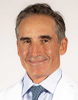 David W. Altchek合作论文数Hospital for Special Surgery;Weill Cornell Medical College;NewYork-Presbyterian Hospital2
 Joshua S Dines合作论文数2Barnes Ronnie P合作论文数2Demetrakopoulos Demetris合作论文数2M. J. Gardner合作论文数2Potter H G合作论文数2Christopher Good合作论文数2Scott W. Wolfe合作论文数2Christopher C. Dodson合作论文数2K.J. Burton合作论文数2Lawrence V. Gulotta合作论文数Sports Medicine/Shoulder Service, Hospital for Special Surgery2
 Stephen Lyman合作论文数Healthcare Research Institute, Hospital for Special Surgery;Kyushu University;Department of Health Policy and Research, Weill Cornell Medical College2Peter D. Asnis合作论文数2Jason Koob合作论文数2
 David L Helfet合作论文数2
 Riley J. Williams合作论文数Department of Orthopaedic Surgery, Hospital for Special Surgery;Weill Cornell Medical College;Institute for Cartilage Repair, Hospital for Special Surgery2Elias C. Papadopoulos合作论文数2




