Purpose:Many children with severe genetic disorders remain undiagnosed despite advanced genomic technologies. Early diagnosis is vital for prognosis, genetic counseling, and targeted treatment development. This study aims to increase diagnostic rates in complex pediatric cases and foster research into disease mechanisms. Methods:Launched in 2016, the Telethon Undiagnosed Diseases Program provides a structured, multicenter approach to rare disease diagnosis. Standardized case submission criteria ensured consistent clinical data collection. Children with severe, multisystemic disorders and prior negative genetic tests were eligible. After case approval, trio-based exome sequencing was performed, with regular reanalysis for unsolved cases until December 2024. Results:Between June 2016 and December 2023, 1338 cases were submitted by 60 clinicians from 22 Italian centers; 1019 were accepted. A definitive genetic diagnosis was achieved in 49% of cases, implicating 330 genes. Most pathogenic variants (70.2%) were de novo, reflecting demographic trends, such as delayed parenthood. The remainder included autosomal recessive or X-linked variants, with homozygosity observed in 9% of patients. Conclusion:The Telethon Undiagnosed Diseases Program significantly shortened the average diagnostic odyssey of ∼8 years. Children born after 2016 benefited from faster diagnoses. This initiative offers a scalable, cost-effective model for improving diagnosis, guiding treatment, and supporting therapeutic innovation in rare pediatric diseases.
Background:Loeys-Dietz syndrome (LDS) is a connective tissue disorder that is characterized by vascular and skeletal abnormalities. LDS, classified into six subtypes (LDS1-6), is caused by mutations in the genes encoding proteins involved in the transforming growth factor-beta (TGF-β) signaling pathway. LDS4 is the mild form of the LDS spectrum, considering that aneurysms are observed in the fourth decade and the disease progression is slower than the other forms. Case Presentation:We reported the case of a novel TGFB2 variant c.280_298del p.(Ser94Profs*7), NM_001135599.2 responsible of LDS4 in a family with sudden death and vascular lesions. Conclusion:The pedigree analysis highlighted the high clinical variability of LDS signs even within the same family. In addition, TGFB2 mutations which do not always lead to vascular phenotype and skeletal signs can be prevalent. We emphasize the relevance of genetic counseling and molecular analysis to identify genetic diseases subtending sudden deaths and to distinguishing heritable connective tissue disorders with phenotypic overlap. Genetic testing can be used to guide clinical management and provide valuable prognostic information.
Chromosomal rearrangements can interfere with the disjunction and segregation of other chromosome pairs not involved in the rearrangements, promoting the occurrence of numerical abnormalities in resulting gametes and predisposition to trisomy in offspring. This phenomenon of interference is known as the interchromosomal effect (ICE). Here we report a prenatal case potentially generated by ICE. The first-trimester screening ultrasound of the pregnant woman was normal, but the NIPT indicated a high risk for three copies of chromosome 21, thus suspecting trisomy 21 (T21). After a comprehensive clinical evaluation and genetic counseling, the couple decided to undergo amniocentesis. The prenatal karyotype confirmed T21 but also showed a balanced translocation between the long arm of chromosome 15 (q22) and the long arm of chromosome 22. The parents’ karyotypes also showed that the mother had the 15;22 translocation. We reviewed T21 screening methods, and we performed a literature review on ICE, a generally overlooked phenomenon. We observed that ours is the first report of a prenatal case potentially due to ICE derived from the mother. The recurrence risk of aneuploidy in the offspring of translocated individuals is likely slightly increased, but it is not possible to estimate to what extent. In addition to supporting observations, there are still open questions such as, how frequent is ICE? How much is the aneuploidy risk altered by ICE?
This comprehensive review provides insights and suggested strategies for the analysis of germline variants using second- and third-generation sequencing technologies (SGS and TGS). It addresses the critical stages of data processing, starting from alignment and preprocessing to quality control, variant calling, and the removal of artifacts. The document emphasized the importance of meticulous data handling, highlighting advanced methodologies for annotating variants and identifying structural variations and methylated DNA sites. Special attention is given to the inspection of problematic variants, a step that is crucial for ensuring the accuracy of the analysis, particularly in clinical settings where genetic diagnostics can inform patient care. Additionally, the document covers the use of various bioinformatics tools and software that enhance the precision and reliability of these analyses. It outlines best practices for the annotation of variants, including considerations for problematic genetic alterations such as those in the human leukocyte antigen region, runs of homozygosity, and mitochondrial DNA alterations. The document also explores the complexities associated with identifying structural variants and copy number variations, underscoring the challenges posed by these large-scale genomic alterations. The objective is to offer a comprehensive framework for researchers and clinicians, ensuring that genetic analyses conducted with SGS and TGS are both accurate and reproducible. By following these best practices, the document aims to increase the diagnostic accuracy for hereditary diseases, facilitating early diagnosis, prevention, and personalized treatment strategies. This review serves as a valuable resource for both novices and experts in the field, providing insights into the latest advancements and methodologies in genetic analysis. It also aims to encourage the adoption of these practices in diverse research and clinical contexts, promoting consistency and reliability across studies.
CELSR3 codes for a planar cell polarity protein. We describe twelve affected individuals from eleven independent families with bi-allelic variants in CELSR3. Affected individuals presented with an overlapping phenotypic spectrum comprising central nervous system (CNS) anomalies (7/12), combined CNS anomalies and congenital anomalies of the kidneys and urinary tract (CAKUT) (3/12) and CAKUT only (2/12). Computational simulation of the 3D protein structure suggests the position of the identified variants to be implicated in penetrance and phenotype expression. CELSR3 immunolocalization in human embryonic urinary tract and transient suppression and rescue experiments of Celsr3 in fluorescent zebrafish reporter lines further support an embryonic role of CELSR3 in CNS and urinary tract formation.
BACKGROUND:Inherited retinal diseases (IRD) are genetically heterogeneous disorders that cause the dysfunction or loss of photoreceptor cells and ultimately lead to blindness. To date, next-generation sequencing procedures fail to detect pathogenic sequence variants in coding regions of known IRD disease genes in about 30-40% of patients. One of the possible explanations for this missing heritability is the presence of yet unidentified transcripts of known IRD genes. Here, we aimed to define the transcript composition of IRD genes in the human retina by a meta-analysis of publicly available RNA-seq datasets using an ad-hoc designed pipeline.RESULTS:We analysed 218 IRD genes and identified 5,054 transcripts, 3,367 of which were not previously reported. We assessed their putative expression levels and focused our attention on 435 transcripts predicted to account for at least 5% of the expression of the corresponding gene. We looked at the possible impact of the newly identified transcripts at the protein level and experimentally validated a subset of them.CONCLUSIONS:This study provides an unprecedented, detailed overview of the complexity of the human retinal transcriptome that can be instrumental in contributing to the resolution of some cases of missing heritability in IRD patients.
OBJECTIVE:Epilepsy with eyelid myoclonia (EEM) spectrum is a generalized form of epilepsy characterized by eyelid myoclonia with or without absences, eye closure-induced seizures with electroencephalographic paroxysms, and photosensitivity. Based on the specific clinical features, age at onset, and familial occurrence, a genetic cause has been postulated. Pathogenic variants in CHD2, SYNGAP1, NEXMIF, RORB, and GABRA1 have been reported in individuals with photosensitivity and eyelid myoclonia, but whether other genes are also involved, or a single gene is uniquely linked with EEM, or its subtypes, is not yet known. We aimed to dissect the genetic etiology of EEM.METHODS:We studied a cohort of 105 individuals by using whole exome sequencing. Individuals were divided into two groups: EEM- (isolated EEM) and EEM+ (EEM accompanied by intellectual disability [ID] or any other neurodevelopmental/psychiatric disorder).RESULTS:We identified nine variants classified as pathogenic/likely pathogenic in the entire cohort (8.57%); among these, eight (five in CHD2, one in NEXMIF, one in SYNGAP1, and one in TRIM8) were found in the EEM+ subcohort (28.57%). Only one variant (IFIH1) was found in the EEM- subcohort (1.29%); however, because the phenotype of the proband did not fit with published data, additional evidence is needed before considering IFIH1 variants and EEM- an established association. Burden analysis did not identify any single burdened gene or gene set.SIGNIFICANCE:Our results suggest that for EEM, as for many other epilepsies, the identification of a genetic cause is more likely with comorbid ID and/or other neurodevelopmental disorders. Pathogenic variants were mostly found in CHD2, and the association of CHD2 with EEM+ can now be considered a reasonable gene-disease association. We provide further evidence to strengthen the association of EEM+ with NEXMIF and SYNGAP1. Possible new associations between EEM+ and TRIM8, and EEM- and IFIH1, are also reported. Although we provide robust evidence for gene variants associated with EEM+, the core genetic etiology of EEM- remains to be elucidated.
Collectively, rare genetic disorders affect a substantial portion of the world's population. In most cases, those affected face difficulties in receiving a clinical diagnosis and genetic characterization. The understanding of the molecular mechanisms of these diseases and the development of therapeutic treatments for patients are also challenging. However, the application of recent advancements in genome sequencing/analysis technologies and computer-aided tools for predicting phenotype-genotype associations can bring significant benefits to this field. In this review, we highlight the most relevant online resources and computational tools for genome interpretation that can enhance the diagnosis, clinical management, and development of treatments for rare disorders. Our focus is on resources for interpreting single nucleotide variants. Additionally, we present use cases for interpreting genetic variants in clinical settings and review the limitations of these results and prediction tools. Finally, we have compiled a curated set of core resources and tools for analyzing rare disease genomes. Such resources and tools can be utilized to develop standardized protocols that will enhance the accuracy and effectiveness of rare disease diagnosis.
The ubiquitin-specific protease USP9X has been found to play a role in multiple aspects of neural development including processes of neuronal migrations. In males, hemizygous partial loss of function variants in USP9X lead to a clinical phenotype primarily characterized by intellectual disability, hypotonia, speech and language impairment, behavioral disturbances accompanied by additional clinical features with variable expressivity. Structural brain abnormalities are reported in all cases where neuro-imaging was performed. The most common radiological features described include hypoplasia/agenesis of the corpus callosum, widened ventricles, white matter disturbances, and cerebellar hypoplasia. Here we report a child harboring a missense variant in USP9X presenting with the classical neurodevelopmental phenotype and a previously unreported radiological picture of periventricular heterotopia. This case expands the phenotypic landscape of this emergent condition and supports the critical role of USP9X in neuronal migration processes.
Eukaryotic initiation factor-4A2 (EIF4A2) is an ATP-dependent RNA helicase and a member of the DEAD-box protein family that recognizes the 5' cap structure of mRNAs, allows mRNA to bind to the ribosome, and plays an important role in microRNA-regulated gene repression. Here, we report on 15 individuals from 14 families presenting with global developmental delay, intellectual disability, hypotonia, epilepsy, and structural brain anomalies, all of whom have extremely rare de novo mono-allelic or inherited bi-allelic variants in EIF4A2. Neurodegeneration was predominantly reported in individuals with bi-allelic variants. Molecular modeling predicts these variants would perturb structural interactions in key protein domains. To determine the pathogenicity of the EIF4A2 variants in vivo, we examined the mono-allelic variants in Drosophila melanogaster (fruit fly) and identified variant-specific behavioral and developmental defects. The fruit fly homolog of EIF4A2 is eIF4A, a negative regulator of decapentaplegic (dpp) signaling that regulates embryo patterning, eye and wing morphogenesis, and stem cell identity determination. Our loss-of-function (LOF) rescue assay demonstrated a pupal lethality phenotype induced by loss of eIF4A, which was fully rescued with human EIF4A2 wild-type (WT) cDNA expression. In comparison, the EIF4A2 variant cDNAs failed or incompletely rescued the lethality. Overall, our findings reveal that EIF4A2 variants cause a genetic neurodevelopmental syndrome with both LOF and gain of function as underlying mechanisms.
ABSTRACTBACKGROUNDConversion of adenosine in RNA to inosine by ADAR enzymes, termed ‘RNA editing’, occurs at thousands of sites across the transcriptome, and is required for healthy development of the central nervous system. RNA editing can modify protein sequences, and dampen the innate immune response. RNA editing is tissue-specific and partly genetically determined. Modifications of RNA editing sites contribute to multiple diseases, particularly neurodevelopmental and neuropsychiatric diseases. Despite the importance of RNA editing in the brain, nothing is known about this process in the human retina. We describe the landscape of retinal editing revealing its importance in key biological processes that underpin vision.METHODS & RESULTSWe analysed the transcriptomes of >500 donor retinae and identified ∼153,000 high-confidence RNA editing sites. Some 80% of editing sites occurred within protein-coding RNA, with the majority in intronicAlurepeats, and 3’ UTR sequence. Novel retina-specific sites were concentrated in genes related to photoreceptor function and which cause retinitis pigmentosa, most notably in PDE6A. Exonic, protein recoding sites were enriched in zinc-finger domains. AMD subjects exhibit relatively few differences in RNA editing compared to controls, consistent with limited gene expression differences. We identified ∼10,000 editing QTLs. The genetic architecture of editing in the retina resembles the brain, whereas editing and expression QTLs in the retina show modest genetic overlap. We report colocalization between edQTLs and retinal disease GWAS peaks for age-related macular degeneration, glaucoma and macular telangiectasia. These findings provide new insights into epi-transcriptomic regulation of genes critical for vision, and elaborate putative genetic disease driver mechanisms that appear to be independent of changes in gene expression.
SRSF1 (also known as ASF/SF2) is a non-small nuclear ribonucleoprotein (non-snRNP) that belongs to the arginine/serine (R/S) domain family. It recognizes and binds to mRNA, regulating both constitutive and alternative splicing. The complete loss of this proto-oncogene in mice is embryonically lethal. Through international data sharing, we identified 17 individuals (10 females and 7 males) with a neuro-developmental disorder (NDD) with heterozygous germline SRSF1 variants, mostly de novo, including three frameshift variants, three nonsense variants, seven missense variants, and two microdeletions within region 17q22 encompassing SRSF1. Only in one family, the de novo origin could not be established. All individuals featured a recurrent phenotype including developmental delay and intellec-tual disability (DD/ID), hypotonia, neurobehavioral problems, with variable skeletal (66.7%) and cardiac (46%) anomalies. To investi-gate the functional consequences of SRSF1 variants, we performed in silico structural modeling, developed an in vivo splicing assay in Drosophila, and carried out episignature analysis in blood-derived DNA from affected individuals. We found that all loss-of-function and 5 out of 7 missense variants were pathogenic, leading to a loss of SRSF1 splicing activity in Drosophila, correlating with a detectable and specific DNA methylation episignature. In addition, our orthogonal in silico, in vivo, and epigenetics analyses enabled the separation of clearly pathogenic missense variants from those with uncertain significance. Overall, these results indicated that haploinsufficiency of SRSF1 is responsible for a syndromic NDD with ID due to a partial loss of SRSF1-mediated splicing activity.
Variants in RAC3, encoding a small GTPase RAC3 which is critical for the regulation of actin cytoskeleton and intracellular signal transduction, are associated with a rare neurodevelopmental disorder with structural brain anomalies and facial dysmorphism. We investigated a cohort of 10 unrelated participants presenting with global psychomotor delay, hypotonia, behavioural disturbances, stereotyped movements, dysmorphic features, seizures and musculoskeletal abnormalities. MRI of brain revealed a complex pattern of variable brain malformations, including callosal abnormalities, white matter thinning, grey matter heterotopia, polymicrogyria/dysgyria, brainstem anomalies and cerebellar dysplasia. These patients harboured eight distinct de novo RAC3 variants, including six novel variants (NM_005052.3): c.34G > C p.G12R, c.179G > A p.G60D, c.186_188delGGA p.E62del, c.187G > A p.D63N, c.191A > G p.Y64C and c.348G > C p.K116N. We then examined the pathophysiological significance of these novel and previously reported pathogenic variants p.P29L, p.P34R, p.A59G, p.Q61L and p.E62K. In vitro analyses revealed that all tested RAC3 variants were biochemically and biologically active to variable extent, and exhibited a spectrum of different affinities to downstream effectors including p21-activated kinase 1. We then focused on the four variants p.Q61L, p.E62del, p.D63N and p.Y64C in the Switch II region, which is essential for the biochemical activity of small GTPases and also a variation hot spot common to other Rho family genes, RAC1 and CDC42. Acute expression of the four variants in embryonic mouse brain using in utero electroporation caused defects in cortical neuron morphology and migration ending up with cluster formation during corticogenesis. Notably, defective migration by p.E62del, p.D63N and p.Y64C were rescued by a dominant negative version of p21-activated kinase 1. Our results indicate that RAC3 variants result in morphological and functional defects in cortical neurons during brain development through variant-specific mechanisms, eventually leading to heterogeneous neurodevelopmental phenotypes.
 Vincenzo Nigro合作论文数Istituto di Patologia Generale ed Oncologia, Seconda Università degli Studi di Napoli33
 Nicola Brunetti-Pierri合作论文数Department of Translational Medicine, University of Naples "Federico II';Telethon Institute of Genetics and Medicine30
 Gerarda Cappuccio合作论文数Department of Paediatrics, University of Naples Federico II26
 Torella Annalaura合作论文数TIGEM (Telethon Institute of Genetics and Medicine), Napoli, Italy.26
 Sergio Cocozza合作论文数Department of Molecular Medicine and Medical Biotechnology, University of Naples Federico II25
 Antonella Monticelli合作论文数Department of Molecular and Cellular Pathology and IEOS, University of Naples “Federico II18Sandro Banfi合作论文数Telethon Institute of Genetics and Medicine (TIGEM), Via Olgettina 58, 20132, Milan, Italy15Olga Vaccaro合作论文数11
 Gabriele Riccardi合作论文数Università degli Studi di Napoli Federico II;Istituto Di Scienza Dell'alimentazione, CNR di Avellino10
 Margherita Mutarelli合作论文数Department of General Pathology, Second University of Naples10
 Giorgio Casari合作论文数Telethon Institute of Genetics and Medicine (TIGEM), San Raffaele Biomedical Science Park, Milan, Italy9Fabio Acquaviva合作论文数Department of Cellular and Molecular Biology, University of Naples “Federico II”9
 Francesco Musacchia合作论文数8
 Imma Castaldo合作论文数Department of Cellular and Molecular Biology, University of Naples “Federico II”8
 Valeria Capra合作论文数U.O. Neurochirurgia, Istituto G. Gaslini8
 Marcello Scala合作论文数Dept Neurosci Rehabil Ophthalmol Genet Maternal &, Univ Genoa7Stefano D'Arrigo合作论文数7
 Andrea Accogli合作论文数7Emanuela Lapice合作论文数7
 Marianna Alagia合作论文数6
 Pasquale Striano合作论文数Center of Experimental and Translational Myology, Dipartimento Di Neuroscienze, Riabilitazione, Oftalmologia, Genetica E Scienze Materno-Infantili, Università Degli Studi Di Genova;IRCCS 'G. Gaslini'6Manuela Giacchetti合作论文数6
 Christian Gilissen合作论文数Department of Human Genetics, UMC St. Radboud Nijmegen6Silvia Maitz合作论文数6
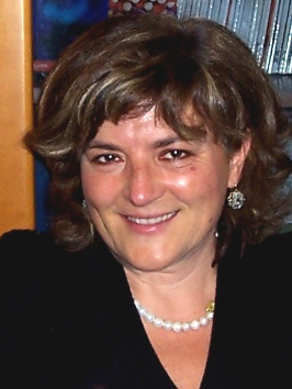 Marcella Zollino合作论文数Dipartimento di Scienze della vita e sanità pubblica, Facolta' di Medicina e Chirurgia, Università Cattolica del Sacro Cuore6E. Lapice合作论文数5Raffaele Castello合作论文数5Claudia Ciaccio合作论文数5
 Manuela Morleo合作论文数5Angela A Rivellese合作论文数5Chiara Pantaleoni合作论文数5Diego Di Bernardo合作论文数Group Leader5
 Mariasavina Severino合作论文数4Amber Begtrup合作论文数Genedx, Inc.4
 Pia Bernardo合作论文数4Del Giudice Ennio合作论文数4
 Pippucci Tommaso合作论文数Policlin S Orsola Malpighi, Univ Bologna4Ciolfi Andrea合作论文数Dipartimento di Ematologia, Oncologia e Medicina Molecolare, Istituto Superiore di Sanità4
 Diego Carrella合作论文数4
 Joris Veltman合作论文数Biosciences Institute, Faculty of Medical Sciences, Newcastle University4
 Gennaro Miele合作论文数Universita " Federico II ";Dipartimento di Scienze Fisiche;Sezione di Napoli;Sezione di Napoli, Universita " Federico II "4
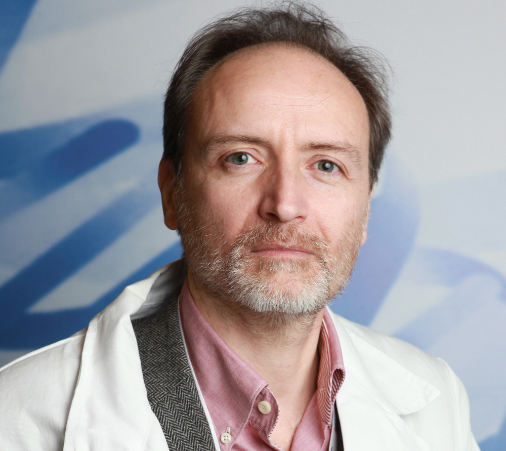 Marco Tartaglia合作论文数Ospedale Pediatrico Bambino Gesù4Angela Giacco合作论文数4Giovanna Donnarumma合作论文数4
 Pietro Chiurazzi合作论文数Catholic University of Rome4Marianthi Karali合作论文数Laboratoire de Biologie Moleculaire de la Cellule, Ecole Normale Supérieure de Lyon4
 L.E.L.M. Vissers (Lisenka)合作论文数Radboudumc;Radboud University4
 A. Hoischen (Alexander)合作论文数Radboud Universiteit;Radboudumc4




