Recent stroke studies have shown that the ipsi-lesional thalamus longitudinally and significantly decreases after stroke in the acute and subacute stages. However, additional considerations in the chronic stages of stroke require exploration including time since stroke, gender, intracortical volume, aging, and lesion volume to better characterize thalamic differences after cortical infarct. This cross-sectional retrospective study quantified the ipsilesional and contralesional thalamus volume from 69 chronic stroke subjects’ anatomical MRI data (age 35–92) and related the thalamus volume to time since stroke, gender, intracortical volume, age, and lesion volume. The ipsi-lesional thalamus volume was significantly smaller than the contra-lesional thalamus volume (t(68) = 13.89, p < 0.0001). In the ipsilesional thalamus, significant effect for intracortical volume (t(68) = 2.76, p = 0.008), age (t(68) = 2.47, p = 0.02), lesion volume (t(68) = − 3.54, p = 0.0008), and age*time since stroke (t(68) = 2.46, p = 0.02) were identified. In the contralesional thalamus, significant effect for intracortical volume (t(68) = 3.2, p = 0.002) and age (t = − 3.17, p = 0.002) were identified. Clinical factors age and intracortical volume influence both ipsi- and contralesional thalamus volume and lesion volume influences the ipsilesional thalamus. Due to the cross-sectional nature of this study, additional research is warranted to understand differences in the neural circuitry and subsequent influence on volumetrics after stroke.
PURPOSERecent investigations into effects of intensity or distribution of aphasia therapy have provided moderate evidence supporting intensive therapy schedules on aphasia treatment response. The purpose of the present study was to investigate the feasibility of creating an intensive therapy session without extending the amount of daily time a person spends in treatment.METHODIndividuals who presented with chronic anomia poststroke (N = 8) participated in 2 weeks of a computerized, therapist-delivered, cued, picture-naming treatment. Dosing parameters for each session were 8 presentations of 50 pictures, totaling 400 teaching episodes per session.RESULTSOf the 8 participants, 6 achieved significant increases from baseline on trained items after 400 teaching episodes (i.e., 1 treatment hr), and the remaining 2 participants achieved significant increases from baseline after 1200 teaching episodes (i.e., 3 treatment hr). Maintenance data from 7 of the participants indicated that 6 participants maintained significant improvement from baseline on trained items.CONCLUSIONSGiven an intensive and saturated context, anomic individuals were surprisingly quick at relearning to produce problematic words successfully. Most participants demonstrated retention of the gains 2 months after treatment ended. The high density of teaching episodes within the treatment session (i.e., the intensive treatment schedule) may have contributed to the behavioral gains.
Purpose In this study, the authors assessed how the addition of intentional left-hand gestures to an intensive treatment for anomia affects 2 types of discourse: picture description and responses to open-ended questions. Method Fourteen people with aphasia completed treatment for anomia comprising 30 treatment sessions over 3 weeks. Seven subjects also incorporated intentional left-hand gestures into each treatment trial. Results Both groups demonstrated significant changes in trained items and improved naming of untrained items but no change in Western Aphasia Battery—Aphasia Quotient (WAB–AQ; Kertesz, 1982) scores. Changes in discourse were limited to the 3-month follow-up assessment. Several discourse measures showed significant improvements in the picture description task and declines during question responses. Additionally, the gesture group produced more words at each assessment, whereas the no gesture group produced fewer words at each assessment. These patterns led to improvements in picture descriptions and minimal declines in question responses in the gesture group. In contrast, the no gesture group showed minimal improvements in picture descriptions and production declines in question responses relative to pretreatment levels. Conclusion The intensive treatment protocol is a successful method for improving picture naming even of untrained items. Further, the authors conclude that the intentional left-hand gesture contributed significantly to the generalization of treatment to discourse.
Background. An aphasia treatment was designed to shift laterality from the left to right lateral frontal lobe during word production by initiating word-finding trials with complex left-hand movements. Previous findings indicated successful relateralization. Objective. The current study was designed to ascertain whether the shift was attributable to the left-hand movement. Methods. Using stratified random sampling, 14 subjects were equally divided between Intention (IT) and Control (CT) treatments. CT was identical to IT, except with no left-hand movements. Both treatments trained picture naming (phases 1 and 2) and category-member generation (phase 3), each phase lasting 10 sessions. Functional magnetic resonance imaging of category member generation occurred at pretreatment, posttreatment, and 3-month follow-up. Results. IT shifted lateral frontal activity rightward compared with pretreatment both at posttreatment ( t = −2.602, df = 6, P < .05) and 3-month follow-up ( t = −2.332, df = 5, P < .05), but CT did not. IT and CT yielded similar changes for all picture-naming and category probes. However, IT patients showed gains for untrained category ( t = 3.33, df = 6, P < .01) and picture-naming probes ( t = 3.77, df = 5, P < .01), but CT patients did not. Conclusions. The rightward shift in lateral frontal activity for IT was because of the left-hand movements. IT evoked greater generalization than CT.
Previous functional imaging studies that compared activity patterns in older and younger adults during nonlinguistic tasks found evidence for 2 phenomena: older participants usually show more pronounced task-related positive activity in the brain hemisphere that is not dominant for the task and less pronounced negative task-related activity in temporo-parietal and midline brain regions. The combined effects of these phenomena and the impact on word retrieval, however, have not yet been assessed. We used functional magnetic resonance imaging to explore task-related positive (active task > baseline) and negative activity (baseline > active task) during semantic and phonemic verbal fluency tasks. Increased right frontal positive activity during the semantic task and reduced negative activity in the right hemisphere during both tasks was associated with reduced performance in older subjects. No substantial relationship between changes in positive and negative activity was observed in the older participants, pointing toward 2 partially independent but potentially co-occurring processes. Underlying causes of the observed functional network inefficiency during word retrieval in older adults need to be determined in the future.
Background: Language fluency is a common diagnostic marker for discriminating among aphasia subtypes and improving clinical inference about site of lesion. Nevertheless, fluency remains a subjective construct that is vulnerable to a number of potential sources of variability, both between and within raters. Moreover, this variability is compounded by distinct neurological aetiologies that shape the characteristics of a narrative speech sample. Previous research on fluency has focused on characteristics of a particular patient population. Less is known about the ways that raters spontaneously weigh different perceptual cues when listening to narrative speech samples derived from a heterogeneous sample of brain-damaged adults. Aim: We examined the weighted contribution of a series of perceptual predictors that influence listeners' judgements of language fluency among a diverse sample of speakers. Our goal was to sample a range of narrative speech representing most fluent (i.e., healthy controls) to potentially least nonfluent (i.e., left inferior frontal lobe stroke). Methods & Procedures: Three raters blind to patient diagnosis made forced choice judgements of fluency (i.e., fluent or nonfluent) for 61 pseudorandomly presented narrative speech samples elicited by the BDAE Cookie Theft picture. Samples were collected from a range of clinical populations, including patients with frontal and temporal lobe pathologies and non-brain-damaged speakers. We conducted a logistic regression analysis in which the dependent measure was the majority judgement of fluency for each speech sample (i.e., fluent or non-fluent). The statistical model contained five predictors: speech rate, syllable type token ratio, speech productivity, audible struggle, and filler ratio. Outcomes & Results: This statistical model fit the data well, discriminating group membership (i.e., fluent or nonfluent) with 95.1% accuracy. The best step of the regression model included the following predictors: speech rate, speech productivity, and audible struggle. Listeners were sensitive to different weightings of these predictors. Conclusions: A small combination of perceptual variables can strongly discriminate whether a listener will assign a judgement of fluent versus nonfluent. We discuss implications for these findings and identify areas of potential future research towards further specifying the construct of fluency among adults with acquired speech and language disorders.
Little is known regarding the differences in active cortical and subcortical systems during opposing movements of an agonist-antagonist muscle group. The objective of this study was to characterize the differences in cortical activation during active ankle dorsiflexion and plantarflexion using functional MRI (fMRI). Eight right-handed healthy adults performed auditorily cued right ankle dorsiflexions and plantarflexions during fMRI. Differences in activity patterns between dorsiflexion and plantarflexion during fMRI were assessed using between- and within-subject voxel-wise t-tests. Results indicated that ankle dorsiflexion recruited significantly more regions in left M1, the supplementary motor area (SMA) bilaterally, and right cerebellum. Both movements activated similar left hemisphere regions in the putamen and thalamus. Dorsiflexion activated additional areas in the right putamen. Results suggest that ankle dorsiflexion and plantarflexion may be controlled by both shared and independent neural circuitry. This has important implications for functional investigations of gait pathology and how rehabilitation may differentially affect each movement.
Event related fMRI was used to investigate age-related changes in BOLD activity during the execution of right hand finger movements in internally or externally guided tasks. All of the younger adults exhibited typical (positive) BOLD responses in supplementary motor areas (SMA) bilaterally, and in the left sensorimotor cortex. Negative BOLD responses were found, however, in the right sensorimotor cortex of the younger adults. In contrast, all but one of the older adults had positive BOLD responses in SMA and sensorimotor cortex of both hemispheres. Across both tasks, older adults showed increased activity (relative to younger adults) in right ventrolateral premotor and medial premotor areas, but more so during the internally guided task. Overall, these results suggest age-related changes in motor control. The younger adults' hemispheric asymmetry and the lack thereof in older adults suggest a fundamental change in interhemispheric communication as part of the normal aging process.
Five nonfluent aphasia patients participated in a picture-naming treatment that used an intention manipulation (opening a box and pressing a button on a device in the box with the left hand) to initiate naming trials and was designed to re-lateralize word production mechanisms from the left to the right frontal lobe. To test the underlying assumption regarding re-lateralization, patients participated in fMRI of category-member generation before and after treatment. Generally, the four patients who improved during treatment showed reduced frontal activity from pre- to post-treatment fMRI with increasing concentration of activity in the right posterior frontal lobe (motor/premotor cortex, pars opercularis), demonstrating a significant shift in lateraliity toward the right lateral frontal lobe, as predicted. Three of these four patients showed no left frontal activity by completion of treatment, indicating that right posterior lateral frontal activity supported category-member generation. Patients who improved in treatment showed no difference in lateralization of lateral frontal activity from normal controls pre-treatment, but post-treatment, their lateral frontal activity during category-member generation was significantly more right lateralized than that of controls. Patterns of activity pre- and post-treatment suggested increasing efficiency of cortical processing as a result of treatment in the four patients who improved. The one patient who did not improve during treatment showed a leftward shift in lateral frontal lateralization that was significantly different from the four patients who did improve. Neither medial frontal nor posterior perisylvian re-lateralization from immediately pre- to immediately post-treatment images was a necessary condition for significant treatment gains or shift in lateral frontal lateralization. Of the three patients who improved and in whom posterior perisylvian activity could be measured at post-treatment fMRI, all maintained equal or greater amounts of left-hemisphere perisylvian activity as compared to right. This finding is consistent with reviews suggesting both hemispheres are involved in recovery of language in aphasia patients.
AbstractRecent findings suggest that neural representations of semantic knowledge contain information about category, modality, and attributes. Although an object’s category is defined according to shared attributes that uniquely distinguish it from other category members, a clear dissociation between visual attribute and category representation has not yet been reported. We investigated the contribution of category (living and nonliving) and visual attribute (global form and local details) to semantic representation in the fusiform gyrus. During functional magnetic resonance imaging (fMRI), 40 adults named pictures of animals, tools, and vehicles. In a preliminary study, identification of objects in these categories was differentially dependent on globalversuslocal visual feature processing. fMRI findings indicate that activation in the lateral and medial regions of the fusiform gyrus distinguished stimuli according to category, that is, livingversusnonliving, respectively. In contrast, visual attributes of global form (animals) were associated with higher activity in the right fusiform gyrus, while local details (tools) were associated with higher activity in the left fusiform gyrus. When both global and local attributes were relevant to processing (vehicles), cortex in both left and right medial fusiform gyri was more active than for other categories. Taken together, results support distinctions in the role of visual attributes and category in semantic representation. (JINS, 2009,15, 169–181.)
Healthy older adults frequently report word-finding difficulties, yet the underlying cause of these problems is not well understood. This study examined whether age-related changes in word retrieval are related to changes in areas of the frontal lobes thought to subserve word retrieval or changes in areas of the inferior temporal lobes thought to be involved in semantic knowledge. Twenty younger and 20 older healthy adults named aloud photographs during event-related fMRI. Results showed that in the face of equivalent naming accuracy, older adults activated a larger frontal network than younger adults during word retrieval, but there were no activity differences between groups in the fusiform gyrus, suggesting that the substrates for word retrieval but not for semantic knowledge change with aging. Additionally, correlations between BOLD response and naming accuracy and response latency were found in several frontal and subcortical regions in older adults. Findings are discussed in the context of possible compensatory mechanisms invoked to maintain performance in healthy aging, and suggest that increased involvement of the right hemisphere is not universally beneficial to performance.
This chapter describes the basal ganglia organization and endeavors to develop a conceptual framework for understanding how the basal ganglia impact semantic and related language functions. At the most general level of discussion, the basal ganglia can be best understood through their role in intention and attention. Actions and cognitions are enhanced through the direct loop of the basal ganglia. Enhancement is modulated by D1 receptor activity, which has the effect of facilitating and prolonging the enhancement. Semantic priming studies hold great promise for continuing to unravel the impact of basal ganglia functions on semantics. The chapter concludes that more research will lead to a greater understanding of how basal ganglia functions influence semantic and other cognitive functions. It will help us to manage or even mitigate the cognitive deficits that occur in Parkinson's disease, in lesions of the basal ganglia, or in other basal ganglia dysfunctions.
Animal analogue studies show that damaged adult brains reorganize to accommodate compromised functions. In the human arena, functional magnetic resonance imaging (fMRI) and other functional neuroimaging techniques have been used to study reorganization of language substrates in aphasia. The resulting controversy regarding whether the right or the left hemisphere supports language recovery and treatment progress must be reframed. A more appropriate question is when left-hemisphere mechanisms and when right-hemisphere mechanisms support recovery of language functions. Small lesions generally lead to good recoveries supported by left-hemisphere mechanisms. However, when too much language eloquent cortex is damaged, right-hemisphere structures may provide the better substrate for recovery of language. Some studies suggest that recovery is particularly supported by homologues of damaged left-hemisphere structures. Evidence also suggests that under some circumstances, activity in both the left and right hemispheres can interfere with recovery of function. Further research will be needed to address these issues. However, daunting methodological problems must be managed to maximize the yield of future fMRI research in aphasia, especially in the area of language production. In this review, we cover six challenges for imaging language functions in aphasia with fMRI, with an emphasis on language production: (1) selection of a baseline task, (2) structure of language production trials, (3) mitigation of motion-related artifacts, (4) the use of stimulus onset versus response onset in fMRI analyses, (5) use of trials with correct responses and errors in analyses, and (6) reliability and stability of fMRI images across sessions. However, this list of methodological challenges is not exhaustive. Once methodology is advanced, knowledge from conceptually driven fMRI studies can be used to develop theoretically driven, mechanism-based treatments that will result in more effective therapy and to identify the best patient candidates for specific treatments. While the promise of fMRI in the study of aphasia is great, there is much work to be done before this technique will be a useful clinical tool.
Individuals with manifest Huntington's disease (HD) were interviewed with regard to the presence, frequency, and severity of depression symptoms to better characterize depressed mood across the disease course in HD. Rates of depression were more than twice that found in the general population. One-half reported that they had sought treatment for depression, and more than 10% reported having at least one suicide attempt. The proportion of HD patients endorsing significant depression diminished with disease progression. Despite the public health impact of depression, available treatments are underutilized in HD, and research is needed to document the efficacy and effectiveness of standard depression treatments in this population.
OBJECTIVE:The degree to which people with psychiatric symptoms and cognitive dysfunction can provide informed consent to participate in research is a controversial issue. This study was designed to examine the capacity of subjects with schizophrenia and subjects with HIV to provide informed consent for research participation and to determine the relationships among cognitive dysfunction, psychiatric symptoms, and decisional capacity.METHOD:Twenty-five men and women with a DSM-IV diagnosis of schizophrenia and 25 men and women with HIV were recruited. The groups were compared in terms of neuropsychological functioning, psychiatric symptoms, and ability to provide informed consent to a hypothetical drug trial.RESULTS:Eighty percent of the subjects with schizophrenia and 96% of the HIV-positive subjects demonstrated adequate capacity to consent to the hypothetical drug trial, but subjects in the schizophrenia group had significantly lower scores on two of the four aspects of decisional capacity. For the subjects with schizophrenia, neuropsychological functioning and psychiatric symptoms (e.g., apathy and avolition), but not psychotic symptoms (e.g., hallucinations and delusions), were significantly associated with decisional capacity.CONCLUSIONS:The majority of subjects who are recruited and willing to participate in schizophrenia or HIV research will have adequate capacity to provide consent. Cognitive dysfunction and the symptoms shown to be associated with impaired decisional capacity are not unique to schizophrenia and may occur with many other forms of illness. These findings underscore the importance of considering how decisional capacity will be assessed in all types of research, regardless of the specific condition being studied.
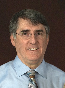 Bruce A. Crosson合作论文数Department of Neurology, Emory School of Medicine14
 Keith M McGregor合作论文数Department of Psychology, University of Florida7
 Jane Paulsen合作论文数Department of Neurology, University of Wisconsin School of Medicine and Public Health6
 Stacy Harnish合作论文数Department of Communication Sciences and Disorders, University of Cincinnati5
 Tim Conway合作论文数McKnight Brain Institute, University of Florida5
 Leslie J. Gonzalez Rothi合作论文数Department of Neurology, College of Medicine, University of Florida Health5
 Christina Wierenga合作论文数Department of Psychiatry, School of Medicine, University of California, San Diego4
 Zvinka Zlatar合作论文数University of California San Diego, University of California4
 Kaundinya Gopinath合作论文数4
 John C. Rosenbek合作论文数4
 Richard W. Briggs合作论文数Center for Neotectonic Studies;University of Nevada;Center for Neotectonic Studies, University of Nevada4Yu-Ling Chang合作论文数3
 David Moser合作论文数3
 Jamie Reilly合作论文数Department of Neurology, University of Pennsylvania School of Medicine3
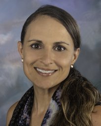 Amy D. Rodriguez合作论文数Department of Neurology, University of Florida3
 Keith D. White合作论文数University of Florida Department of Psychology3Amanda Garcia合作论文数2Hyejin Park合作论文数2Yvonne Rogalski合作论文数2
 Christiana M. Leonard合作论文数Department of Neuroscience, College of Medicine, University of Florida2
 Jonathan Trinastic合作论文数2
 Stephan Arndt合作论文数University of Iowa2
 M. Allison Cato Jackson合作论文数Veterans Affairs Brain Rehabilitation Research Center, University of Florida2Megan E. Sherod合作论文数2
 Susan K. Schultz合作论文数University of Iowa2Carissa Nehl合作论文数2Atchar Sudhyadhom合作论文数Department of Neurosurgery, University of Florida2Jason E. Kanz合作论文数Department of Psychiatry, University of Iowa Carver College of Medicine2
 Chris M. Gregory合作论文数Department of Physical Therapy, University of Florida2
 Karin F. Hoth合作论文数Department of Psychiatry and Human Behavior, Brown Medical School, Providence, RI USA2
 Steven A. Kautz合作论文数Department of Veteran Affairs Rehabilitation Research and Development Brain Rehabilitation Research Center, the Malcom Randall VA Medical Center2Anna Bacon Moore合作论文数2Lauren Seeds合作论文数2
 William Michael Perlstein合作论文数McKnight Brain Institute, University of Florida2William G. Haynes合作论文数1Norman C. Reynolds合作论文数1CATHERINE L. LEVERONI合作论文数1Pamela J Carvajal合作论文数1Jodi Morgan合作论文数1
 Gabriell Champion合作论文数1CS Brems合作论文数1Simone Renee Roberts合作论文数Ctr Visual & Neurocognit Rehabil, Atlanta Vet Affairs Med Ctr VAMC1
 Jonathan H Drucker合作论文数Aptima, Inc.1P Como合作论文数1Nocera Joe R合作论文数1Park Hyejin合作论文数1
 Borich Michael R合作论文数Department of Physical Medicine and Rehabilitation, University of Minnesota1Reese Rebecca L合作论文数1
 Cohen Matthew L合作论文数Department of Clinical &College of Public Health and Health, University of Florida;Department of Clinical & Health Psychology, College of Public Health and Health University of Florida1




