Access to rigorous optical microscopy education remains unevenly distributed across the globe despite widespread use of optical methods. While lower-resourced settings certainly feel this burden, it is far from a foregone conclusion that such opportunities are ubiquitous, even at well-funded institutions. Despite a growing number of online educational resources and other tools, many biomedical researchers learn microscopy in a task-specific manner, and without the conceptual foundation to maximise the potential of its capabilities, robustly interpret data, avoid bias, and/or troubleshoot effectively. Given microscopy's significant impact in the discovery process and status as a cornerstone scientific tool, we developed a remotely accessible, global, and highly interactive program to help address the education gap with respect to the fundamentals of microscopy, irrespective of one's location or ability to access high-end platforms. Here we reflect on the design and evolution of the resulting 'Widening the Lens' (WtL) program: what worked, what didn't, and how the course adapted in response, with the intent of enabling others to leverage our experience to develop programs of their own. We also describe our partnership with critical network organisations, including the African BioImaging Consortium (ABIC) to implement a WtL based train-the-trainer model across the African continent. WtL demonstrates that equitable microscopy education is not only achievable, but that closing education gaps involves building a community in concert with likeminded educational opportunities and networks across the globe.
Epithelial cells from diverse systems assemble apical microvilli to mediate biochemical and physical interactions with the external environment. Despite the central importance of these actin-based protrusions in epithelial physiology, how microvilli grow during differentiation remains poorly understood. Here, we report that an epithelial cell's potential for growing microvilli of normal size is limited by an adjacent actin-dependent process: clathrin-mediated endocytosis. Time-lapse imaging of individual microvillar growth events revealed tight spatial and temporal coupling to sites of apical clathrin-mediated endocytosis. Moreover, ultrastructural characterization of differentiating epithelia showed that most nascent microvilli are in contact with an endocytic pit. Finally, inhibition of the Arp2/3 complex, which drives actin polymerization on coated pits, significantly reduced the accumulation of new microvilli on the cell surface. We conclude that clathrin-mediated endocytosis and its associated Arp2/3-based actin nucleation activity control the timing and location of microvillar growth as well as the dimensions of the resulting protrusions.
When dealing with giga-pixel digital pathology in whole-slide imaging, a notable proportion of data records holds relevance during each analysis operation. For instance, when deploying an image analysis algorithm on whole-slide images (WSI), the computational bottleneck often lies in the input-output (I/O) system. This is particularly notable as patch-level processing introduces a considerable I/O load onto the computer system. However, this data management process could be further paralleled, given the typical independence of patch-level image processes across different patches. This paper details our endeavors in tackling this data access challenge by implementing the Adaptable IO System version 2 (ADIOS2). Our focus has been constructing and releasing a digital pathology-centric pipeline using ADIOS2, which facilitates streamlined data management across WSIs. Additionally, we've developed strategies aimed at curtailing data retrieval times. The performance evaluation encompasses two key scenarios: (1) a pure CPU-based image analysis scenario ("CPU scenario"), and (2) a GPU-based deep learning framework scenario ("GPU scenario"). Our findings reveal noteworthy outcomes. Under the CPU scenario, ADIOS2 showcases an impressive two-fold speed-up compared to the brute-force approach. In the GPU scenario, its performance stands on par with the cutting-edge GPU I/O acceleration framework, NVIDIA Magnum IO GPU Direct Storage (GDS). From what we know, this appears to be among the initial instances, if any, of utilizing ADIOS2 within the field of digital pathology. The source code has been made publicly available at https://github.com/hrlblab/adios.
In the dynamic landscape of scientific research, imaging core facilities are vital hubs propelling collaboration and innovation at the technology development and dissemination frontier. Here, we present a collaborative effort led by Global BioImaging (GBI), introducing international recommendations geared towards elevating the careers of Imaging Scientists in core facilities. Despite the critical role of Imaging Scientists in modern research ecosystems, challenges persist in recognising their value, aligning performance metrics and providing avenues for career progression and job security. The challenges encompass a mismatch between classic academic career paths and service-oriented roles, resulting in a lack of understanding regarding the value and impact of Imaging Scientists and core facilities and how to evaluate them properly. They further include challenges around sustainability, dedicated training opportunities and the recruitment and retention of talent. Structured across these interrelated sections, the recommendations within this publication aim to propose globally applicable solutions to navigate these challenges. These recommendations apply equally to colleagues working in other core facilities and research institutions through which access to technologies is facilitated and supported. This publication emphasises the pivotal role of Imaging Scientists in advancing research programs and presents a blueprint for fostering their career progression within institutions all around the world.
Epithelial cells from diverse contexts assemble apical specializations to serve tissue-specific functions. In virtually all cases, these features consist of arrays of microvilli: micron-scale, actin bundle-supported protrusions that mediate biochemical and physical interactions with the external environment. Despite their importance for epithelial physiology, how microvilli grow during cellular differentiation remains poorly understood. Using genetic and small molecule perturbations, we found that an epithelial cell[&prime]s potential for growing microvilli of normal size is limited by an adjacent actin-dependent process: apical clathrin-mediated endocytosis. Timelapse imaging of individual microvillar growth events revealed tight spatial and temporal coupling to sites of clathrin-mediated endocytosis. Finally, ultrastructural analysis of undifferentiated epithelial monolayers showed that most nascent microvilli are immediately adjacent to or in contact with an apical endocytic pit. Based on these discoveries, we conclude that clathrin-mediated endocytosis controls the timing and location of microvillar growth, as well as the dimensions of the resulting protrusions. ### Competing Interest Statement The authors have declared no competing interest.
Objective: Ultrasound is being researched as a method to modulate the brain. Studies of the interaction of sound with neurons support the hypothesis that mechanosensitive ion channels play an important role in ultrasound neuromodulation. The response of cells other than neurons (e.g., astrocytes, pericytes and endothelial cells) have not been fully characterized, despite playing an important role in brain function. Methods: To address this gap in knowledge, we examined cultured murine primary cortical neurons, astrocytes, endothelial cells and pericytes in an in vitro widefield microscopy setup during application of a 500 ms burst of 250 kHz focused ultrasound over a pressure range known to elicit neuromodulation. We examined cell membrane health in response to a range of pulses and used optical calcium indicators in conjunction with pharmacological antagonists to selectively block different groups of thermo- and mechanosensitive ion channels known to be responsive to ultrasound. Results: All cell types experienced an increase in calcium fluorescence in response to ultrasound. Gadolinium (Gad), 2-aminoethoxydiphenyl borate (2 -APB) and ruthenium red (RR) reduced the percentage of responding neurons and magnitude of response. The percentage of astrocytes responding was significantly lowered only by Gad, whereas both 2 -APB and Gad decreased the amplitude of the fluorescence response. 2 -APB decreased the percentage of responding endothelial cells, whereas only Gad reduced the magnitude of responses. Pericytes exposed to RR or Gad were less likely to respond to stimulation. RR had no detectable effect on the magnitude of the pericyte responses while 2 -APB and Gad significantly decreased the fluorescence intensity, despite not affecting the percentage responding. Conclusion: Our study highlights the role of non -neuronal cells during FUS neuromodulation. All of the investigated cell types are sensitive to mechanical ultrasound stimulation and rely on mechanosensitive ion channels to undergo ultrasound neuromodulation.
To maximize solute transport, epithelial cells build an apical "brush border,"where thousands of microvilli are linked to their neighbors by protocadherin-containing intermicrovillar adhesion complexes (IMACs). Previous studies established that the IMAC is needed to build a mature brush border, but how this complex contributes to the accumulation of new microvilli during differentiation remains unclear. We found that early in differentiation, mouse, human, and porcine epithelial cells exhibit a marginal accumulation of microvilli, which span junctions and interact with protrusions on neighboring cells using IMAC protocadherins. These transjunctional IMACs are highly stable and reinforced by tension across junctions. Finally, long-term live imaging showed that the accumulation of microvilli at cell margins consistently leads to accumulation in medial regions. Thus, nascent microvilli are stabilized by a marginal capture mechanism that depends on the formation of transjunctional IMACs. These results may offer insights into how apical specializations are assembled in diverse epithelial systems.
RNA transfer via extracellular vesicles (EVs) influences cell phenotypes; however, lack of information regarding biogenesis of RNA-containing EVs has limited progress in the field. Here, we identify endoplasmic reticulum membrane contact sites (ER MCSs) as platforms for the generation of RNA-containing EVs. We identify a subpopulation of small EVs that is highly enriched in RNA and regulated by the ER MCS linker protein VAP-A. Functionally, VAP-A-regulated EVs are critical for miR-100 transfer between cells and in vivo tumor formation. Lipid analysis of VAP-A-knockdown EVs revealed reductions in the EV biogenesis lipid ceramide. Knockdown of the VAP-A-binding ceramide transfer protein CERT led to similar defects in EV RNA content. Imaging experiments revealed that VAP-A promotes luminal filling of multivesicular bodies (MVBs), CERT localizes to MVBs, and the ceramide-generating enzyme neutral sphingomyelinase 2 colocalizes with VAP-A-positive ER. We propose that ceramide transfer via VAP-A-CERT linkages drives the biogenesis of a select RNA-containing EV population.
Contrastive learning, a recent family of self-supervised learning, leverages pathological image analysis by learning from large-scale unannotated data. However, the state-of-the-art contrastive learning methods (e.g., SimCLR, BYOL) are typically limited by the more expensive computational hardware (with large GPU memory) as compared with traditional supervised learning approaches in achieving large training batch size. Fortunately, recent advances in the machine learning community provide multiple approaches to reduce GPU memory usage, such as (1) activation compressed training, (2) In-place activation, and (3) mixed precision training. Yet, such approaches are currently deployed independently without systematical assessments for contrastive learning. In this work, we applied these memory-efficient approaches into a self-supervised framework. The contribution of this paper is three-fold: (1) We combined previously independent GPU memory-efficient methods with self-supervised learning framework; (2) Our experiments are to maximize the memory efficiency via limited computational resources (a single GPU); (3) The self-supervised learning framework with GPU memory-efficient method allows a single GPU to triple the batch size that typically requires three GPUs. From the experimental results, contrastive learning model with larger batch size leads to higher accuracy enabled by GPU memory-efficient method on single GPU.
ABSTRACT Leigh syndrome (LS) is a rare, inherited neurometabolic disorder that presents with bilateral brain lesions caused by defects in the mitochondrial respiratory chain and associated nuclear-encoded proteins. We generated human induced pluripotent stem cells (iPSCs) from three LS patient-derived fibroblast lines. Using whole-exome and mitochondrial sequencing, we identified unreported mutations in pyruvate dehydrogenase (GM0372, PDH; GM13411, MT-ATP6/PDH) and dihydrolipoyl dehydrogenase (GM01503, DLD). These LS patient-derived iPSC lines were viable and capable of differentiating into progenitor populations, but we identified several abnormalities in three-dimensional differentiation models of brain development. LS patient-derived cerebral organoids showed defects in neural epithelial bud generation, size and cortical architecture at 100 days. The double mutant MT-ATP6/PDH line produced organoid neural precursor cells with abnormal mitochondrial morphology, characterized by fragmentation and disorganization, and showed an increased generation of astrocytes. These studies aim to provide a comprehensive phenotypic characterization of available patient-derived cell lines that can be used to study Leigh syndrome.
Recent advances in bioimaging have provided scientists a superior high spatial-temporal resolution to observe dynamics of living cells as 3D volumetric videos. Unfortunately, the 3D biomedical video analysis is lagging, impeded by resource insensitive human curation using off-the-shelf 3D analytic tools. Herein, biologists often need to discard a considerable amount of rich 3D spatial information by compromising on 2D analysis via maximum intensity projection. Recently, pixel embedding based cell instance segmentation and tracking provided a neat and generalizable computing paradigm for understanding cellular dynamics. In this work, we propose a novel spatial-temporal voxel-embedding (VoxelEmbed) based learning method to perform simultaneous cell instance segmenting and tracking on 3D volumetric video sequences. Our contribution is in four-fold: (1) The proposed voxel embedding generalizes the pixel embedding with 3D context information; (2) Present a simple multi-stream learning approach that allows effective spatial-temporal embedding; (3) Accomplished an end-toend framework for one-stage 3D cell instance segmentation and tracking without heavy parameter tuning; (4) The proposed 3D quantification is memory efficient via a single GPU with 12 GB memory. We evaluate our VoxelEmbed method on four 3D datasets (with different cell types) from the ISBI Cell Tracking Challenge. The proposed VoxelEmbed method achieved consistent superior overall performance (OP) on two densely annotated datasets. The performance is also competitive on two sparsely annotated cohorts with 20.6% and 2% of data-set having segmentation annotations. The results demonstrate that the VoxelEmbed method is a generalizable and memory-efficient solution.
The unsupervised segmentation is an increasingly popular topic in biomedical image analysis. The basic idea is to approach the supervised segmentation task as an unsupervised synthesis problem, where the intensity images can be transferred to the annotation domain using cycle-consistent adversarial learning. The previous studies have shown that the macro-level (global distribution level) matching on the number of the objects (e.g., cells, tissues, protrusions etc.) between two domains resulted in better segmentation performance. However, no prior studies have exploited whether the unsupervised segmentation performance would be further improved when matching the exact number of objects at micro-level (mini-batch level). In this paper, we propose a deep learning based unsupervised segmentation method for segmenting highly overlapped and dynamic sub-cellular microvilli. With this challenging task, both micro-level and macro-level matching strategies were evaluated. To match the number of objects at the micro-level, the novel fluorescence-based micro-level matching approach was presented. From the experimental results, the micro-level matching did not improve the segmentation performance, compared with the simpler macro-level matching.
Quantitative analysis of microscope videos often requires instance segmentation and tracking of cellular and subcellular objects. Traditional method is composed of two stages: (1) instance object segmentation of each frame, and (2) associate objects frame by frame. Recently, pixel embedding based deep learning approaches provide single stage holistic solutions to tackle instance segmentation and tracking simultaneously. However, the deep learning methods require consistent annotations not only spatially (for segmentation), but also temporally (for tracking). In computer vision, annotated training data with consistent segmentation and tracking is resource intensive, which can be more severe in microscopy imaging owing to (1) dense objects (e.g., overlapping or touching), and (2) high dynamics (e.g., irregular motion and mitosis). To alleviate the lack of such annotations in dynamics scenes, adversarial simulations have provided successful solutions in computer vision, such as using simulated environments (e.g., computer games) to train real-world self-driving systems. In this paper, we proposed an annotation-free synthetic instance segmentation and tracking (ASIST) method with adversarial simulation and single-stage pixel-embedding based learning. The contribution is three-fold: (1) the proposed method aggregates adversarial simulations and single-stage pixel-embedding based deep learning; (2) the method is assessed with both cellular (i.e., HeLa cells) and subcellular (i.e., microvilli) objects; and (3) to the best of our knowledge, this is the first study to explore annotation-free instance segmentation and tracking study for microscope videos. From the results, our ASIST method achieved promising results compared with fully supervised approaches.
BACKGROUND:The quantitative analysis of microscope videos often requires instance segmentation and tracking of cellular and subcellular objects. The traditional method consists of two stages: (1) performing instance object segmentation of each frame, and (2) associating objects frame-by-frame. Recently, pixel-embedding-based deep learning approaches these two steps simultaneously as a single stage holistic solution. Pixel-embedding-based learning forces similar feature representation of pixels from the same object, while maximizing the difference of feature representations from different objects. However, such deep learning methods require consistent annotations not only spatially (for segmentation), but also temporally (for tracking). In computer vision, annotated training data with consistent segmentation and tracking is resource intensive, the severity of which is multiplied in microscopy imaging due to (1) dense objects (e.g., overlapping or touching), and (2) high dynamics (e.g., irregular motion and mitosis). Adversarial simulations have provided successful solutions to alleviate the lack of such annotations in dynamics scenes in computer vision, such as using simulated environments (e.g., computer games) to train real-world self-driving systems.METHODS:In this paper, we propose an annotation-free synthetic instance segmentation and tracking (ASIST) method with adversarial simulation and single-stage pixel-embedding based learning.CONTRIBUTION:The contribution of this paper is three-fold: (1) the proposed method aggregates adversarial simulations and single-stage pixel-embedding based deep learning (2) the method is assessed with both the cellular (i.e., HeLa cells); and subcellular (i.e., microvilli) objects; and (3) to the best of our knowledge, this is the first study to explore annotation-free instance segmentation and tracking study for microscope videos.RESULTS:The ASIST method achieved an important step forward, when compared with fully supervised approaches: ASIST shows 7%-11% higher segmentation, detection and tracking performance on microvilli relative to fully supervised methods, and comparable performance on Hela cell videos.
Recently, single-stage embedding based deep learning algorithms gain increasing attention in cell segmentation and tracking. Compared with the traditional "segment-then-associate" two-stage approach, a single-stage algorithm not only simultaneously achieves consistent instance cell segmentation and tracking but also gains superior performance when distinguishing ambiguous pixels on boundaries and overlaps. However, the deployment of an embedding based algorithm is restricted by slow inference speed (e.g., around 1-2 mins per frame). In this study, we propose a novel Faster Mean-shift algorithm, which tackles the computational bottleneck of embedding based cell segmentation and tracking. Different from previous GPU-accelerated fast mean-shift algorithms, a new online seed optimization policy (OSOP) is introduced to adaptively determine the minimal number of seeds, accelerate computation, and save GPU memory. With both embedding simulation and empirical validation via the four cohorts from the ISBI cell tracking challenge, the proposed Faster Mean-shift algorithm achieved 7-10 times speedup compared to the state-of-the-art embedding based cell instance segmentation and tracking algorithm. Our Faster Mean-shift algorithm also achieved the highest computational speed compared to other GPU benchmarks with optimized memory consumption. The Faster Mean-shift is a plug-and-play model, which can be employed on other pixel embedding based clustering inference for medical image analysis. (Plug-and-play model is publicly available: https://github.com/masqm/Faster-Mean-Shift)
Rationale: Bronchopulmonary dysplasia (BPD) is a leading complication of preterm birth that affects infants born in the saccular stage of lung development at <32 weeks of gestation. Although the mechanisms driving BPD remain uncertain, exposure to hyperoxia is thought to contribute to disease pathogenesis.Objectives: To determine the effects of hyperoxia on epithelial-mesenchymal interactions and to define the mediators of activated Wnt/β-catenin signaling after hyperoxia injury.Methods: Three hyperoxia models were used: A three-dimensional organotypic coculture using primary human lung cells, precision-cut lung slices (PCLS), and a murine in vivo hyperoxia model. Comparisons of normoxia- and hyperoxia-exposed samples were made by real-time quantitative PCR, RNA in situ hybridization, quantitative confocal microscopy, and lung morphometry.Measurements and Main Results: Examination of an array of Wnt ligands in the three-dimensional organotypic coculture revealed increased mesenchymal expression of WNT5A. Inhibition of Wnt5A abrogated the BPD transcriptomic phenotype induced by hyperoxia. In the PCLS model, Wnt5A inhibition improved alveolarization following hyperoxia exposure, and treatment with recombinant Wnt5a reproduced features of the BPD phenotype in PCLS cultured in normoxic conditions. Chemical inhibition of NF-κB with BAY11-7082 reduced Wnt5a expression in the PCLS hyperoxia model and in vivo mouse hyperoxia model, with improved alveolarization in the PCLS model.Conclusions: Increased mesenchymal Wnt5A during saccular-stage hyperoxia injury contributes to the impaired alveolarization and septal thickening observed in BPD. Precise targeting of Wnt5A may represent a potential therapeutic strategy for the treatment of BPD.
Instance object segmentation and tracking provide comprehensive quantification of objects across microscope videos. The recent single-stage pixel-embedding based deep learning approach has shown its superior performance compared with "segment-then-associate" two-stage solutions. However, one major limitation of applying a supervised pixel-embedding based method to microscope videos is the resource-intensive manual labeling, which involves tracing hundreds of overlapped objects with their temporal associations across video frames. Inspired by the recent generative adversarial network (GAN) based annotation-free image segmentation, we propose a novel annotation-free synthetic instance segmentation and tracking (ASIST) algorithm for analyzing microscope videos of sub-cellular microvilli. The contributions of this paper are three-fold: (1) proposing a new annotation-free video analysis paradigm is proposed. (2) aggregating the embedding based instance segmentation and tracking with annotation-free synthetic learning as a holistic framework; and (3) to the best of our knowledge, this is first study to investigate microvilli instance segmentation and tracking using embedding based deep learning. From the experimental results, the proposed annotation-free method achieved superior performance compared with supervised learning.
The methyltransferase SET domain-containing 2 (SETD2) was originally identified as Huntingtin (HTT) yeast partner B. However, a SETD2 function associated with the HTT scaffolding protein has not been elucidated, and no linkage between HTT and methylation has yet been uncovered. Here, we show that SETD2 is an actin methyltransferase that trimethylates lysine-68 (ActK68me3) in cells via its interaction with HTT and the actin-binding adapter HIP1R. ActK68me3 localizes primarily to the insoluble F-actin cytoskeleton in cells and regulates actin polymerization/depolymerization dynamics. Disruption of the SETD2-HTT-HIP1R axis inhibits actin methylation, causes defects in actin polymerization, and impairs cell migration. Together, these data identify SETD2 as a previously unknown HTT effector regulating methylation and polymerization of actin filaments and provide new avenues for understanding how defects in SETD2 and HTT drive disease via aberrant cytoskeletal methylation.
ABSTRACT SET-domain-containing-2 (SETD2) was identified as the methyltransferase responsible for the histone 3 lysine 36 trimethyl (H3K36me3) mark of the histone code. Most recently, SETD2 has been shown to be a dual-function remodeler that regulates genome stability via methylation of dynamic microtubules during mitosis and cytokinesis. Here we show that actin is a bona fide target for methylation by SETD2 in vitro and in cells. Antibodies against the SETD2 trimethyl lysine epitope recognize methylated actin, with this methyl mark localizing to areas of active actin cytoskeleton reorganization in migrating cells. Disruption of this methylation activity causes defects in actin polymerization and impairs collective cell migration. Together, these data identify SETD2 as a multifunctional cytoskeletal remodeler regulating methylation and polymerization of actin filaments, and provide new avenues for understanding how defects in SETD2 drive disease via aberrant cytoskeletal methylation.
Transporting epithelial cells, like those that line the intestinal tract, are specialized for solute processing and uptake. One defining feature is the brush border, an array of microvilli that serves to amplify apical membrane surface area and increase functional capacity. During differentiation, upon exit from stem-cell-containing crypts, enterocytes build thousands of microvilli, each supported by a parallel bundle of actin filaments several microns in length. Given the high concentration of actin residing in mature brush borders, we sought to determine whether enterocytes were resource (i.e., actin monomer) limited in assembling this domain. To examine this possibility, we inhibited Arp2/3, the ubiquitous branched actin nucleator, to increase G-actin availability during brush border assembly. In native intestinal tissues, Arp2/3 inhibition led to increased microvilli length on the surface of crypt, but not villus, enterocytes. In a cell culture model of brush border assembly, Arp2/3 inhibition accelerated the growth and increased the length of microvilli; it also led to a redistribution of F-actin from cortical lateral networks into the brush border. Effects on brush border growth were rescued by treatment with the G-actin sequestering drug, latrunculin A. G-actin binding protein, profilin-1, colocalized in the terminal web with G-actin, and knockdown of this factor compromised brush border growth in a concentration-dependent manner. Finally, the acceleration in brush border assembly induced by Arp2/3 inhibition was abrogated by profilin-1 knockdown. Thus, brush border assembly is limited by G-actin availability, and profilin-1 directs unallocated actin monomers into microvillar core bundles during enterocyte differentiation.
 Matthew J. Tyska合作论文数Department of Cell and Developmental Biology, Vanderbilt University21
 Yuankai Huo合作论文数Southeast University, Southeast Univ9
 Quan Liu合作论文数Vanderbilt Univ, Dept Elect Engn & Comp Sci, Nashville, TN 37235 USA8
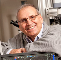 Bechara Kachar合作论文数Section on Structural Cell Biology, National Institute on Deafness and Other Communications Disorders, National Institutes of Health8
 Ruining Deng合作论文数Vanderbilt University6
 Aadarsh Jha合作论文数Vanderbilt Univ, Dept Elect Engn & Comp Sci, Nashville, TN 37235 USA6
 Anita Mahadevan-Jansen合作论文数Biophotonics Center, Vanderbilt University6Taissia G. Popova合作论文数6Isabella M Gaeta合作论文数5
 Mengyang Zhao合作论文数Thayer School of Engineering, Dartmouth College, Hanover, NH 03755, USA5Krystofiak Evan S合作论文数5
 Seham Ebrahim合作论文数National Institutes on Deafness and Other Communication Disorders;National Institutes of Health;Bethesda;National Institutes on Deafness and Other Communication Disorders, National Institutes of Health5
 Charles G. Bailey合作论文数Gene and Stem Cell Therapy Unit, Centenary Institute5
 Serguei Popov合作论文数Instituto de Matematica e Estat ´ õstica;Universidade de Sao Paulo;Instituto de Matematica e Estat ´ õstica, Universidade de Sao Paulo4Leslie M. Meenderink合作论文数4
 Lakshmi Sundararajan合作论文数3Jennifer M S Sucre合作论文数3Benjamin John T合作论文数Department of Pediatrics, Vanderbilt University Medical Center3
 Cody J. Smith合作论文数Department of Biological Sciences, College of Science, University of Notre Dame3Cui Runjia合作论文数National Institute for Deafness and Communications Disorders, National Institute of Health3
 Timothy S. Blackwell合作论文数Department of Internal Medicine, The University of Michigan3Jetter Christopher S合作论文数3
 Vikas Chandhoke合作论文数Center for the Study of Genomics in Liver Diseases, George Mason University3Susan H. Guttentag合作论文数3Svetlana Nazarenko合作论文数3
 Namasivayam Ambalavanan合作论文数Department of Pediatrics, Heersink School of Medicine, The University of Alabama at Birmingham3
 Christopher Bradburne合作论文数Advanced Biosystems, George Mason University3
 Myung-Chul Chung合作论文数Korea Research Institute of Bioscience and Biotechnology3s g popov合作论文数2Caroline S Cencer合作论文数2Sarah Kearns合作论文数<label>*</label><p>Equal contributions</p>2
 Rahul K Jangid合作论文数Ctr Precis Environm Hlth, Baylor Coll Med2Tk Maugel合作论文数2Joseph T. Roland合作论文数Vanderbilt University Medical Center, Nashville TN 37215, USA.2Taro Fujikawa合作论文数2
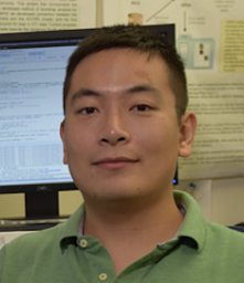 Shunxing Bao (鲍舜行)合作论文数Department of Electrical and Computer Engineering, School of Engineering, Vanderbilt University2
 Thiago Gomes De Castro合作论文数Federal University of Rio Grande do Sul2
 Kristen Verhey合作论文数Department of Cell & Developmental Biology, Medical School, University of Michigan2
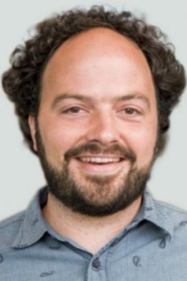 Michael Cianfrocco合作论文数Biophysics, College of Literature, Science, and the Arts, University of Michigan-Ann Arbor2Vickers Kasey C合作论文数Med Ctr, Vanderbilt Univ2Gleaves Linda A合作论文数2Corrotte Matthias合作论文数2Kropski Jonathan A合作论文数2Baird Michelle A合作论文数Natl High Magnet Field Lab, Florida State Univ2Young Lisa R合作论文数2Miller Heather合作论文数2Deutsch Gail H合作论文数2Mao Suli合作论文数2Almeida Patricia E合作论文数2




