Functional vascularization remains a critical challenge in the regeneration of full-thickness skin defects. Here, we present a 3D embedded bioprinting strategy for engineering vascularized biopatches through the precise integration of prevascularized multicellular spheroids within a skin-derived decellularized extracellular matrix (SdECM) bioink. Rheological results reveal that a 1.0% (w/v) SdECM formulation exhibits yield-stress behavior, pronounced shear-thinning, and rapid shear recovery, enabling high-fidelity of spheroids while preserving cell viability and structural integrity. Using this platform, endothelial spheroids with tunable diameters are fabricated to investigate size-dependent vascular self-organization. Intermediate-sized spheroids (∼700 µm) demonstrate robust endothelial network formation, with enhanced vascular coverage, branching complexity, and angiogenic gene expression while avoiding excessive hypoxia. Spatial organization of prevascularized spheroids critically regulates paracrine-mediated wound repair, with enhanced fibroblast migration observed when inter-spheroid distances were within 1.8 mm. Furthermore, incorporation of mesenchymal stem cells (MSCs) provides perivascular-like support, significantly stabilizing microvascular networks, with an optimized endothelial cell-to-MSC ratio of 1:0.5. In a full-thickness murine wound model, multicellular spheroids-laden SdECM biopatches significantly accelerate wound closure, enhance re-epithelialization, promoted angiogenesis, and improve blood perfusion. Collectively, this study establishes a scalable embedded bioprinting for engineering prevascularized biopatches and provides design principles for spheroid-based soft tissue regeneration.
The development of models for neurodegenerative and neurovascular diseases has become crucial for understanding complex biological processes, including the progression of Alzheimer’s disease, stroke, and glioblastoma. Traditional in vitro systems have significant limitations in replicating the intricate interactions between the blood-brain barrier (BBB), endothelial cells, and surrounding tissues. This review explores a novel strategy to enhance disease modeling technology through the integration of 3D bioprinting with microfluidic chips. This advanced approach enables the precise creation of dynamic tissue constructs that emulate the physiological microenvironment of the neurovascular unit, facilitating studies on drug cytotoxicity, cell differentiation, and intercellular signaling. Furthermore, the platform offers promising applications for identifying therapeutic targets and designing miniaturized biosensor-embedded MEMS (micro-electro-mechanical systems) and NEMS (nano-electro-mechanical systems) for the sensitive detection of disease biomarkers. The integrated platform further incorporates design modifications, including optimized microchannel geometry, bioprinted three-dimensional cellular architectures, and improved spatial organization of biological components within the microfluidic chip. These engineering advancements enhance cell viability, physiological cell-cell and cell-matrix interactions, nutrient and oxygen diffusion, and overall biomimetic functionality, while clearly demonstrating the contribution of 3D bioprinting to improved device performance, experimental reproducibility, and biological relevance for neurovascular disease modeling and therapeutic evaluation. The system is particularly suited for modeling the pathophysiological conditions of neurovascular and neurodegenerative diseases, such as Alzheimer’s disease, stroke, and brain cancer (glioblastoma), in a controlled and reproducible manner. This capability was demonstrated through preclinical and co-clinical drug trials, generating functional data to support effective therapeutic interventions. Additionally, the study highlights the creation of pathophysiological models incorporating neural, endothelial, and adjacent cell types to investigate neural–microvascular interactions. The integrated platform serves as a powerful tool for evaluating drug efficacy, deciphering disease mechanisms, and advancing personalized medicine approaches. These findings underscore the transformative potential of combining 3D bioprinting and microfluidic technologies to develop highly functional models for neurodegenerative and neurovascular diseases, offering significant value for translational biomedical research.
Increased matrix stiffness within tumor microenvironments (TMEs) significantly influences cancer progression and gene expression, contributing to drug resistance and poor clinical outcomes. Studies demonstrate a strong correlation between nuclear factor kappa B (NF-κB) upregulation and prostate cancer malignancy. However, the mechanisms by which the mechanical stress within the TME activates NF-κB remain underexplored. This study developed a prostate cancer spheroid model using an in-bath 3D bioprinting technique. Cancer spheroids were printed within a bespoke hydrogel bath with tunable stiffness, facilitating the investigation of the relationship between mechanical cues and oncogenic behavior. Increased hydrogel stiffness promoted spheroid compaction, induction of epithelial-mesenchymal transition (EMT) and stemness programs, and elevated drug resistance. Transcriptomic analysis revealed that the phosphoinositide 3-kinase (PI3K) pathway is most enriched under mechanical stress. Findings demonstrated that increased extracellular matrix stiffness activated PI3K/NF-κB signaling through mechanotransduction. Pharmacological inhibition of PI3K suppressed NF-κB nuclear translocation and enhanced chemotherapy efficacy. The bespoke hydrogel effectively recapitulated the mechanical environment of prostate cancer, indicating the pivotal role of PI3K/NF-κB signaling in regulating prostate cancer malignancy under mechanical stimulation. This suggests a promising therapeutic avenue for improving treatment outcomes.
Ischemic diseases, characterized by impaired blood flow and progressive tissue necrosis, remain a major challenge in regenerative medicine. Surgical revascularization remains the gold standard for restoring blood flow in major vessels, but still shows limited effects on microvascular regeneration. To address this unmet need, various strategies in therapeutic angiogenesis have been explored to induce microvascular formation, including delivery of bioactive molecules, stem cells, or pre-vascularized grafts. However, injection-based approaches often suffer from off-target effects, and conventional implantable grafts lack sufficient angiogenic secretory activity. To address these limitations, we aimed to develop a dual-function prevascularized graft that accelerates neovascularization and promotes vascular integration to restore tissue perfusion in ischemic conditions. The graft was engineered by combining a microvascular pattern (µVP) with spatially organized mesenchymal stem cell (MSC) spheroids, fabricated via high-precision coprinting of endothelial cells and MSCs. Optimization of spheroid density and spatial arrangement enhanced VEGF secretion and increased host capillary infiltration nearly two-fold. In a murine critical limb ischemia model, implantation of these engineered grafts achieved a 60
Development of physiologically functional skeletal muscle constructs is vital for regenerative therapies and drug-screening applications. Herein, we present an integrated strategy that combines viscoelastic muscle-derived extracellular matrix (MdECM) bioinks with a 3D printing-assisted cyclic strain bioreactor. To enhance mechanical performance, an improved decellularization protocol incorporating isopropanol was introduced to effectively remove residual lipids. The resulting bioinks exhibited superior viscoelasticity, structural stability, and thixotropic recovery compared with conventional formulations. The viscoelastic bioinks enabled stable myoblast encapsulation and alignment within the 3D-printed pillar frames. These counterforces guided robust myotube formation, while optimized ECM concentration and cell density further promoted alignment and myogenic differentiation. Spatial regulation of myotube orientation was achieved by tuning the pillar geometry and spacing. Muscle blocks were matured in a custom-designed multi-chamber bioreactor capable of applying cyclic uniaxial strain, yielding constructs with uniaxial cellular alignment, elevated myogenic marker expression, and contractile responses confirmed by calcium imaging and electrical stimulation. This platform supports scalable production of structurally and functionally mature muscle microtissues, offering a promising model for therapeutic implantation and high-throughput drug screening targeting sarcopenia and muscle-wasting diseases.
Skin injuries including burns, chronic wounds, and diabetic ulcers present significant clinical challenges due to impaired healing, high risk of infection, and limited efficacy of conventional transdermal patches. Owing to recent advances in biomaterials and regenerative medicine, skin-repair strategies are currently focused on engineered patches that better mimic the native tissue structure and function. Particularly, digital light processing (DLP)-based 3D printing is a powerful platform for fabricating high-resolution personalized skin patches with precise architectural control than other conventional approaches. The rapid layer-by-layer photopolymerization of hydrogel-based materials enables the creation of mechanically compliant biocompatible constructs that support cell proliferation, immunomodulation, angiogenesis, and extracellular matrix remodeling. Furthermore, DLP technology enables controlled therapeutic delivery by facilitating the integration of advanced functionalities such as microneedle arrays, vascularized architectures, and stimuli-responsive systems. This review systematically discusses the fundamental principles of DLP printing, key parameters that govern printing fidelity, recent progress in bioink development, and emerging applications of DLP-fabricated skin patches in wound healing and skin regeneration. Additionally, current challenges and future perspectives are highlighted to guide the continued development and clinical translation of DLP-based regenerative skin therapies.
Airway epithelial dysfunction is a hallmark of chronic airway diseases; however, current clinical interventions fail to restore functional airway mucosa. We aimed to develop a bioink derived from airway mucosa-derived decellularized extracellular matrix (MudECM) and evaluate its potential for airway tissue engineering throughin vitroandin vivostudies. Airway mucosal tissue was decellularized to preserve tissue-specific extracellular matrix components and processed into a thermo-responsive bioink. The biochemical composition, rheological properties, and printability of the bioink were characterized. Human tracheal epithelial cells (HTEpCs) were cultured on MudECM hydrogels under air-liquid interface conditions to assess epithelial differentiation and mucociliary function. A bilayered airway construct was fabricated by 3D bioprinting using MudECM-based bioinks and evaluated for epithelial-stromal organizationin vitro. Functional regeneration was tested in a rat tracheal defect model by implanting 3D-printed MudECM scaffolds. MudECM retained collagen and glycosaminoglycans while achieving >98% decellularization. The bioink exhibited shear-thinning and thermal gelation properties suitable for bioprinting. HTEpCs cultured on MudECM hydrogelsin vitroshowed enhanced mucociliary differentiation, tight junction formation, goblet cell development, and directional mucus transport, compared to collagen controls. Bioprinted bilayered airway constructs supported fibroblast viability, growth factor expression, and epithelial differentiation with upregulation of Trp63, FoxJ1, and mucin 5AC. Implanting MudECM scaffolds into tracheal defectsin vivoresulted in complete re-epithelialization, restoration of ciliary beating, and improved mucociliary clearance. By contrast, collagen controls showed only partial regeneration. Thus, MudECM bioink provides tissue-specific biochemical and mechanical cues that promote mucociliary epithelial regeneration. By enabling structural and functional restorationin vitroandin vivo, this bioink represents a clinically translatable biomaterial for airway reconstruction. Beyond regenerative graft fabrication, it offers a robust platform for disease modeling and drug testing in airway research.
Connective tissues display distinct mechanical behaviors, ranging from unidirectional stiffness in regular tissues to multidirectional compliance in irregular tissues. Replicating these biomechanical characteristics in engineered constructs remains a key challenge in regenerative medicine. This study presents a novel biofabrication platform for hybrid biopatches composed of tonsil-derived mesenchymal stem cell (TMSC)-laden collagen bioink reinforced with 3D printed polymeric patterns. Two distinct geometries, chiral and chevron, are designed to emulate the mechanical behavior of irregular and regular connective tissues, respectively. Mechanical testing shows that the chiral pattern exhibits quasi-isotropic behavior with balanced stiffness and extensibility, whereas the chevron pattern demonstrate anisotropic mechanical properties. These mechanical features are maintained within the hybrid biopatches, leading to enhanced tensile strength and fatigue resistance compared with constructs composed solely of TMSC-laden collagen. In a porcine mucosal defect model, the chiral-patterned hybrid biopatch promoted superior epithelial repair, evidenced by narrower wound margins, continuous epithelial layers, and elevated expression of epithelial markers. These results suggest that mechanical compatibility with host tissue influences regenerative outcomes. Collectively, this study highlights the potential of incorporating geometric polymer patterns as a strategy for engineering tissue-specific mechanics and improving regenerative performance, offering a promising platform for soft connective tissue repair.
Despite the growing recognition of adipose tissue as an endocrine organ, engineering it remains a challenge owing to difficulties in replicating its native structure and densely packed lipid droplets. Furthermore, integrating adipose tissue with other tissues, though critical for its endocrine function, remains underexplored, limiting the understanding of its roles in metabolic homeostasis and tissue repair. This study introduces a rapid tissue printing method that constructs adipose units by extruding preadipocyte-laden bioink within 0.3 s using a modular polycaprolactone framework optimized through rheological and computational analyses. In standard, cell-friendly environments, preadipocytes typically proliferate and migrate, inhibiting the formation of dense lipid droplets. To address this issue, a hybrid bioink that limits cell migration and promotes adipocyte maturation is developed. The optimal adipose unit diameter (<= 600 mu m) is calculated, with adipogenic markers evaluated in various spatial configurations. Tissue assembly integrates the adipose module and dermis module, validating its functionality as endocrine tissue. In vivo studies show that the endocrine activity of the adipose units significantly enhances wound closure, vascularization, and re-epithelialization. These findings highlight the regenerative capabilities of the proposed tissue assembly strategy for fabricating large-scale, multicellular, 3D composite tissues.
Microneedle (MN) patches have emerged as a promising drug delivery technology for wound healing treatments, offering several advantages over traditional administration methods, including minimal invasiveness, precise drug delivery, and minimal pain. This review explores the basic principles of MN patch technology, three-dimensional (3D) printing-assisted fabrication techniques, and the key considerations in designing effective MN patches for regenerative medicine. Moreover, the in vivo applications of the MN patches in wound healing, tissue regeneration, and drug delivery are discussed in detail. Finally, the challenges and future research directions of the MN patch technology are discussed, highlighting its potential to revolutionize personalized medication.
The lack of functional vasculature in skin substitutes leads to ischemia and delayed regeneration, highlighting the need for effective prevascularization strategies. This study employs in-bath 3D bioprinting to fabricate prevascularized skin patches by patterning endothelial cells (ECs) within light-activated decellularized extracellular matrix (dECM) bioink. ECs were printed in line, grid, and dot patterns, rapidly crosslinked under visible light to achieve high shape fidelity, even with low dECM concentrations. Integration of human adipose-derived stem cells further enhanced vascular sprouting. Among the patterns, the grid configuration demonstrated the highest microvessel formation and significantly improved neovascularization and wound healing in a murine model. A mechanistic analysis suggests that differences in surface area, linked to integrin-mediated and vascular endothelial growth factor-related pathways, drive the superior outcomes of the grid pattern. This study underscores the potential of tailored vascular patterning in advancing bioprinted skin patches as innovative solutions for skin regeneration.
Stenotic regions in cerebral vessels are implicated in diseases such as atherosclerosis, where shear-responsive endothelial function is critical to disease progression. However, studying flow-induced inflammation remains challenging due to the complexity of in vivo conditions, highlighting the need for a well-engineered in vitro model. A physiologically relevant in vitro model of stenotic brain vessels using 3D-coaxial bioprinting and a mechanically enhanced extracellular matrix (ECM) bioink is developed to investigate flow-induced endothelial inflammation. The hybrid bioink, composed of vascular decellularized ECM, collagen, and alginate, exhibits an approximately 65-fold increase in dynamic modulus, enabling stable formation of perfusable structures. Printing parameter optimization facilitates precise fabrication of stenotic vessels with a luminal diameter of 250-500 mu m. Computational fluid dynamics simulations under an inlet flow rate of 3 mL min-1 predict disturbed fluid flow in stenotic regions. The bioprinted vessels exhibit continuous endothelial coverage, expression of junction proteins (CD31, ZO-1, and VE-cadherin), and size-dependent permeability, indicating a mature vascular barrier formation. Under disturbed flow conditions, ICAM-1 (approximately 2.2-fold) and VCAM-1 (approximately 1.5-fold) are upregulated, confirming the hemodynamic stress-induced inflammation. These findings highlight the potential of 3D bioprinting for modeling cerebrovascular disease in vitro and paving the way for future therapeutic innovation.
The skin is composed of many cells that are organized into different layers and connected by dense and complex vascular networks. This creates a dynamic microenvironment in which cells interact within the matrix. Significant advancements have been made in this field over the past decade, and various strategies have been developed for accelerating and enhancing skin regeneration. The primary challenge for successful skin grafts is the integration of the functional vasculature, which can supply essential nutrients and oxygen to cell-laden structures and damaged native tissues. An inadequate vascular network can lead to ischemia, which can cause slow wound healing—particularly in the case of chronic skin conditions. Therefore, blood vessel formation remains one of the most significant obstacles that skin tissue engineering must overcome to create vascularized skin tissue substitutes with specific living cells. Technological advances can augment effective vascularization. The three-dimensional (3D) bioprinting platform is a promising technology that allows precise deposition of living cells and bioactive materials. The application of this technology to skin tissue engineering can provide solutions for augmenting pre-vascularization in engineered in vitro skin models and in vivo skin substitutes. This review presents the significance of skin vascularization in in vitro modeling and in vivo wound healing. Various strategies and related applications involving 3D bioprinting technology are introduced for the biofabrication of enhanced vascularized skin in vitro and in vivo, followed by a discussion of their limitations and future research directions.
Gelatin methacrylate (GelMA) bioink has been widely used in bioprinting because it is a printable and biocompatible biomaterial. However, it is difficult to print GelMA bioink without any temperature control because it has a thermally-sensitive rheological property. Therefore, in this study, we developed a temperature-controlled printing system in real time without affecting the viability of the cells encapsulated in the bioink. In addition, a skin-derived decellularized extracellular matrix (SdECM) was printed with GelMA to better mimic the native tissue environment compared with solely using GelMA bioink with the enhancement of structural stability. The temperature setting accuracy was calculated to be 98.58 ± 1.8 % for the module and 99.48 ± 1.33 % for the plate from 5 °C to 37 °C. The group of the temperature of the module at 10 °C and the plate at 20 °C have 93.84 % cell viability with the printable range in the printability window. In particular, the cell viability and proliferation were increased in the encapsulated fibroblasts in the GelMA/SdECM bioink, relative to the GelMA bioink, with a morphology that significantly spread for seven days. The gene expression and growth factors related to skin tissue regeneration were relatively upregulated with SdECM components. In the bioprinting process, the rheological properties of the GelMA/SdECM bioink were successfully adjusted in real time to increase printability, and the native skin tissue mimicked components providing tissue-specific biofunctions to the encapsulated cells. The developed bioprinting strategies and bioinks could support future studies related to the skin tissue reconstruction, regeneration, and other medical applications using the bioprinting process.
Cancer vasculogenesis is a pivotal focus of cancer research and treatment given its critical role in tumor development, metastasis, and the formation of vasculogenic microenvironments. Traditional approaches to investigating cancer vasculogenesis face significant challenges in accurately modeling intricate microenvironments. Recent advancements in three-dimensional (3D) bioprinting technology present promising solutions to these challenges. This review provides an overview of cancer vasculogenesis and underscores the importance of precise modeling. It juxtaposes traditional techniques with 3D bioprinting technologies, elucidating the advantages of the latter in developing cancer vasculogenesis models. Furthermore, it explores applications in pathological investigations, preclinical medication screening for personalized treatment and cancer diagnostics, and envisages future prospects for 3D bioprinted cancer vasculogenesis models. Despite notable advancements, current 3D bioprinting techniques for cancer vasculogenesis modeling have several limitations. Nonetheless, by overcoming these challenges and with technological advances, 3D bioprinting exhibits immense potential for revolutionizing the understanding of cancer vasculogenesis and augmenting treatment modalities.
Vascular diseases are complex conditions orchestrated by multiple factors, including cellular components, biochemical stimuli, and mechanical forces. Despite the advancement of numerous therapeutic approaches, the global mortality associated with the diseases continues to escalate owing to a lack of understanding of the underlying pathologies. Tissue engineering and computational strategies have been recently developed to investigate diseased blood vessels from multifactorial perspective, enabling more accurate prediction of disease progression and opening new avenues for preclinical advances. This review focuses onin vitroand in silico blood vessel models to elucidate the pathomechanisms of vascular diseases. Following a discussion of biofabrication and computational modeling strategies, the recent research that utilizes the models of various blood vessel diseases, such as atherosclerosis, aneurysms, varicose veins, and thrombosis, are introduced. Finally, current breakthroughs, existing challenges, and outlooks in the field are described.
Extracellular matrix (ECM) stiffening is a common occurrence during the progression of many diseases, such as breast cancer. To accurately mimic the pathophysiological context of disease within 3D in vitro models, there is high demand for smart biomaterials which replicate the dynamic and temporal mechanical cues of diseased states. This study describes a preclinical disease model, using breast cancer as an example, which replicates the dynamic plasticity of the tumour microenvironment by incorporating temporal (3-week progression) biomechanical cues within a tissue-specific hydrogel microenvironment. The composite hydrogel formulation, integrating adipose-derived decellularised ECM (AdECM) and silk fibroin, was initially crosslinked using a visible light-mediated system, and then progressively stiffened through spontaneous secondary structure interactions inherent between the polymer chains (∼10–15 kPa increase, with a final stiffness of 25 kPa). When encapsulated and cultured in vitro, MCF-7 breast cancer cells initially formed numerous, large spheroids (>1000 μm2 in area), however, with progressive temporal stiffening, cells demonstrated growth arrest and underwent phenotypic changes resulting in intratumoral heterogeneity. Unlike widely-investigated static mechanical models, this stiffening hydrogel allowed for progressive phenotypic changes to be observed, and fostered the development of mature organoid-like spheroids, which mimicked both the organisation and acinar-structures of mature breast epithelium. The spheroids contained a central population of cells which expressed aggressive cellular programs, evidenced by increased fibronectin expression and reduction of E-cadherin. The phenotypic heterogeneity observed using this model is more reflective of physiological tumours, demonstrating the importance of establishing temporal cues within preclinical models in future work. Overall, the developed model demonstrated a novel strategy to uncouple ECM biomechanical properties from the cellular complexities of the disease microenvironment and offers the potential for wide applicability in other 3D in vitro disease models through addition of tissue-specific dECM materials.
Human skin is an organ located in the outermost part of the body; thus, it frequently exhibits visible signs of physiological health. Ethical concerns and genetic differences in conventional animal studies have increased the need for alternative in vitro platforms that mimic the structural and functional hallmarks of natural skin. Despite significant advances in in vitro skin modeling over the past few decades, different reproducible biofabrication strategies are required to reproduce the pathological features of diseased human skin compared to those used for healthy-skin models. To explain human skin modeling with pathological hallmarks, we first summarize the structural and functional characteristics of healthy human skin. We then provide an extensive overview of how to recreate diseased human skin models in vitro, including models for wounded, diabetic, skin-cancer, atopic, and other pathological skin types. We conclude with an outlook on diseased-skin modeling and its technical perspective for the further development of skin engineering.
Advanced Healthcare MaterialsVolume 12, Issue 27 2370171 Back CoverFree Access Engineering of Uniform Epidermal Layers via Sacrificial Gelatin Bioink-Assisted 3D Extrusion Bioprinting of Skin (Adv. Healthcare Mater. 27/2023) Minjun Ahn, Minjun AhnSearch for more papers by this authorWon-Woo Cho, Won-Woo ChoSearch for more papers by this authorHanju Lee, Hanju LeeSearch for more papers by this authorWonbin Park, Wonbin ParkSearch for more papers by this authorSeok-Hyeon Lee, Seok-Hyeon LeeSearch for more papers by this authorJae Woo Back, Jae Woo BackSearch for more papers by this authorQiqi Gao, Qiqi GaoSearch for more papers by this authorGe Gao, Ge GaoSearch for more papers by this authorDong-Woo Cho, Dong-Woo ChoSearch for more papers by this authorByoung Soo Kim, Byoung Soo KimSearch for more papers by this author Minjun Ahn, Minjun AhnSearch for more papers by this authorWon-Woo Cho, Won-Woo ChoSearch for more papers by this authorHanju Lee, Hanju LeeSearch for more papers by this authorWonbin Park, Wonbin ParkSearch for more papers by this authorSeok-Hyeon Lee, Seok-Hyeon LeeSearch for more papers by this authorJae Woo Back, Jae Woo BackSearch for more papers by this authorQiqi Gao, Qiqi GaoSearch for more papers by this authorGe Gao, Ge GaoSearch for more papers by this authorDong-Woo Cho, Dong-Woo ChoSearch for more papers by this authorByoung Soo Kim, Byoung Soo KimSearch for more papers by this author First published: 27 October 2023 https://doi.org/10.1002/adhm.202370171AboutPDF ToolsRequest permissionExport citationAdd to favoritesTrack citation ShareShare Give accessShare full text accessShare full-text accessPlease review our Terms and Conditions of Use and check box below to share full-text version of article.I have read and accept the Wiley Online Library Terms and Conditions of UseShareable LinkUse the link below to share a full-text version of this article with your friends and colleagues. Learn more.Copy URL Share a linkShare onEmailFacebookTwitterLinkedInRedditWechat Graphical Abstract Extrusion Bioprinting Article 2301015 by Ge Gao, Dong-Woo Cho, Byoung Soo Kim, and co-workers highlights the first study to use sacrificial gelatin-assisted extrusion bioprinting to reproduce a uniform and stratified epidermal layer. Various evaluations, including rheological properties, bioprintability. cell viability, initial adhesion of keratinocytes, and finally dermal–epidermal maturation are demonstrated to show the potential of our methodology. The findings reveal that the gelatin-assisted approach is advantageous for recreating reliable full-thickness skin models with significant consistency for mass production. Volume12, Issue27October 27, 20232370171 RelatedInformation
To reconstruct an ideal full‐thickness skin model, basal keratinocytes must be distributed as a confluent monolayer on the dermis. However, the currently available extrusion bioprinting method for the skin is limited when producing an air‐exposed cellular monolayer because the cells are encapsulated within a bioink. This is the first study to use sacrificial gelatin‐assisted extrusion bioprinting to reproduce a uniform and stratified epidermal layer. Experimental analyses of the rheological properties, printability, cell viability, and initial keratinocyte adhesion shows that the optimal gelatin bioink concentration is 4 wt.%. The appropriate thickness of the bioprinted gelatin structure for achieving a confluent keratinocyte layer is determined to be 400 µm. The suggested strategy generates a uniform keratinocyte monolayer with tight junctions throughout the central and peripheral regions, whereas manual seeding generates non‐uniform cellular aggregates and vacancies. These results influence gene expression, exhibiting a propensity for epidermal differentiation. Finally, the gelatin‐assisted keratinocytes are bioprinted onto a dermis composed of gelatin methacryloyl and dermis‐derived decellularized extracellular matrix to establish a full‐thickness skin model. Thus, this strategy leads to significant improvements in epidermal differentiation/stratification. The findings demonstrate that the gelatin‐assisted approach is advantageous for recreating reliable full‐thickness skin models with significant consistency for mass production.
 Byoung Soo Kim合作论文数26
 Dong-Woo Cho合作论文数Department of Mechanical Engineering, Pohang University of Science and Technology;Center for Rapid Prototyping Based 3D Tissue/Organ Printing, Pohang University of Science and Technology20
 Wonwoo Cho合作论文数13
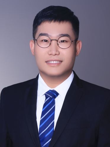 Ge Gao (高戈)合作论文数Institute of Engineering Medicine, Beijing Institute of Technology11
 Jinah Jang合作论文数Biofabrication & Translational Medicine Laboratory, Pohang University of Science and Technology;School of Convergence Science and Technology, Pohang University of Science and Technology;School of Interdisciplinary Bioscience and Bioengineering, Pohang University of Science and Technology;Yonsei Institute of Convergence Technology, Yonsei University;BioBricks Co., Ltd8Dongjun Lee合作论文数5
 Wonbin Park合作论文数Department of Mechanical Engineering, Pohang University of Science and Technology5
 Sik Yoon合作论文数College of Medicine, Pusan National University5
 Arvind Kumar Shukla合作论文数4Ju Young Park合作论文数3Jae-Seong Lee合作论文数3Qiqi Gao合作论文数3Jeong Sik Kong合作论文数POSTECH, Pohang Univ Sci & Technol3Suhun Chae合作论文数EDmicBio Inc.3Jae Yeon Lee合作论文数3
 Jongwon Rhie合作论文数3
 Dong-Heon Ha合作论文数3
 Sung Won Kim合作论文数2
 Jaeho Kim合作论文数Department of Physiology, School of Medicine, Pusan National University2
 Jongmin Kim合作论文数Harvard Univ, Wyss Inst Biol Inspired Engn, Boston, MA 02115 USA2Ryu Yeon-Hee合作论文数2Sae-Ock Oh合作论文数2Kye-Sung Lee合作论文数1Hee-Gyeong Yi合作论文数1Inn Chul Nam合作论文数1donguk kim合作论文数1Dahong Kim合作论文数1
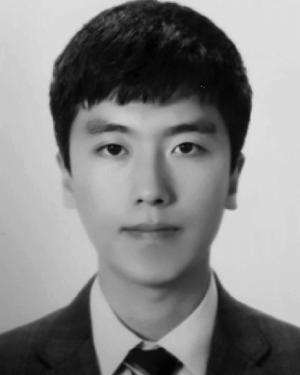 Chan Bae Jeong合作论文数1sungjun ahn合作论文数1DW Cho合作论文数1
 Elisabeth Phillips合作论文数1
 Kangwon Lee合作论文数1Jeonghyun Son合作论文数1Dongjin Lee合作论文数1Suryong Kim合作论文数Seoul National University1Wonil Han合作论文数Division of Integrative Bioscience and Biotechnology, POSTECH1Gyu Tae Park合作论文数1
 Gretel Major合作论文数Christchurch Regenerat Med & Tissue Engn CReaTE G, Univ Otago Christchurch1
 Ji Min Seok合作论文数1Keunchul Lee合作论文数Dept Phys Educ, Changwon Natl Univ1Hyeonji Kim合作论文数Department of Mechanical Engineering, Pohang University of Science and Technology (POSTECH),1Jisoo Kim合作论文数POSTECH, Pohang Univ Sci & Technol1Jin Woo Jung合作论文数POSTECH, Pohang Univ Sci & Technol1Steven Wise合作论文数School of Medicine Sciences, Faculty of Medicine and Health, The University of Sydney1Choi Tae Hyun合作论文数1
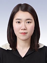 Yeo Seon Ju合作论文数School of Chemical Engineering, Sungkyunkwan University1Kang Hyun-Wook合作论文数Wake Forest Institute for Regenerative Medicine, Wake Forest University School of Medicine1Choi Seok Hwa合作论文数1
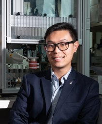 Khoon S Lim合作论文数1




