Small axons far outnumber larger fibers in the corticospinal tract, but the function of these small axons remains poorly understood. This is because they are difficult to identify, and therefore their physiology remains obscure. To assess the extent of the mismatch between anatomic and physiological measures, we compared conduction time and velocity in a large number of macaque corticospinal neurons with the distribution of axon diameters at the level of the medullary pyramid, using both light and electron microscopy. At the electron microscopic level, a total of 4,172 axons were sampled from 2 adult male macaque monkeys. We confirmed that there were virtually no unmyelinated fibers in the pyramidal tract. About 14% of pyramidal tract axons had a diameter smaller than 0.50 μm (including myelin sheath), most of these remaining undetected using light microscopy, and 52% were smaller than 1 μm. In the electrophysiological study, we determined the distribution of antidromic latencies of pyramidal tract neurons, recorded in primary motor cortex, ventral premotor cortex, and supplementary motor area and identified by pyramidal tract stimulation (799 pyramidal tract neurons, 7 adult awake macaques) or orthodromically from corticospinal axons recorded at the mid-cervical spinal level (192 axons, 5 adult anesthetized macaques). The distribution of antidromic and orthodromic latencies of corticospinal neurons was strongly biased toward those with large, fast-conducting axons. Axons smaller than 3 μm and with a conduction velocity below 18 m/s were grossly underrepresented in our electrophysiological recordings, and those below 1 μm (6 m/s) were probably not represented at all. The identity, location, and function of the majority of corticospinal neurons with small, slowly conducting axons remains unknown.
A possible role of the cerebellum in cognitive function might be revealed through its anatomical connections with specific regions of the cerebral cortex. To understand the kind of information transmitted between the cortex and cerebellum, we studied the connections from six subdivisions of frontal and prefrontal cortex using diffusion imaging tractography. Cortico-pontine fibers travel through the cerebral peduncles and reach the cerebellum by way of a synaptic link in the pontine nuclei. In 19 human data sets, we tracked connections between the cerebral peduncle and left hemispheric masks of the superior frontal gyrus (SFG), precentral gyrus (PcG), middle frontal gyrus (MFG), orbital frontal cortex, and two regions of inferior frontal gyrus, including pars opercularis and pars triangularis. Cortico-pontine fibers arose from the PcG, the caudal/medial SFG and a small region of the MFG in a majority of the subjects analyzed. While these regions do have known roles in cognitive and executive functions, all three are strongly associated with the planning and execution of eye movements. Connections from more ventral prefrontal cortex were negligible, indicating that these regions are only sparsely represented in the circuit. Based on this pattern of connectivity, it is likely that the prefrontal connections to the cerebellum are involved in covert motor operations and the control of eye movements.
In this chapter, the authors pay tribute to Mike Gazzaniga, who graduated from Dartmouth College in Hanover, New Hampshire, and pioneered human split-brain research along with Roger W. Sperry and Joseph Bogen. They briefly discuss Gazzaniga’s life and work, and their association with him. Gazzaniga received his Ph.D. in psychobiology from the California Institute of Technology (Cal Tech), where he worked under the guidance of Roger Sperry. His work in the field of human split-brain research is considered a remarkable contribution to the discipline of psychobiology, which helps to explain the neural basis of the human mind. The chapter further focuses on the structural and functional analysis of the corpus callosum and how it works in hemispheric communication to help explain cognition and behavioral control.
 BE MERCIER合作论文数5A Gibson合作论文数5
 Farrel Richard Robinson合作论文数5G Mower合作论文数4C.R. Legg合作论文数4Jan Voogd合作论文数4Kralj-Hans Ines合作论文数3Christopher H Yeo合作论文数3
 Giovanni Berlucchi合作论文数Department of Neurosciences, Biomedicine and Movement Sciences, University of Verona;Medical Faculty, University of Verona3Janet Lee Cohen合作论文数3MJ HARDIMAN合作论文数3J G May合作论文数2M A Maier合作论文数2Hilperath F合作论文数2
 Giacomo Rizzolatti合作论文数University of Parma2
 John Stein合作论文数Department of Physiology, Anatomy and Genetics, Medical Sciences Division, University of Oxford2
 Gottfried Schlaug合作论文数Neuroimaging Laboratory, Beth Israel Deaconess Medical Center;Music and Neuroimaging Laboratory, Harvard Medical School;Department of Neurology, University of Massachusetts Medical School2Peter Thier合作论文数Hertie Institute for Clinical Brain Research, University of Tübingen2
 Karl W. Doron合作论文数Harold Goodglass Boston University Aphasia Research Center, Boston University School of Medicine and the Veterans Affairs Boston Healthcare System2Joan S. Baizer合作论文数2
 Elisabetta Vaudano合作论文数Department of Human;Anatomy and Physiology;University of Turin2Peter a Kirkwood合作论文数2
 Joseph Classen合作论文数Clinic and Policlinic of Neurology, University Hospital of Leipzig;Department of Neurology, University Hospital Leipzig2
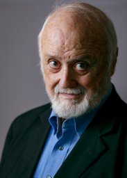 William H. Calvin合作论文数Department of Neurological Surgery, University of Washington School of Medicine2J. S. Baizer合作论文数2
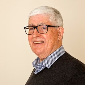 Roger Lemon合作论文数Queen Square Institute of Neurology, University College London2Erwin Kunesch合作论文数2S Barash合作论文数1adam w jackson合作论文数1Jg May合作论文数1H.-J. Freund合作论文数1Hj Freund合作论文数1R A Johnson合作论文数1Mark Hollins合作论文数1
 Jye-Chang Lee合作论文数1
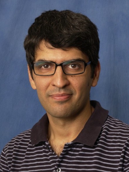 Ehsan Arabzadeh合作论文数John Curtin School of Medical Research, College of Health and Medicine, The Australian National University1
 Rudiger Seitz合作论文数1
 Johannes Mayrhofer合作论文数University of Zurich, University of Zurich1Jenkinson E W合作论文数1Sivakov A合作论文数1Melikyan A合作论文数1
 Sasha Kraskov合作论文数Faculty of Medical Sciences, Newcastle University1
 Fritzie Arce-McShane合作论文数Department of Oral Health Sciences, School of Dentistry, University of Washington1Bruno Weber合作论文数PET Center, University Hospital Zurich1
 Karoline Spang合作论文数1
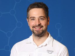 Musall Simon合作论文数1
 von der Behrens Wolfger合作论文数1Skreb Vida合作论文数1James M. Sprague合作论文数1




