Extracellular vesicles (EVs) are lipid membrane-bound nanoscale (20 nm – 1000 nm) objects shed by all cells. As EVs play an important role in intercellular signaling, these have emerged as promising biomarkers for many diseases including cancer and neurodegenerative disorders. A major bottleneck in research into EVs and their smooth translation to clinic as disease biomarkers is a lack of access to affordable and user-friendly technologies to quickly isolate EVs from complex biological and clinical samples with acceptable purity and yield. Here, we report two different designs of inertial microfluidic devices (e.g. LoAR-Exo and HiAR-Exo) that can isolate EVs from different kinds of biological samples in a single label-free step using a spiral microchannel design with different aspect ratios. LoAR-Exo has a height-to-width aspect ratio ∼ 1, while HiAR-Exo has a height-to-width aspect ratio of 2.5, optimized by COMSOL simulations. We tested both designs by separating polystyrene particles of size < 1 μm from a heterogeneous mixture. We then benchmarked the performance of the microfluidic chips against ultracentrifugation and a precipitation kit by isolating EVs from the cell culture-conditioned media (CCM) of MDA-MB-231 cells. We also compared the microchip with SEC by using the CCM from H1975/OR cells. The size distribution of EVs isolated by the microfluidic chip was comparable with ultracentrifugation, precipitation and SEC. In summary, both devices isolated EVs from as little as 1 mL of sample volume using a label-free technique in a continuous manner and without any user intervention. Both microfluidic platforms offer a simple, efficient, and scalable alternative to conventional methods for EV isolation. ### Competing Interest Statement DP, SA and AA have filed an Indian patent application (202421016839) and a PCT application (WO 2025/186844 A1) titled Device and method for label-free isolation of extracellular vesicles. The authors have no other conflicts of interest to declare. Wadhwani Research Centre for Bioengineering, IIT Bombay, DO/2022-WRCB002-082, DO/2025-WRCB002-105 Blockchain for Impact-BIOME, DO/2024-INCO002-003 VAIBHAV fellowship, INAE/DST-VF/2024/I/17 Seed Funding for Collaboration and Partnership Projects, IOE cell, IIT Bombay, RD/0523-IOE00I0-053 WRCB-HiMedia Entrepreneur-in-Residence fellowship, DO/2025-HIME004
The elastic properties of nanoscale extracellular vesicles (EVs) are believed to influence their cellular interactions, thus having a profound implication in intercellular communication. However, accurate quantification of their elastic modulus is challenging due to their nanoscale dimensions and their fluid-like lipid bilayer. We show that the previous attempts to develop atomic force microscopy-based protocol are flawed as they lack theoretical underpinning as well as ignore important contributions arising from the surface adhesion forces and membrane fluctuations. We develop a protocol comprising a theoretical framework, experimental technique, and statistical approach to accurately quantify the bending and elastic modulus of EVs. The method reveals that membrane fluctuations play a dominant role even for a single EV. The method is then applied to EVs derived from human embryonic kidney cells and their genetically engineered classes altering the tetraspanin expression. The data show a large spread; the area modulus is in the range of 4 to 19 mN/m and the bending modulus is in the range of 15 to 33 k B T , respectively. Surprisingly, data for a single EV, revealed by repeated measurements, also show a spread that is attributed to their compositionally heterogeneous fluid membrane and thermal effects. Our protocol uncovers the influence of membrane protein alterations on the elastic modulus of EVs.
Detection of analytes using streaming current has previously been explored using both experimental approaches and theoretical analyses of such data. However, further developments are needed for establishing a viable microchip that can be exploited to deliver a sensitive, robust, and scalable biosensor device. In this study, we demonstrated the fabrication of such a device on silicon wafer using a scalable silicon microfabrication technology followed by characterization and optimization of this sensor for detection of small extracellular vesicles (sEVs) with sizes in the range of 30 to 200 nm, as determined by nanoparticle tracking analyses. We showed that the sensitivity of the devices, assessed by a common protein-ligand pair and sEVs, significantly outperforms previous approaches using the same principle. Two versions of the microchips, denoted as enclosed and removable-top microchips, were developed and compared, aiming to discern the importance of high-pressure measurement versus easier and better surface preparation capacity. A custom-built chip manifold allowing easy interfacing with standard microfluidic connections was also constructed. By investigating different electrical, fluidic, morphological, and fluorescence measurements, we show that while the enclosed microchip with its robust glass-silicon bonding can withstand higher pressure and thus generate higher streaming current, the removable-top configuration offers several practical benefits, including easy surface preparation, uniform probe conjugation, and improvement in the limit of detection (LoD). We further compared two common surface functionalization strategies and showed that the developed microchip can achieve both high sensitivity for membrane protein profiling and low LoD for detection of sEV detection. At the optimum working condition, we demonstrated that the microchip could detect sEVs reaching an LoD of 104 sEVs/mL (when captured by membrane-sensing peptide (MSP) probes), which is among the lowest in the so far reported microchip-based methods.
High heterogeneity in the membrane protein expression of small extracellular vesicles (sEVs) means that bulk methods relying on antibody-based capture for expression analysis have a drawback that each type of antibody may capture a different sub-population. An improved approach is to capture a representative sEV population, without any bias, and then perform a multiplexed protein expression analysis on this population. However, such a possibility has been largely limited to fluorescence-based methods. Here, we present a novel electrostatic labelling strategy and a microchip-based all-electric method for membrane protein analysis of sEVs. The method allows us to profile multiple surface proteins on the captured sEVs using alternating charge labels. It also permits the comparison of expression levels in different sEV-subtypes. The proof of concept was tested by capturing sEVs both non-specifically (unbiased) as well as via anti-CD9 capture probes (biased), and then profiling the expression levels of various surface proteins using the charge labelled antibodies. The method is the first of its kind, demonstrating an all-electrical and microchip based method that allows for unbiased analysis of sEV membrane protein expression, comparison of expression levels in different sEV subsets, and fractional estimation of different sEV sub-populations. These results were also validated in parallel using a single-sEV fluorescence technique.
Detection of analyte using streaming current has been previously explored using both experimental and theoretical approaches. However, little has been done to develop a viable microchip which can be exploited to deliver a sensitive, robust, and scalable biosensor device. In this study, we demonstrate the fabrication of such a device on silicon wafer using a scalable silicon microfabrication technology followed by their characterization and optimization for highly sensitive detection of small extracellular vesicles (sEVs). We show that the sensitivity of the devices, estimated using a common protein-ligand pair and sEVs significantly outperforms previous reports using the same principle. Two versions of the microchips, denoted as enclosed and open-top microchip, were developed and compared aiming to discern the importance of high-pressure measurement vs easier and better surface preparation capacity. A custom-built chip-manifold allowing easy interfacing with standard microfluidic connections was also developed. By investigating different electrical, fluidic, morphological, and fluorescence measurements, we show that while the enclosed microchip with its robust glass-silicon bonding can withstand higher pressure and thus generate higher streaming current, the open-top configuration offers several practical benefits including easy surface preparation, uniform probe conjugation, and improvement in the limit of detection (LoD). We further compare two common surface functionalization strategies and show that the proposed microchip can achieve both high sensitivity for membrane protein profiling and low LoD for sEV detection. At the optimum condition, we demonstrate that the microchip can detect sEVs reaching a LoD of 10 4 sEV/mL, which is among the lowest in the reported microchip-based methods.
During the last decade, hydrodynamic cavitation has been implemented in various applications such as energy harvesting and biomedical applications. Facile hydrodynamic cavitation methods are required for fulfilling the requirements in these applications. In this study, a new generation microfluidic device containing eight parallel micro-orifices with a new design was fabricated and tested with the purpose of intensifying the cavitating flows and early cavitation inception. The roughness elements in the micro-orifices facilitated cavitation inception. This study presents a general perspective of occurrence of different cavitating flow patterns in microscale and addresses the ambiguities about the conditions for the formation of a specific flow pattern. Cavitation inception occurred with the appearance of small bubbles emerging from roughness elements at a rather low upstream pressure in the open loop experimental setup. A reduction in the cavitation number resulted in the formation of different flow patterns such as cavitation clouds, twin cavities, sheet cavities, and bubbly flows. Having several flow patterns with different intensities all together within a single microfluidic device is the main advantage of the proposed device over the state of the art microfluidic devices. Generation of flow patterns with various released energy levels makes this proposed device a unique multi-functional platform, which can be implemented to a lab on a chip platform for applications such as nanoparticle synthesis and wound healing.
This study presents a method based on carpet bombardment of immobilized cells with cavitating flows. For this, immobilized cancer cell lines are exposed to micro scale cavitating flows from the tip of a micro nozzle under the effect of cavitation microbubbles. The deformation as a result of cavitation bubbles on exposed cells differs from one cell type to another. Therefore, the difference in cell deformation upon cavitation exposure (carpet bombardment) acts as a valuable indicator for cancer diagnosis. The developed system is tested on HCT-116 (Human Colorectal Carcinoma), MDA-MB-231 (Breast Adenocarcinoma), ONCO-DG-1 (Ovarian Adenocarcinoma) cell lines due to their clinical importance. The mechanical effects of cavitation are examined by considering the single-cell lysis effect (the cell membrane is ruptured, and the cell is destroyed) with the help of the Scanning Electron Microscopy (SEM) technique. Our study proposes a promising label-free method for the potential use in cancer diagnosis with cavitation bubble collapse, where microbubbles could be precisely controlled and directed to the desired locations, as well as the characterization of the biophysical properties of cancer cells. The proposed approach tool has the advantages of label-free approach, simple structure and low cost and is a substantial alternative for the existing tools.
Hydrodynamic cavitation is one of the major phase change phenomena and occurs with a sudden decrease in the local static pressure within a fluid. With the emergence of microelectromechanical systems (MEMS), high-speed microfluidic devices have attracted considerable attention and been implemented in many fields, including cavitation applications. In this study, a new generation of ‘cavitation-on-a-chip’ devices with eight parallel structured microchannels is proposed. This new device is designed with the motivation of decreasing the upstream pressure (input energy) required for facile hydrodynamic cavitation inception. Water and a poly(vinyl alcohol) (PVA) microbubble (MB) suspension are used as the working fluids. The results show that the cavitation inception upstream pressure can be reduced with the proposed device in comparison with previous studies with a single flow restrictive element. Furthermore, using PVA MBs further results in a reduction in the upstream pressure required for cavitation inception. In this new device, different cavitating flow patterns with various intensities can be observed at a constant cavitation number and fixed upstream pressure within the same device. Moreover, cavitating flows intensify faster in the proposed device for both water and the water–PVA MB suspension in comparison to previous studies. Due to these features, this next-generation ‘cavitation-on-a-chip’ device has a high potential for implementation in applications involving microfluidic/organ-on-a-chip devices, such as integrated drug release and tissue engineering.
Tremendous research efforts have recently focused on the synthesis of graphene from graphitic materials, while environmental issues, scalability, and cost are some of the major challenges to be surmounted. Liquid phase exfoliation (LPE) of graphene is one of the principal methods for this synthesis. Nevertheless, sufficient information about the mechanisms of exfoliation has yet to emerge. Here, a microreactor based on the hydrodynamic cavitation (HC) on a chip concept is introduced to exfoliate graphite in a totally green process which involves only natural graphite flakes and water. HC-treated graphitic materials were characterized by UV-Vis and Raman spectroscopy, DLS (Dynamic Light Scattering), AFM (Atomic Force Microscopy), and SEM (Scanning Electron Microscopy) analyses. The present sustainable reactor system was found to exfoliate thick and large graphite particles to nano-sized sheets (∼1.2 nm) with a lateral size of ∼500 nm to 5 μm.
Hydrodynamic cavitation is considered an effective tool to be used in different applications, such as surface cleaning, ones in the food industry, energy harvesting, water treatment, biomedical applications, and heat transfer enhancement. Thus, both characterization and intensification of cavitation phenomenon are of great importance. This study involves design and optimization of cavitation on chip devices by utilizing wall roughness elements and working fluid alteration. Seven different microfluidic devices were fabricated and tested. In order to harvest more energy from cavitating flows, different roughness elements were used to decrease the inlet pressure (input to the system), at which cavitation inception occurs. The implemented wall roughness elements were engineered structures in the shape of equilateral triangles embedded in the design of the microfluidic devices. The cavitation phenomena were also studied using ethanol as the working fluid, so that the fluid behavior differences in the tested cavitation on chip devices were explained and compared. The employment of the wall roughness elements was an effective approach to optimize the performances of the devices. The experimental results exhibited entirely different flow patterns for ethanol compared to water, which suggests the dominant effect of the surface tension on hydrodynamic cavitation in microfluidic channels.
Due to the potential of significant energy release in cavitating flows, early cavitation inception and intensification of cavitating flows are of great importance. To use this potential, we investigated the deagglomeration of nanoparticle clusters with the implementation of hydrodynamic cavitation in a microfluidic device. For this purpose, a microfluidic device with a micro-orifice geometry was designed and fabricated using standard microfabrication processes. The system was tested with distilled water in the assembled experimental setup. The flow patterns were characterized using the cavitation number and inlet pressure. Titania nanoparticles were utilized to prepare nanoparticle suspensions. The suspensions were heated to allow agglomeration of nanoparticles. The system was operated with the new working fluid (nanoparticle clusters) at different inlet pressures. After characterizing flow patterns, the flow patterns were compared with those of pure water. The deagglomeration effects of hydrodynamic cavitation on nanoparticle clusters showed the possibility to apply this method for the stabilization of nanoparticles, which paves way to the implementation of nanoparticle suspensions to thermal fluid systems for increased energy efficiency as well as to drug delivery. Our results also indicate that the presence of nanoparticles in the working fluid enhanced cavitation intensity due to the increase in the number of heterogeneous nucleation sites.
In this study, three microfluidic devices with different geometries are fabricated on silicon and are bonded to glass to withstand high-pressure fluid flows in order to observe bacteria deactivation effects of micro cavitating flows. The general geometry of the devices was a micro orifice with macroscopic wall roughness elements. The width of the microchannel and geometry of the roughness elements were varied in the devices. First, the thermophysical property effect (with deionized water and phosphate-buffered saline (PBS)) on flow behavior was revealed. The results showed a better performance of the device in terms of cavitation generation and intensity with PBS due to its higher density, higher saturation vapor pressure, and lower surface tension in comparison with water. Moreover, the second and third microfluidic devices were tested with water and Salmonella typhimurium bacteria suspension in PBS. Accordingly, the presence of the bacteria intensified cavitating flows. As a result, both devices performed better in terms of the intensity of cavitating flow with the presence of bacteria. Finally, the deactivation performance was assessed. A decrease in the bacteria colonies on the agar plate was detected upon the tenth cycle of cavitating flows, while a complete deactivation was achieved after the fifteenth cycle. Thus, the proposed devices can be considered as reliable hydrodynamic cavitation reactors for "water treatment on chip" applications.
In this study, the mixing performances of passive micromixers with sharp edges were investigated. First, a validation study was conducted by using experimental results on a passive micromixers in the literature. The arrangement of the triangle obstacles was then changed to optimize the mixing performance. The numerical simulation results showed an acceptable agreement with the experimental results. The new geometry of the mixer exhibited a significantly better performance compared to the original reference microfluidic device. The reason for this enhancement was analysed and discussed detail. The doubled vortex region in the optimized micromixer was responsible for the performance enhancement.
Cavitation is one of the phase change phenomena, which occurs as a result of a sharp static pressure drop in a fluidic system. The required pressure drop could be obtained either by acoustic wave propagation, known as acoustic cavitation, or by hydrodynamic pressure drop, known as hydrodynamic cavitation. There are different parameters affecting flow patterns in hydrodynamic cavitation such as geometry of the flow path, thermophysical properties of the working fluid, and roughness elements. In this thesis, high pressure resistive microfluidic devices are fabricated and tested at high inlet pressures. The flow behavior is characterized using cavitation number, Reynolds number, and cavitation flowrate. Different working fluids such as ethanol, PBS, and PFC5 droplets suspension in water are used and the performances of the devices are compared with the case of water. The effect of different lateral wall roughness elements on the flow pattern is studied and the general geometry of the devices is optimized. In addition, applications of hydrodynamic cavitation are considered. The energy harvesting application of hydrodynamic cavitation is implemented and the amount of the possible power generation of the microfluidic devices in the case of coupling with μ−TEGs is analytically investigated. Finally, the bacteria removal performances of the microfluidic devices are assessed. The advantages of the designed and fabricated microfluidic devices are presented and explored as well
Centrifugal pumps are among the most applicable machines in a wide variety of industrial systems for fluid pumping and transportation. Therefore their optimization has always been of great importance. Pump impellers play an important role in these machines as the energy transfer takes place in this part. In the present study, the impeller of a centrifugal pump is optimized by investigating the effect of adding splitter blades and modifying their geometry. A centrifugal pump is experimentally tested and numerically simulated and the characteristic curves are obtained. In the first stage, two different sets of splitter blades with different lengths are added to the impeller and the effect of splitter blade lengths on the results are explored. The case with the highest total head and overall efficiency is selected for the optimization process. The main blade and the splitter blade leading edge position and also the splitter blade distance between two successive blades are selected for the optimization process in the second stage. Efficiency and total head of the pump are considered as the optimization objectives. Using Design of Experiment (DoE) technique, the design space is created and response surface method is utilized to find the optimum geometry. The results show adding splitters can improve total head by about 10.6% and by modifying the geometry using DoE technique it could increase further by 4.4% with the negligible effect on the pump overall efficiency.
The tremendous potential of "hydrodynamic cavitation on microchips" has been highlighted during recent years in various applications. Cavitating flow patterns, substantially depending upon thermophysical and geometrical characteristics, promote diverse industrial and engineering applications, including food and biomedical treatment. Highly vaporous and fully developed patterns in microfluidic devices are of particular interest. In this study, the potential of a new approach, which includes cellulose nanofiber (CNF)- stabilized perfluorodroplets (PFC5s), was assessed inside microfluidic devices. The surfaces of these devices were modified by assembling various sizes of silica nanoparticles, which facilitated in the generation of cavitation bubbles. To examine the pressure effects on the stabilized droplets in the microfluidic devices, the upstream pressure was varied, and the cavitation phenomenon was characterized under different experimental conditions. The results illustrate generation of interesting, fully developed, cavitating flows at low pressures for the stabilized droplets, which has not been previously observed in the literature. Supercavitation flow pattern, filling the entire microchannel, were recorded at the upstream pressure of 1.7 MPa for the case of CNF-stabilized PFC5s, which hardly corresponds to cavitation inception for pure water in the same microfluidic device.
The phase change phenomenon in fluids as a result of low local pressure under a critical value is known as cavitation. Acoustic wave propagation or hydrodynamic pressure drop of the working fluid are the main reasons for inception of this phenomenon. Considering the released energy from the collapsing cavitation bubbles as a reliable source has led to its implementation to different fields, namely, heat transfer, surface cleaning and fouling, water treatment, food industry, chemical reactions, energy harvesting. A considerable amount of energy in the mentioned industries is required for thermal applications. Cavitation could serve for minimizing the energy demand and optimizing the processes. Thus, the energy efficiency of the systems could be significantly enhanced. This review article focuses on the direct and indirect thermal applications of hydrodynamic and acoustic cavitation. Relevant studies with emerging applications are discussed, while developments in cavitation, which have given rise to thermal applications during the last decade, are also included in this review.
In this study, energy harvesting with micro scale hydrodynamic cavitation-thermoelectric generation coupling is investigated. For this, three micro orifices with different geometrical dimensions are fabricated. The hydraulic diameter of the micro orifices are 66.6 μm, 75.2 μm, and 80 μm, while their length is the same (2000μm). Two different working fluids, namely water and Perfluoropentane droplet-water suspension, are utilized for cavitating flows in the fabricated micro orifices. The flow patterns at different upstream pressures are recorded using the high-speed camera system, and the experimental results are analyzed and compared. Thereafter, energy harvesting perspectives of cavitating flows are considered. The released heat from collapsing bubbles and the subsequent temperature rise on the end wall of the microchannel, which can be used as the source for the power generation, is calculated over time. Finally, a miniature energy harvesting system with cavitation system and thermoelectric generator coupling is presented. The maximum power corresponding to two different thermoelectric generators is estimated for with both working fluids and is compared with the required power to run miniature daily used electronics components.
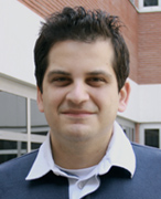 Ali Koşar合作论文数Faculty of Engineering and Natural Sciences, Sabanci University11
 Morteza Ghorbani合作论文数10
 Apurba Dev合作论文数Division of Photonics, Department of Applied Physics, School of Engineering Sciences, KTH Royal Institute of Technology5
 Petra Haag合作论文数4
 Kristina Viktorsson合作论文数Karolinska Biomics Center, Department of Oncology/Pathology, Stockholm, Sweden4Fredrik Stridfeldt合作论文数3
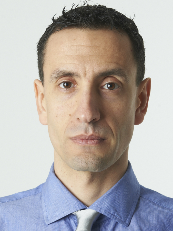 Guillermo Villanueva合作论文数School of Engineering, École Polytechnique Fédérale de Lausanne3
 Rolf Lewensohn合作论文数Department of Oncology-Pathology, Karolinska Institutet;Oncology Clinic, Karolinska Hospital3
 Dmitry Grishenkov合作论文数Division of Biomedical Imaging, Department of Biomedical Engineering and Health Systems, School of Engineering Sciences in Chemistry, Biotechnology and Health, KTH Royal Institute of Technology3
 Ali Hosseinpour Shafaghi合作论文数Fac Engn & Nat Sci, Sabanci Univ2
 Soroush Niazi合作论文数Faculty of Engineering and Natural Science, Sabanci University2
 Siddharth Sourabh Sahu合作论文数School of Engineering Sciences, KTH Royal Institute of Technology2
 Farzad Rokhsar Talabazar合作论文数Nanotechnol Res & Applicat Ctr, Sabanci Univ2
 Mohammad Jafarpour合作论文数Institute of Materials Science and Engineering, Ecole Polytechnique Federale de Lausanne (EPFL),2
 Araz Sheibani Aghdam合作论文数Fac Engn & Nat Sci, Sabanci Univ2
 Zuvin Merve合作论文数2Anna Hanner合作论文数Division of Polymeric Materials, Department of Fibre and Polymer Technology, School of Engineering Sciences in Chemistry, Biotechnology and Health, KTH Royal Institute of Technology2
 Dhrubaditya Mitra合作论文数QMUL2Ábel Nagy合作论文数1Vikash Pandey合作论文数1
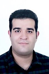 M. Namazizadeh合作论文数Fac Mech & Energy Engn, Shahid Beheshti Univ1Taher Abbasiasl合作论文数1
 Ghazaleh Gharib合作论文数Fac Engn & Nat Sci FENS, Sabanci Univ1Ayhan Parlar合作论文数Faculty of Engineering and Natural Sciences, Sabanci University1
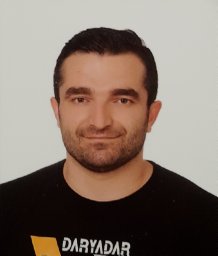 Milad Torabfam合作论文数Faculty of Engineering and Natural Science, Sabanci University1
 Maxime Gestin合作论文数1
 Vipin Agrawal合作论文数Stockholm Univ1
 Gizem Aydemir合作论文数Department of Mechatronics Engineering, Yildiz Technical University1
 Hongjian Chen合作论文数KTH Royal Inst Technol, Dept Biomed Engn & Hlth Syst, SE-14157 Stockholm, Sweden1
 Mohammad Vajdi合作论文数Dept Mech Engn, Univ Mohaghegh Ardabili1
 Ozlem Kutlu合作论文数Nanotechnology Research and Applications Center, Sabanci University1
 Mohammad Mojaddam合作论文数Fac Mech & Energy Engn, Shahid Beheshti Univ1
 Mehrdad Karimzadehkhouei合作论文数Dept Mech Engn, Koc Univ1
 Margaret Holme合作论文数University of Basel, University Hospital Basel1
 Andre Gorgens合作论文数University Hospital Essen, University of Duisburg-Essen;German Cancer Consortium (DKTK).;University of Duisburg-Essen, University Hospital Essen1
 Samir El Andaloussi合作论文数1
 Debjani Paul合作论文数MRC Cancer Cell Unit, Hutchison/MRC Research Centre1
 Prakriti Tayalia合作论文数School of Engineering and Applied Sciences, Harvard University1
 Mustafa K. Bayazit合作论文数Department of Chemistry, Durham University1
 Jan Linnros合作论文数KTH Royal Institute of Technology1
 Fevzi Çakmak Cebeci合作论文数Istanbul International Center for Energy and Climate, Sabanci University1Meral Yüce合作论文数Sabanci Univ, SUNUM Nanotechnol Res & Applicat Ctr, TR-34956 Istanbul, Turkey.1
 Kursat Sendur合作论文数Seagate Technology1
 Murat Kaya Yapici合作论文数Department of Electrical and Computer Engineering, Texas A&M University1
 Luigi de Petris合作论文数Department of Oncology-Pathology, Karolinska Institutet1
 M. Pinar Menguc合作论文数Department of Mechanical Engineering, University of Kentucky1
 Marina Cretich合作论文数Institute of Chemistry of Molecular Recognition (ICRM),, CNR1
 Amelie Eriksson Karlström合作论文数Division of Protein Engineering, Department of Protein Science, School of Engineering Sciences in Chemistry, Biotechnology and Health, KTH Royal Institute of Technology1
 Huseyin Uvet合作论文数Graduate School of Engineering Science, Osaka University1




