Non-myeloablative hematopoietic cell transplantation (HCT) is a curative option for individuals with sickle cell disease (SCD). Our traditional goal with this approach has been to achieve a state of mixed donor/recipient chimerism. Recently, we reported an increased risk of hematologic malignancies (HMs) in adults with SCD following graft failure or mixed chimerism. To evaluate the origin of HMs, we performed chimerism analyses of 5 patients with SCD who developed HMs after non-myeloablative HCT. DNA was extracted from sorted peripheral blood or bone marrow cells representing mature cell lineages or leukemic blasts and subjected to chimerism analysis by PCR amplification of polymorphic short tandem repeats. Unlike mature cell lineages in patients with mixed chimerism, which still showed a donor-derived fraction of cells, leukemic blast cells were found to be 99-100% recipient-derived in all patients. Non-myeloablative conditioning allows for the survival of patients' cells that might harbor pre-leukemic clones that possess the capacity to evolve under genotoxic or environmental stress into malignancies; therefore, we have modified our HCT protocols with the goal of full donor chimerism to mitigate the risk of HM development.
Haploidentical hematopoietic cell transplantation (haplo-HCT) is associated with an increased risk of allograft rejection. Here, we employed a major histocompatibility complex (MHC)-mismatched allogeneic HCT (allo-HCT) murine model to better understand the role of Gal-1 in immune tolerance. Transplanted mice were classified into either rejected or engrafted based on donor chimerism levels. We noted significantly higher frequencies of CD4+ T cells, CD8+ T cells, natural killer cells, IFN-γ and TNF-α producing CD4+ T cells, and IFN-γ producing dendritic cells and macrophages in rejected mice. Conversely, we found significantly increased frequencies of regulatory T cells (Tregs), predominantly Helios+, IL-10-producing CD4+ T cells, type 1 regulatory (Tr1) cells, and the proportion of Tr1+Gal-1+ cells in engrafted mice. Further, Gal-1 specific blockade in Tregs reduced suppression of effector T cells in engrafted mice. Lastly, effector T cells from engrafted mice were more prone to undergo apoptosis. Collectively, we have shown that Gal-1 may favor HSC engraftment in an MHC-mismatched murine model. Our results demonstrate that Gal-1-expressing Tregs, especially at earlier time points post-transplant, are associated with inducing immune tolerance and stable mixed chimerism after HCT.
Background: Hematopoietic stem cells (HSCs) with multilineage potential are critical for effective de novo T-cell generation and thymic recovery, restoring the adaptive immune system after Hematopoietic Cell Transplantation (HCT). Recent studies have brought new insights into functional heterogeneity within HSCs, revealing an organized and predictable framework governing the adoption of lineage-restricted fates. However, the gene-regulatory networks underlying lymphoid determination and their conservation with aging in HSCs need to be elucidated. This study aims to comprehensively investigate the molecular profile of young and old HSCs and identify HSC subsets with multilineage transcriptional programs. Results: We conducted single-cell multiomic RNA and ATAC sequencing of young and old HSCs defined by Lineage-CD34-CD48-CD150+Sca-1+Kit+. We identified two CD117 (Kit) HSC subsets, Kit lo and Kit hi, with unique transcriptional profiles. Kit hi HSCs were characterized by quiescence, platelet bias, and low-output gene signatures, while Kit lo HSCs enriched for multi-lineage and high-output gene signatures. We observed a decreased frequency of Kit lo HSCs in aged mice. Although Kit lo HSCs have been previously described to exhibit increased self-renewal (Shin et al. JEM, 2014), their T-cell potential and the molecular underpinnings governing lymphoid differentiation programs are unknown. We performed in vitro studies, S17 stromal assay, and artificial thymic organoids (ATOs), which revealed an enhanced output of lymphoid progenitors and T-cells from Kit lo HSCs, consistent across all age cohorts. We next compared Kit lo vs. Kit hi HSCs in a competitive allogeneic HCT model. Kit lo HSCs demonstrated improved thymic recovery and post-HCT T-cell reconstitution, independent of age. To orthogonally evaluate post-HCT thymic function, we assessed Recent Thymic Emigrants (RTEs) output, which further substantiated superior recovery in mice receiving Kit lo HSCs. We further observed that Kit lo HSC-derived T-cells exhibited better proliferation in response to an acute Listeria monocytogenes infection. Next, we transplanted middle-aged mice to evaluate the rejuvenation potential of different Kit HSC subsets. We observed that Kit lo HSCs partly mitigated age-related changes in the thymic microenvironment, specifically by enhancing the regeneration of thymic epithelial cells (TECs) in middle-aged mice, thereby reversing the decline in T-cell production. We found differential expression and activity of key transcription factors (TFs), including Runx3, Zbtb7a, and Ezh1, associated with lymphoid differentiation through integrative transcriptional and chromatin accessibility analyses of Kit lo HSCs. These TFs notably showed distinct patterns in Kit lo HSCs independent of age, indicating their potential role in driving their enhanced T-cell potential. To establish the existence of a comparable human subset, we interrogated a human BM CITE-Seq dataset (Sommarin et al. Biorxiv 2021) for the mouse Kit lo gene signature. In concordance with our findings in mice, we observed an enrichment of the Kit lo gene program in young HSCs and decreased frequency of Kit lo HSCs in old BM. Next, we used ATOs to validate differential lymphoid potential. We found increased T-cell output originating from Kit lo HSCs, thus underscoring their enhanced lymphoid capacity and potential clinical relevance. Conclusion: Collectively, we demonstrate a distinct HSC subset, Kit lo HSC, with an age-conserved lymphoid gene program that enhances T-cell production and facilitates thymic regeneration. Importantly, we demonstrate the presence of an analogous HSC subset in humans, revealing the relevance of these insights to human health and disease. Thus, Kit lo HSCs have therapeutic potential to counteract age-associated immune senescence and treatment-related immune suppression.
Allogeneic hematopoietic cell transplantation (allo-HCT) offers a curative option for patients with certain non-malignant hematological diseases. High-dose post-transplant cyclophosphamide (PT-Cy) (200 mg/kg) and sirolimus (3 mg/kg), (HiC) synergistically induce stable mixed chimerism. Further, sirolimus and cytotoxic T lymphocyte-associated antigen-4 immunoglobulin (CTLA4-Ig), also known as Abatacept (Aba), promote immune tolerance and allograft survival. Here, in a major histocompatibility complex (MHC)-mismatched allo-HCT murine model, we combined Aba and/or T-cell depleting anti-Thy1.2 (Thy) with a lower dose of PT-Cy (50 mg/kg) and Sirolimus (3 mg/kg), (LoC). While mice in the LoC group showed graft rejection, the addition of Thy to LoC induced similar donor chimerism levels when compared to the HiC group. However, the addition of Aba to LoC led to graft acceptance only in younger mice. When Thy was added to the LoC+Aba setting, graft acceptance was restored in both age groups. Engrafted groups displayed significantly reduced frequencies of recipient-specific interferon-γ-producing T cells as well as an increased frequency in regulatory T cells (Tregs) except in the LoC+Aba group. Splenocytes from engrafted mice showed no proliferation upon restimulation with Balb/c stimulators. Collectively, in combination with Aba or Thy, LoC may be considered to reduce graft rejection in patients who undergo allo-HCT.
Roux-en-Y gastric bypass (RYGB) leads to the improvement of many obesity-associated conditions. The degree to which post-operative macronutrient composition contributes to metabolic improvement after RYGB is understudied. A mouse model of RYGB was used to examine the effects of diet on the post-operative outcomes of RYGB. Obese mice underwent either Sham or RYGB surgery and were administered either chow or HFD and then monitored for an additional 8 weeks. After RYGB, reductions to body weight, fat mass, and lean mass were similar regardless of diet. RYGB and HFD were independently detrimental to bone mineral density and plasma vitamin D levels. Independent of surgery, HFD accelerated hematopoietic stem and progenitor cell proliferation and differentiation and exhibited greater myeloid lineage commitment. Independent of diet, systemic iron deficiency was present after RYGB. In both Sham and RYGB groups, HFD increased energy expenditure. RYGB increased fecal energy loss, and HFD after RYGB increased fecal lipid content. RYGB lowered fasting glucose and liver glycogen levels but HFD had an opposing effect. Indices of insulin sensitivity improved independent of diet. HFD impaired improvements to dyslipidemia, NAFLD, and fibrosis. Post-operative diet plays a significant role in determining the degree to which RYGB reverses obesity-induced metabolic abnormalities such as hyperglycemia, dyslipidemia, and NAFLD. Diet composition may be targeted in order to assist in the treatment of post-RYGB bone mineral density loss and vitamin D deficiency as well as to reverse myeloid lineage commitment. HFD after RYGB continues to pose a significant multidimensional health risk.
Skeletal stem and progenitor cells (SSPCs) perform bone maintenance and repair. With age, they produce fewer osteoblasts and more adipocytes leading to a loss of skeletal integrity. The molecular mechanisms that underlie this detrimental transformation are largely unknown. Single-cell RNA sequencing revealed that Notch signaling becomes elevated in SSPCs during aging. To examine the role of increased Notch activity, we deleted Nicastrin, an essential Notch pathway component, in SSPCs in vivo. Middle-aged conditional knockout mice displayed elevated SSPC osteo-lineage gene expression, increased trabecular bone mass, reduced bone marrow adiposity, and enhanced bone repair. Thus, Notch regulates SSPC cell fate decisions, and moderating Notch signaling ameliorates the skeletal aging phenotype, increasing bone mass even beyond that of young mice. Finally, we identified the transcription factor Ebf3 as a downstream mediator of Notch signaling in SSPCs that is dysregulated with aging, highlighting it as a promising therapeutic target to rejuvenate the aged skeleton.
Background: Aging hematopoiesis is characterized by increased numbers of hematopoietic stem cells (HSCs) that exhibit impaired self-renewal and long-term reconstitution potential. We previously demonstrated that differences in surface expression of c-Kit in young mouse HSCs could identify distinct functional HSC subsets, with c-Kithi HSCs exhibiting reduced self-renewal and megakaryocytic biased differentiation. We therefore sought to determine if similar functional and molecular differences may be observed in old HSCs. Methods: We evaluated c-Kithi and c-Kitlo subsets of HSCs (CD34-CD150+LSK) from young (8-12 wk), and old (18-20 mo) C57BL/6J mice. For competitive transplantation assays, 250 double sorted c-Kithi or c-Kitlo HSCs from aged CD45.2 mice were mixed with 5x10⁵ unfractionated bone marrow mononuclear cells (BMMCs) from young CD45.1 mice. In parallel, we generated the chromatin accessibility and transcriptional profiles of c-Kithi and c-Kitlo HSCs from young and old mice. Results: We first performed competitive transplants of old c-Kithi and c-Kitlo HSCs into young recipients. These studies demonstrated age-related myeloid bias exhibited by both HSC subsets. In addition, similar to young HSCs, old c-Kithi HSCs exhibit significantly reduced long-term reconstitution capacity compared to old c-Kitlo HSCs. Next, to evaluate age-related functional differences, we performed equal competitive transplants in which equal numbers of c-Kithi or c-Kitlo old and young HSCs were transplanted into the same young recipient. These studies demonstrated that c-Kithi HSCs exhibit similar reconstituting capacity independent of age. In contrast, old c-Kitlo HSCs exhibited significantly reduced long-term reconstitution compared to their young counterparts, though with better-preserved self-renewal capacity, as reflected in their self-renewal quotients (SRQ). However, when we quantified the capacity of transplanted HSCs to give rise to downstream committed progenitors and mature hematopoietic cells, calculated as their differentiation quotient (DQ), old c-Kitlo HSCs exhibited markedly reduced DQ compared to their young counterparts. In contrast, c-kithi recipients showed that old c-Kithi HSCs gave rise to mature cell lineages more efficiently. To investigate molecular mechanisms driving heterogeneity in HSC aging, we evaluated the transcriptional profiles of young and old HSCs. Old c-Kitlo HSCs showed significant enrichment of genes associated with inflammation, interferon responses, and the quiescent state in comparison to old c-Kithi HSCs. On the other hand, old c-Kithi HSCs were enriched for cell cycle related and MYC target genes compared to old c-Kitlo HSCs and young HSCs. Old c-Kithi HSCs also exhibited increased expression of genes associated with high cell output state and high mitochondrial membrane potential, which is associated with a more active HSC cell state. To investigate the potential role of chromatin accessibility in determining the observed transcriptional changes, we performed ATAC-seq of young and old HSC subsets. Old HSCs exhibited more open chromatin peaks than young HSCs, with the number of peaks decreasing in the order of old c-Kitlo > old c-Kithi > young c-Kithi > young c-Kitlo HSCs. Evaluation of predicted transcription factor binding sites showed enrichment for CTCF and PU.1 sites in young c-Kitlo and c-Kithi HSCs, respectively, with PU.1 associated with high cell output states. In contrast, old c-Kitlo and c-Kithi HSCs were enriched for ERG and CTCF sites, respectively, with predicted binding sites enriched in genes associated with low and high cell output, respectively. These data suggest that differential ERG binding of quiescence and cellular output-related gene signatures regulate functional differences between old c-Kitlo and old c-Kithi HSCs. Conclusion: Overall, our studies demonstrate functional heterogeneity among old HSCs and identify a novel strategy to identify old HSCs with preserved self-renewal and long-term reconstitution capacity. Identifying and prospectively fractionating old HSCs offers a novel approach for investigating the molecular mechanisms underlying HSC aging.
T-cell acute lymphoblastic leukemia (T-ALL) is an aggressive form of leukemia that currently requires intensive chemotherapy. While childhood T-ALL is associated with high cure rates, adult T-ALL is not, and both are associated with significant short- and long-term morbidities. Thus, less toxic and effective strategies to treat T-ALL are needed. CD99 is overexpressed on T-ALL blasts at diagnosis and at relapse. Although targeting CD99 with cytotoxic antibodies has been proposed, the molecular features required for their activity are undefined. We identified human antibodies that selectively bound to the extracellular domain of human CD99, and the most potent clone, 10A1, shared an epitope with a previously described cytotoxic IgM antibody. We engineered clone 10A1 in bivalent, trivalent, tetravalent, and dodecavalent formats. Increasing the antibody valency beyond two had no effects on binding to T-ALL cells. In contrast, a valency of ≥3 was required for cytotoxicity, suggesting a mechanism of action in which an antibody clusters ≥3 CD99 molecules to induce cytotoxicity. We developed a human IgG-based tetravalent version of 10A1 that exhibited cytotoxic activity to T-ALL cells but not to healthy peripheral blood cells. The crystal structure of the 10A1 Fab in complex with a CD99 fragment revealed that the antibody primarily recognizes a proline-rich motif (PRM) of CD99 in a manner reminiscent of SH3-PRM interactions. This work further validates CD99 as a promising therapeutic target in T-ALL and defines a pathway toward the development of a selective therapy against T-ALL.
In this issue of Blood, Kanayama et al have provided a novel solution to a problem that has long vexed the hematopoiesis field: how to identify hematopoietic stem and progenitor cell (HSPC) populations reliably, even under conditions of inflammation.(1)
Hematopoietic stem cells (HSCs) require highly regulated rates of protein synthesis, but it is unclear if they or lineage-committed progenitors preferentially recruit transcripts to translating ribosomes. We utilized polysome profiling, RNA sequencing, and whole-proteomic approaches to examine the translatome in LSK (Lin−Sca-1+c-Kit+) and myeloid progenitor (MP; Lin−Sca-1−c-Kit+) cells. Our studies show that LSKs exhibit low global translation but high translational efficiencies (TEs) of mRNAs required for HSC maintenance. In contrast, MPs activate translation in an mTOR-independent manner due, at least in part, to proteasomal degradation of mTOR by the E3 ubiquitin ligase c-Cbl. In the near absence of mTOR, CDK1 activates eIF4E-dependent translation in MPs through phosphorylation of 4E-BP1. Aberrant activation of mTOR expression and signaling in c-Cbl-deficient MPs results in increased mature myeloid lineage output. Overall, our data demonstrate that hematopoietic stem and progenitor cells (HSPCs) undergo translational reprogramming mediated by previously uncharacterized mechanisms of translational regulation.
Prior studies in numerous biological systems have shown that alterations in mRNA expression frequently fail to predict changes in protein expression. This may be due to many regulatory mechanisms that occur post-transcriptionally including mRNA recruitment to ribosomes, translational initiation, ribosome processivity, and protein stability, among others. Indeed, several examples of selective translation of mRNAs has been described both in malignant and normal cells. To determine the extent and potential impact of translational reprogramming on early hematopoietic development, we performed an integrated analysis of total RNA, polysome RNA, and whole proteome data generated from HSC-enriched LSK (Lin-Sca-1+c-Kit+) and MP (Lin-Sca-1-c-Kit+) cells from mouse. Our studies revealed that although LSK cells show lower global translation than MPs, they exhibited significantly higher translational efficiency (TE = polysome/total RNA abundance) of mRNAs supporting processes required for HSC maintenance (e.g. glycolysis, fatty acid metabolism, oxidative phosphorylation, mTOR signaling) (Fig 1A). Additionally, integrated analysis of proteomic and RNA expression data showed that, TE changes better predicted protein expression changes for these pathways, than total RNA expression (Fig1B). Biochemical characterization of MP cells revealed markedly decreased mTOR protein expression and signaling in MP cells, especially in GMP and MEP. This is mediated through proteasomal degradation of mTOR protein. An E3 ligase prediction algorithm, identified c-Cbl as a potential candidate, targeting mTOR, which was confirmed by demonstrating the aberrant expression of mTOR in MPs in c-Cbl KO mice. In vitro and in vivo mTOR inhibition studies confirm that the MPN-like phenotype of c-Cbl KO mice, is due to aberrant activation of mTOR signaling in committed myeloid progenitors. Intriguingly, despite decreased expression of mTOR protein in MP cells, 4E-BP1, a known target of mTOR, was still phosphorylated at Ser-65- a critical step for initiating cap-dependent translation. Through a combination of prediction algorithms and candidate gene experimental approaches, we show that the critical phosphorylation event at Ser-65 is mediated by , as immunoprecipitation studies show physical association between CDK1 and 4E-BP, and pharmacological inhibition of CDK1 activity, reduced 4E-BP P-Ser-65 levels. Overall, our data provide the first comprehensive characterization of the translatome in early hematopoiesis and demonstrated that the LSK to MP transition is characterized by significant translational reprogramming. This is, in part, mediated by the activation of a unique, mTOR-independent pathway to activate cap-dependent translation through the concerted action of c-Cbl and CDK1 to induce degradation of mTOR and phosphorylate 4E-BP to activation translation, respectively. Abrogation of the downregulation of mTOR signaling in myeloid progenitors, results in expansions of numerous myeloid lineages including neutrophils, monocytes and platelets (Fig 1C). Thus, our studies demonstrate the importance of proper translational reprogramming in early hematopoiesis. Figure legend. (A) Heatmap showing pathways significantly enriched in LSK and or MP cells based on TE. (B) Comparison of TE to protein expression in LSK cells for genes involved in the indicated biological processes (Blue dots: mRNAs that showed an anticorrelation between total RNA and protein expression; Red dots: mRNAs that showed a positive correlation between total RNA and protein expression). (C) Model for translational reprogramming in early hematopoiesis. Despite lower rates of global translation, LSK cells show preferential translation of mRNAs sensitive to mTOR inhibition and required for HSC maintenance. In contrast, in highly translating MP cells, loss of mTOR expression is mediated by the E3 ubiquitin ligase c-Cbl. When c-Cbl is deleted and mTOR protein is aberrantly expressed, this results in increased mature myeloid output. In the absence of mTOR, eIF4E-cap-dependent translation is maintained through the action of CDK1, which phosphorylates the S65 residue of 4E-BP1 to release eIF4E. Figure Disclosures No relevant conflicts of interest to declare.
Aging hematopoiesis is characterized by increased numbers of immunophenotypic HSCs that exhibit impaired self-renewal and long-term reconstitution potential, both in competitive and noncompetitive settings. We previously demonstrated that normal young mouse HSCs (CD34-CD150+LSK) can be fractionated into subsets based on expression of c-Kit surface expression, with c-Kithi HSCs exhibiting reduced self-renewal and megakaryocytic biased differentiation (Shin et al., 2014). We therefore hypothesized that the expansion of c-Kithi HSCs in old mice could potentially explain the age-related decline in immunophenotypically defined old HSC function. Evaluation of the bone marrow of 24-month-old C57Bl/6 mice revealed that the frequency of c-KithiHSCs (out of total HSCs) is 1.5-fold higher in old mice than in 3-month old mice (P=0.04), while the frequency of c-Kitlo HSCs was 1.5-fold lower in old mice (P=0.007; Fig 1A). This finding is consistent with our previous observation of a megakaryocytic-bias in c-KithiHSCs, since peripheral blood analysis of old mice revealed a 2.1-fold increase in platelets compared to young mice (p<0.01) (Fig 1B). To test the long-term reconstitution potential of aging HSCs, we competitively transplanted 400 c-Kitloor c-Kithi HSCs from 24-month old mice, along with 300,000 competitor bone marrow cells, into lethally irradiated young recipients. Sixteen weeks post-transplantation, mice receiving old c-Kithi HSCs exhibited significantly lower donor peripheral blood chimerism levels compared to old c-Kitlo HSC recipients (9.4% vs 57.1%, P=0.02) (Fig 1C). Both old c-Kithiand old c-Kitlo HSCs exhibited similar myeloid-reconstituting potential (Fig 1D). Furthermore, mice transplanted with old c-Kitlo HSCs exhibited 78% donor HSC chimerism, achieving 6.4-fold higher chimerism levels than mice transplanted with old c-Kithi HSCs, this was comparable to the differences observed with young c-Kitlo and c-Kithi transplanted HSCs (Fig 1E). To quantify the self-renewal capacity of old HSCs, we calculated the "self-renewal quotient" (Challen et al., 2010). This analysis showed that the self-renewal potential in old c-Kithi and c-Kitlo HSCs were 0.8 and 7.8 respectively, indicating higher self-renewal potential in c-Kitlo than c-Kithi HSCs (Fig 1F). Collectively, these data suggest that myeloid-biased differentiation is an age-associated change in hematopoiesis that may not be associated with decreased self-renewal in all HSCs. To gain mechanistic insights underlying these qualitative differences, we interrogated transcriptional profiles of microarray data from c-Kitlo and c-Kithi HSCs, to identify potential pathways critical for HSC maintenance. Gene Ontology and pathway analyses showed several differentially expressed pathways between c-Kithiand c-KitloHSCs, of which genes related to protein translation and mitochondrial activity was significantly enriched in c-Kithi HSCs (Fig 1G). Given the underrepresentation of translation-related genes in c-Kitlo HSCs, we tested whether they exhibit reduced global translation using OP-Puro incorporation assays. These studies confirmed that old c-Kitlo HSCs show lower global translation levels than c-KithiHSCs (Fig 1H). Overall, our studies demonstrate functional heterogeneity among old HSCs and identify a novel strategy to identify old HSCs with preserved self-renewal and long-term reconstitution capacity. The ability to identify and prospectively fractionate old HSCs offers a novel approach to investigate the molecular mechanisms underlying HSC aging. Figure legend. (A) Frequency of c-Kithior c-Kitlo HSCs was assessed by flow cytometry. (B) Circulating platelet numbers were assessed using a Hemavet counter. Competitive transplants of old c-Kitlo and c-Kithi HSCs into lethally irradiated recipients (C-F). Donor chimerism (C) and lineage potential (D) was evaluated in the peripheral blood of primary recipients. Bone marrow was analyzed at 16 weeks, for donor-derived HSC chimerism (E) and self-renewal quotient (F). (G) Enrichment plots comparing microarray data generated from c-Kithiand c-Kitlo HSCs, using pathways translation-related gene sets. (H) OP-Puro incorporation assays in 24-month old mice. Results are representative of three independent experiments, and shown as mean ± SEM. n = 4-5 mice. *, P < 0.05; **, P < 0.01. Figure 1 Disclosures No relevant conflicts of interest to declare.
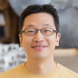 Christopher Y. Park合作论文数Department of Pathology, Stanford University8
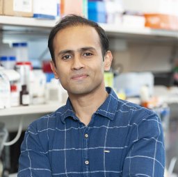 Elias Harold Kunal合作论文数Division of Internal Medicine, St Lukes Roosevelt, New York, NY.5
 Maria Lopez-Ocasio合作论文数3
 Courtney Fitzhugh合作论文数National Heart, Lung, and Blood Institute, National Institutes of Health3
 Katherine Almengo合作论文数2Caryn J. Ha合作论文数2William S Eng合作论文数2Guangjie Zhao合作论文数2Ahmad Shaikh合作论文数Dept Clin Lab Sci, King Khalid Univ2Amanda Ernlund合作论文数NYU, Sch Med, New York, NY USA2
 Ankit Saxena合作论文数NHLBI, NIH2Woolthuis Carolien M合作论文数2Gaëlle H. Martin合作论文数2
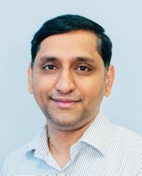 Gangaplara Arunakumar合作论文数School of Veterinary Medicine and Biomedical Sciences, University of Nebraska-Lincoln2
 Selvaraj Shanmugapriya合作论文数Department of Medicine, Yale School of Medicine2
 Chakraborty Sohini合作论文数Bioinformatics Centre, Bose Institute2
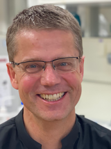 Robert Schneider合作论文数Institute for Functional Epigenetics, Helmholtz Center Munich2Lavanya Kannan合作论文数Texas A&M University2
 J Philip Mccoy合作论文数National Heart, Lung, and Blood Institute, National Institutes of Health2
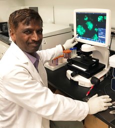 Vinagolu Rajasekhar合作论文数Memorial Sloan-Kettering Cancer Center2
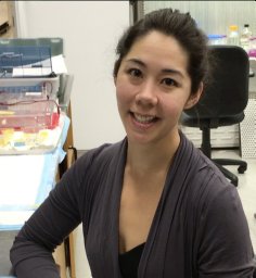 Isabel Beerman合作论文数Immune Disease Institute, Harvard Medical School2Christina Spevak合作论文数Department of Environmental and Biomolecular Systems, Oregon Health & Science University2Larizbeth A. Romero合作论文数1Cameron Fisher合作论文数1Brianna Gipson合作论文数1Abdoul Kone合作论文数1Malissa Ramsukh合作论文数1Paulo El Parente合作论文数1Chaohui Lisa Zhao合作论文数1Margaux Sambon合作论文数1Nicole Lee合作论文数1Emily Limerick合作论文数1Ziyan Lin合作论文数Appl Bioinformat Labs, NYU1Kevin Leclerc合作论文数1Lindsey H Remark合作论文数1
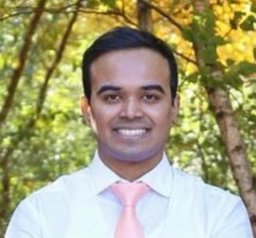 Vasudev Vivekanand Nayak合作论文数Department of Mechanical and Aerospace Engineering, New York University Tandon School of Engineering1Gayatri Ketavarapu合作论文数1Florencia Schlamp合作论文数Department of Medicine, NYU Langone Health1Susan DeWolf合作论文数1Matthew Stevenson合作论文数1
 Sneha Mitra合作论文数Duke Univ, Dept Comp Sci, Durham, NC 27708 USA1Roshan Sharma合作论文数Memorial Sloan Kettering Cancer Center, Sloan Kettering Institute1Melody Smith合作论文数1
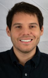 Pablo J Atria合作论文数Dept Biomat, Univ Andes1
 Jenny H Lee合作论文数Department of Medical Oncology, Chris O'Brien Lifehouse1
 Alesha Castillo合作论文数Department of Biomedical Engineering, Tandon School of Engineering, New York University1
 Aristotelis Tsirigos合作论文数Department of Pathology, Grossman School of Medicine, New York University1
 Sean P. Heffron合作论文数Division of Cardiology, Department of Medicine, Grossman School of Medicine, New York University1




