The extracellular signal-regulated kinase (ERK) is a component of the mitogen-activated protein kinase cascade. Exon 2 of erk2 was deleted by homologous recombination and resulted in embryonic lethality at embryonic day 6.5. erk2 mutant embryos did not form mesoderm and showed increased apoptosis but comparable levels of BrdUrd incorporation, indicating a defect in differentiation. erk2 null embryonic stem (ES) cells exhibited reduced total ERK activity upon serum stimulation, augmented ERK1 phosphorylation, and decreased downstream p90Rsk phosphorylation and activity; yet ES cell proliferation was unaffected. Mutant ES cells were capable of forming mesoderm; however, treatment of mutant ES cells with the mitogen-activated protein kinase kinase inhibitor PD184352 decreased total ERK activity and expression of the mesodermal marker brachyury, suggesting that ERK1 can compensate for ERK2 in vitro. Normal embryos at embryonic day 6.5 expressed activated ERK1/2 in the extraembryonic ectoderm, whereas erk2 mutant embryos had no detectable activated ERK1/2 in this region, suggesting that activated ERK1 was not expressed, and therefore cannot compensate for loss of ERK2 in vivo. These data indicate that ERK2 plays an essential role in mesoderm differentiation during embryonic development.
Thrombin, the terminal serine protease in the coagulation cascade, is a proinflammatory molecule in vivo and induces endothelial activation in vitro. The cellular signaling mechanisms involved in this function are unknown. The role of the p38 mitogen-activated protein kinase (MAPK) signaling pathway in thrombin-induced chemokine production was studied. Phosphorylation of both p38 MAPK and its substrate, ATF-2, was observed in human umbilical vein endothelial cells (HUVECs) stimulated with thrombin, with a maximum after 5 minutes of stimulation. Using the selective p38 MAPK inhibitor SB203580, there was a significant decrease in thrombin-induced interleukin-8 (IL-8) and monocyte chemotactic protein-1 (MCP-1) protein production and messenger RNA steady-state levels. In addition, SB203580 decreased IL-8 and MCP-1 production induced by the thrombin receptor-1 agonist peptide (TRAP), suggesting functional links between the thrombin G protein-coupled receptor and the p38 MAPK pathway. Furthermore, endothelial activation in the presence of SB203580 decreased the chemotactic activity of thrombin-stimulated HUVEC supernatant on neutrophils and monocytic cells. In contrast, the p42/p44 MAPK pathway did not appear to be involved in thrombin- or TRAP-induced endothelial chemokine production, because there was no reduction in the presence of the p42/p44-specific inhibitor PD98059. These results demonstrate that the p38 rather than p42/44 MAPK signaling pathway plays an important role in thrombin-induced endothelial proinflammatory activation and suggest that inhibition of p38 MAPK may be an interesting target for anti-inflammatory strategies in vascular diseases combining thrombosis and inflammation. (Blood. 2001;98:667-673)
Activated p38gamma MAP kinase exhibited significant basal ATPase activity in the absence of a kinase substrate, and addition of a phosphoacceptor substrate increased k(cat)/K(m)20-fold. AMP-PCP was competitive with ATP binding and non-competitive with phosphoacceptor substrate binding. The nucleotide binding site affinity label 5'-(p-fluorosulfonylbenzoyl)adenosine (FSBA) bound stoichiometrically at Lys-56 in the ATP site of both unphosphorylated and activated p38gamma. AMP-PCP only protected the activated enzyme from FSBA inactivation, implying that AMP-PCP does not bind unphosphorylated p38gamma. Basal ATPase activities were also observed for activated p38alpha, ERK2 and JNK3 suggesting that the enzymatic mechanism may be similar for all classes of MAP kinases.
p38 MAP kinase is a member of the family of kinases which mediate intracellular transduction pathways. The activation of this particular MAP kinase pathway is in response to a broad variety of extracellular stimuli. Subsequent downstream events triggered by p38 activation result in the production of IL-1 and TNF-a, suggesting that inhibition of this enzyme may provide a useful therapeutic target for intervention in various diseases mediated by these cytokines. Understanding the biological consequences of p38 activation and inhibition has been the subject of intensive research over the past several years and there is now ample evidence to suggest that inhibition of this enzyme represents a valid approach for target intervention in various cytokine-mediated diseases. Crystal structures of both apo enzyme and enzyme bound to various ligands in conjunction with site specific mutagenesis studies have provided a wealth of information regarding the interactions necessary to result in potent inhibition and selectivity from other kinases. This information has proven useful towards the analysis of previously reported compounds and will provide additional insight towards the design of new compounds and building upon existing SAR.
Caspases are essential components of the mammalian cell death machinery. Here we test the hypothesis that Caspase 9 (Casp9) is a critical upstream activator of caspases through gene targeting in mice. The majority of Casp9 knockout mice die perinatally with a markedly enlarged and malformed cerebrum caused by reduced apoptosis during brain development. Casp9 deletion prevents activation of Casp3 in embryonic brains in vivo, and Casp9-deficient thymocytes show resistance to a subset of apoptotic stimuli, including absence of Casp3-like cleavage and delayed DNA fragmentation. Moreover, the cytochrome c-mediated cleavage of Casp3 is absent in the cytosolic extracts of Casp9-deficient cells but is restored after addition of in vitro-translated Casp9. Together, these results indicate that Casp9 is a critical upstream activator of the caspase cascade in vivo.
Signal transduction via MAP kinase pathways plays a key role in a variety of cellular responses, including growth factor‐induced proliferation, differentiation and cell death. In mammalian cells, p38 MAP kinase can be activated by multiple stimuli, such as pro‐inflammatory cytokines and environmental stress. Although p38 MAP kinase is implicated in the control of inflammatory responses, the molecular mechanisms remain unclear. Upon activation, CD4+ T cells differentiate into Th2 cells, which potentiate the humoral immune response or pro‐inflammatory Th1 cells. Here, we show that pyridinyl imidazole compounds (specific inhibitors of p38 MAP kinase) block the production of interferon‐γ (IFNγ) by Th1 cells without affecting IL‐4 production by Th2 cells. These drugs also inhibit transcription driven by the IFNγ promoter. In transgenic mice, inhibition of the p38 MAP kinase pathway by the expression of dominant‐negative p38 MAP kinase results in selective impairment of Th1 responses. In contrast, activation of the p38 MAP kinase pathway by the expression of constitutivelyactivated MAP kinase kinase 6 in transgenic mice caused increased production of IFNγ during the differentiation and activation of Th1 cells. Together, these data demonstrate that the p38 MAP kinase is relevant for Th1 cells, not Th2 cells, and that inhibition of p38 MAP kinase represents a possible site of therapeutic intervention in diseases where a predominant Th1 immune response leads to a pathological outcome. Moreover, our study provides an additional mechanism by which the p38 MAP kinase pathway controls inflammatory responses.
Mitogen-activated protein (MAP) kinases are serine/threonine kinases that mediate intracellular signal transduction pathways. Pyridinyl imidazole compounds block pro-inflammatory cytokine production and are specific p38 kinase inhibitors. ERK2 is related to p38 in sequence and structure, but is not inhibited by pyridinyl imidazole inhibitors. Crystal structures of two pyridinyl imidazoles complexed with p38 revealed these compounds bind in the ATP site. Mutagenesis data suggested a single residue difference at threonine 106 between p38 and other MAP kinases is sufficient to confer selectivity of pyridinyl imidazoles. We have changed the equivalent residue in human ERK2, Q105, into threonine and alanine, and substituted four additional ATP binding site residues. The single residue change Q105A in ERK2 enhances the binding of SB202190 at least 25,000-fold compared to wild-type ERK2. We report enzymatic analyses of wild-type ERK2 and the mutant proteins, and the crystal structure of a pyridinyl imidazole, SB203580, bound to an ERK2 pentamutant, I103L, Q105T, D106H, E109G, T110A. These ATP binding site substitutions induce low nanomolar sensitivity to pyridinyl imidazoles. Furthermore, we identified 5-iodotubercidin as a potent ERK2 inhibitor, which may help reveal the role of ERK2 in cell proliferation.
BACKGROUND:The c-Jun N-terminal kinases (JNKs) are members of the mitogen-activated protein (MAP) kinase family, and regulate signal transduction in response to environmental stress. Activation and nuclear localization of JNK3, a neuronal-specific isoform of JNK, has been associated with hypoxic and ischemic damage of CA1 neurons in the hippocampus. Knockout mice lacking JNK3 showed reduced apoptosis of hippocampal neurons and reduced seizure induced by kainic acid, a glutamate-receptor agonist. Thus, JNK3 may be important in the pathology of neurological disorders and is of significant medical interest.RESULTS:We report here the structure of unphosphorylated JNK3 in complex with adenylyl imidodiphosphate, an ATP analog. JNK3 has a typical kinase fold, with the ATP-binding site situated within a cleft between the N- and C-terminal domains. In contrast to other known MAP kinase structures, the ATP-binding site of JNK3 is well ordered; the glycine-rich nucleotide-binding sequence forms a beta-strand-turn-beta-strand structure over the nucleotide. Unphosphorylated JNK3 assumes an open conformation, in which the N- and C-terminal domains are twisted apart relative to their positions in cAMP-dependent protein kinase. The rotation leads to the misalignment of some of the catalytic residues. The phosphorylation lip of JNK3 partially blocks the substrate-binding site.CONCLUSIONS:This is the first JNK structure to be determined, providing a unique opportunity to compare structures from the three MAP kinase subfamilies. The structure reveals atomic-level details of the shape of JNK3 and the interactions between the kinase and the nucleotide. The misalignment of catalytic residues and occlusion of the active site by the phosphorylation lip may account for the low activity of unphosphorylated JNK3. The structure provides a framework for understanding the substrate specificity of different JNK isoforms, and should aid the design of selective JNK3 inhibitors.
The cytokine interleukin (IL) 18 (formerly interferon gamma-inducing factor) induces the T helper type 1 response. In the present studies, IL-18 increased HIV type 1 (HIV-1) production from 5- to 30-fold in the chronically infected U1 monocytic cell line. Inhibition of tumor necrosis factor (TNF) activity by the addition of TNF-binding protein reduced IL-18-stimulated HIV-1 production by 48%. In the same cultures, IL-18-induced IL-8 was inhibited by 96%. Also, a neutralizing anti-IL-6 mAb reduced IL-18-induced HIV-1 by 63%. Stimulation of U1 cells with IL-18 resulted in increased production of IL-6, and exogenous IL-6 added to U1 cells increased HIV-1 production 4-fold over control. A specific inhibitor of the p38 mitogen-activated protein kinase reduced IL-18-induced HIV-1 by 73%, and a 50% inhibition was observed at 0.05 microM. In the same cultures, IL-8 was inhibited by 87%. By gel-shift and supershift analyses, increased binding activity of the transcription factor NF-kappaB was measured in nuclear extracts from U1 cells 1 h after exposure to IL-18. These results demonstrate induction of HIV-1 by IL-18 in a monocyte target associated with an intermediate role for TNF and IL-6, activation of p38 mitogen-activated protein kinase, and nuclear translocation of NF-kappaB.
IL-18 is synthesized as a precursor molecule without a signal peptide but requires the IL-1 beta converting enzyme (ICE, caspase-1) for cleavage into a mature peptide, Human precursor IL-18 was expressed, purified, and cleaved by ICE into a 18-kD mature form, Mature IL-18 induced IL-8, macrophage inflammatory protein-1 alpha, and monocyte chemotactic protein-1 in human peripheral blood mononuclear cells in the absence of any co-stimuli, Blocking IL-1 with IL-1 receptor antagonist resulted in a 50% reduction in IL-8, Neutralization of TNF with TNF binding protein resulted in a 66% reduction in IL-1 beta, an 80% reduction of IL-8, and an 88% reduction in mean TNF alpha mRNA, In purified CD14(+) cells but not CD3(+)/CD4(+), IL-18 induced gene expression and synthesis of IL-8 and IL-1 beta, TNF alpha production was induced in the non-CD14(+) population and there was no induction of TNF beta by IL-18, In purified natural killer cells, IL-18 induced IL-8 that was also inhibited by TNF binding protein, IL-18 did not induce antiinflammatory cytokines, IL-1Ra, or IL-10, although IL-18 induction of TNF alpha was inhibited by IL-10, In the presence of IFN gamma, IL-18-induced TNF alpha was enhanced and there was an increase in the mature form of IL-1 beta, We conclude that IL-18 possesses proinflammatory properties by direct stimulation of gene expression and synthesis of TNF alpha from CD3(+)/CD4(+) and natural killer cells with subsequent production of IL-1 beta and IL-8 from the CD14(+) population.
Recently, human interleukin 18 (hIL-18) cDNA was cloned, and the recombinant protein with a tentatively assigned NH2-terminal amino acid sequence was generated. However, natural hIL-18 has not yet been isolated, and its cellular processing is therefore still unclear. To clarify this, we purified natural hIL-18 from the cytosolic extract of monocytic THP.1 cells. Natural hIL-18 exhibited a molecular mass of 18.2 kDa, and the NH2-terminal amino acid was Tyr(37). Biological activities of the purified protein were identical to those of recombinant hIL-18 with respect to the enhancement of natural killer cell cytotoxicity and interferon-gamma production by human peripheral blood mononuclear cells. We also found two precursor hIL-18 (prohIL-18)-processing activities in the cytosol of THP.1 cells, These activities were blocked separately by the caspase inhibitors Ac-YVAD-CHO and Ac-DEVD-CHO. Further analyses of the partially purified enzymes revealed that one is caspase-1, which cleaves prohIL-18 at the Asp(36)-Tyr(37) Site to generate the mature hIL-18, and the other is caspase-3, which cleaves both precursor and mature hIL-18 at Asp(71)-Ser(72) and Asp(76)-Asn(77) to generate biologically inactive products, These results suggest that the production and processing of natural hIL-18 are regulated by two processing enzymes, caspase-1 and caspase-3, in THP.1 cells.
The short-chain fatty acid butyrate has been shown to elevate fetal hemoglobin (HbF) by inducing expression of the gamma-globin gene. Regulation of gene expression by butyrate is thought to proceed via inhibition of the enzyme histone deacetylase, leading to elevated levels of core histone acetylation which affect chromatin structure and transcription rates. To determine whether changes in histone acetylation are critical for the regulation of the gamma-globin gene, we tested three potent and specific inhibitors of histone deacetylase, the cyclic tetrapeptides trapoxin and Helminthsporium carbonum toxin (HC toxin), and the antifungal antibiotic trichostatin A for their ability to induce fetal hemoglobin expression in erythroid cells. These compounds induced fetal hemoglobin in both primary erythroid cell cultures and human erythroleukemia (K562) cells. A butyrate-responsive element spanning the duplicated CCAAT box region of the gamma-globin promoter has been identified in transient transfection assays using a reporter construct in K562 cells, and we show that the same promoter region is required for response to trapoxin and trichostatin. Mutational analysis of the gamma-globin promoter indicates that the distal CCAAT box and 3' flanking sequence (CCAATAGCC) is critical for activation by butyrate, trapoxin, and trichostatin, whereas the proximal element (CCAATAGTC) plays a less important role. These results show that inhibition of histone deacetylase can lead to transcriptional activation of gamma-globin promoter reporter gene constructs through proximal promoter elements, and suggest that butyrate induces gamma-globin expression via such changes in histone acetylation.
BACKGROUND:The p38 mitogen-activated protein (MAP) kinase regulates signal transduction in response to environmental stress. Pyridinylimidazole compounds are specific inhibitors of p38 MAP kinase that block the production of the cytokines interleukin-1beta and tumor necrosis factor alpha, and they are effective in animal models of arthritis, bone resorption and endotoxin shock. These compounds have been useful probes for studying the physiological functions of the p38-mediated MAP kinase pathway.RESULTS:We report the crystal structure of a novel pyridinylimidazole compound complexed with p38 MAP kinase, and we demonstrate that this compound binds to the same site on the kinase as does ATP. Mutagenesis showed that a single residue difference between p38 MAP kinase and other MAP kinases is sufficient to confer selectivity among pyridinylimidazole compounds.CONCLUSIONS:Our results reveal how pyridinylimidazole compounds are potent and selective inhibitors of p38 MAP kinase but not other MAP kinases. It should now be possible to design other specific inhibitors of activated p38 MAP kinase using the structure of the nonphosphorylated enzyme.
To determine whether interleukin-1β converting enzyme (ICE) plays a role in the programmed cell death of neuroblastoma, we studied ICE expression in primary tumours. In patients in stages I, II and IVS, ICE mRNA was detected in 22 of 32 (69%) tumours, while only 5 of 26 (19%) tumours expressed ICE in stages III and IV (P<0.001). ICE mRNA was expressed in 27 of 47 (57%) tumours without MYCN amplification, but it was not detected in any tumours with MYCN amplification (P<0.01). Immunohistochemically, the cytoplasm was stained in all 15 neuroblastomas examined. The nuclei were stained in 12 neuroblastomas without MYCN amplification, whereas only 1 of 3 tumours with MYCN amplification had positive staining in the nuclei. In ganglioneuromas, high levels of ICE mRNA were expressed, but immunostaining showed that the protease expression was confined to the cytoplasm. These observations suggest that ICE may be associated with the spontaneous regression often seen in favourable neuroblastomas and that localisation of ICE protease in the cell may be important for the cell death pathway. Double staining for ICE and TUNEL showed that they were co-localised in some nuclei, but the distribution of ICE protease expression was not necessarily the same as that of DNA fragmentation, suggesting that the protease expression probably preceded DNA fragmentation during the apoptotic process. ICE may play an important role in regulating the apoptotic process of neuroblastoma.
The hepatitis C virus (HCV) nonstructural 3 protein (NS3) is a 70-kDa multifunctional enzyme with three known catalytic activities segregated in two somewhat independent domains. The essential machinery of a serine protease is localized in the N-terminal one-third of the protein, and nucleoside triphosphatase (NTPase) and helicase activities reside in the remaining C-terminal region. NS4A is a 54-residue protein expressed immediately downstream of NS3 in the viral polyprotein, and a central stretch of hydrophobic residues in NS4A form an integral structural component of the NS3 serine protease domain. There is no evidence to suggest that the two domains of NS3 are separated by proteolytic processing in vivo. This may reflect economical packaging of essential viral replicative components, but it could also mean that there is functional interdependence between the two domains. In this study, a full-length NS3-NS4A complex was isolated after expression and autoprocessing in transiently transfected COS cells. The protein was used to examine the effects of polynucleotides on the NTPase, helicase, and protease activities. Unlike the previously reported behavior of a separately expressed NS3 helicase domain, the full NS3-NS4A complex demonstrated optimal NTPase activity between pH 7.5 and 8.5. All three NS3-NS4A activities were modulated by polynucleotides, with poly(U) having the most remarkable effect. These findings suggest that the domains within NS3 may influence the activity of one another and that the interplay of HCV genomic elements may regulate the enzyme activities of this complex HCV replicase component.
Hepatitis C virus encodes a large polyprotein precursor that is proteolytically processed into at least 10 distinct products, in the order NH2-C-E1-E2-p7-NS2-NS3-NS4A-NS4B-NS5A-NS5B -COOH. A serine proteinase encoded in the N-terminal 181 residues of the NS3 nonstructural protein is responsible for cleavage at four sites (3/4A, 4A/4B, 4B/5A, and 5A/5B) in the nonstructural region. NS4A, a 54-residue nonstructural protein which forms a stable complex with the NS3 proteinase, is required as a cofactor for cleavage at the 3/4A and 4B/5A sites and enhances processing at the 4A/4B and 5A/5B sites. Recently reported crystal structures demonstrated that NS4A forms an integral part of the NS3 serine proteinase. In this report, we present evidence that NS4A forms a nonionic-detergent-stable complex with the NS4B5A polyprotein substrate, which may explain the requirement of NS4A for the 4B/5A cleavage. Isoleucine-29 of NS4A, which has been previously shown to be essential for its proteinase cofactor activity and formation of the NS3 complex, was found to be important for the interaction between NS4A and the NS4B5A substrate. In addition, two more hydrophobic residues in the NS4A central region (valine-23 and isoleucine-25) were also shown to be essential for the cofactor activity and for the interaction with either the NS3 proteinase or the NS4B5A polyprotein substrate. Finally, the possible mechanisms by which these viral proteins interact with each other are discussed.
The interleukin-1β (IL-1β) converting enzyme (ICE) processes the inactive IL-1β precursor to the proinflammatory cytokine. ICE was also shown to cleave the precursor of interferon-γ inducing factor (IGIF) at the authentic processing site with high efficiency, thereby activating IGIF and facilitating its export. Lipopolysaccharide-activated ICE-deficient (ICE −/− ) Kupffer cells synthesized the IGIF precursor but failed to process it into the active form. Interferon-γ and IGIF were diminished in the sera of ICE −/− mice exposed to Propionibacterium acnes and lipopolysaccharide. The lack of multiple proinflammatory cytokines in ICE −/− mice may account for their protection from septic shock.
Dynamic NMR methods, such as differential line broadening and transferred NOE spectroscopy, are normally reserved for the study of small molecule ligand interactions with large protein receptors. Using a combination of isotope labeling and isotope edited NMR, we have extended these techniques to characterize interactions of a much larger protein/drug complex, FKBP-12/ FK506 with its receptor protein, calcineurin. In order to examine this multicomponent system by dynamic NMR methods, the 93 kDa, tightly bound FKBP-12/FK506/Cn complex was replaced with a lower affinity, rapidly exchanging system consisting of FKBP-12/FK506 (13 kDa), recombinant calcineurin subunit B (CnB) (20 kDa), and a synthetic peptide (4 kDa) corresponding to the B binding domain (BBD) of calcineurin catalytic subunit A (CnA). Analysis of 1H-13C HSQC data acquired for the FKBP-12/ 13C-FK506 and FKBP-12/13C-FK506/CnB/BBD complexes indicates that FKBP-12/FK506 and CnB/BBD are in fast exchange in the quaternary complex. Comparison of proton line widths shows significant broadening of resonances along the macrocycle backbone at 13-CH, 13-OMe, 15-OMe, 18-CH2, 20-CH, 21-CH, and 25-Me, as well as moderate broadening on the macrocycle backbone at 17-Me, 24-CH, and the pyranose 12-CH2 protons. The tri-substituted olefin and cyclohexyl groups also show moderate broadening at the 27-Me, 28-CH, and 30-CH2 positions, respectively. Unexpectedly, little line broadening was observed for the allyl resonances of FK506 in the quaternary complex, although 13C longitudinal relaxation measurements suggest this group also makes contacts with calcineurin. In addition, intermolecular transfer NOE peaks were observed for the allyl 37-CH2, 21-CH, 30-CH2, 13-OMe, 15-OMe, 17-Me, 25-Me, and 27-Me groups, indicating that these are potential sites on the FK506 molecule that interact with calcineurin.
p38 mitogen-activated protein kinase is activated by environmental stress and cytokines and plays a role in transcriptional regulation and inflammatory responses. The crystal structure of the apo, unphosphorylated form of p38 kinase has been solved at 2.3 Å resolution. The fold and topology of p38 is similar to ERK2 (Zhang, F., Strand, A., Robbins, D., Cobb, M. H., and Goldsmith, E. J. (1994) Nature 367, 704-711). The relative orientation of the two domains of p38 kinase is different from that observed in the active form of cAMP-dependent protein kinase. The twist results in a misalignment of the active site of p38, suggesting that the orientation of the domains would have to change before catalysis could proceed. The residues that are phosphorylated upon activation of p38 are located on a surface loop that occupies the peptide binding channel. Occlusion of the active site by the loop, and misalignment of catalytic residues, may account for the low enzymatic activity of unphosphorylated p38 kinase.
 Keith P. Wilson合作论文数6MA Fleming合作论文数5Dj Livingston合作论文数5K Kuida合作论文数5Yong Gu合作论文数5Ted Fox合作论文数5Pg Mccaffrey合作论文数4
 Richard Flavell合作论文数Flavell Lab, Department of Immunobiology, Howard Hughes Medical Institute, School of Medicine, Yale University4Kathy Hsiao合作论文数4J A Lippke合作论文数3Y Gu合作论文数3Sarnecki C合作论文数3
 Charles Dinarello合作论文数Division of Infectious Diseases, School of Medicine, University of Colorado Denver - Anschutz Medical Campus;University of Colorado Health Sciences Center3Ursula A. Germann合作论文数3Matthew J. Fitzgibbon合作论文数3Guy W. Bemis合作论文数3A J Puren合作论文数2RA ALDAPE合作论文数2MW HARDING合作论文数2
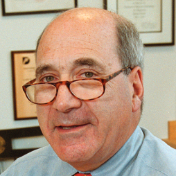 David M. Livingston合作论文数Department of Cancer Biology, Dana-Farber Cancer Institute2Masashi Kurimoto合作论文数2S. Pazhanisamy合作论文数2Vincent Galullo合作论文数2Xiaoling Xie合作论文数2Tadao Tanimoto合作论文数2Joyce T. Coll合作论文数2Paul R. Caron合作论文数2George Ku合作论文数2L A Penix合作论文数1Y Nukada合作论文数1J L Lalanne合作论文数1K Akita合作论文数1L Shapiro合作论文数1K Higashino合作论文数1V Marin合作论文数1KA Morgenstern合作论文数1John A. Thomson合作论文数1C Lin合作论文数1C Faucheu合作论文数1Yp Luong合作论文数1Sa Raybuck合作论文数1JAF BLACK合作论文数1YMC WANG合作论文数1Joel Raingeaud合作论文数1M Fujii合作论文数1M Namba合作论文数1H Ikeda合作论文数1S Grès合作论文数1




