Graph signal processing (GSP) is becoming a major tool in biomedical signal and image analysis. In most GSP techniques, graph structures and edge weights have been typically set via statistical and computational methods. More recently, graph structure learning methods offered more reliable and flexible data representations. In this work, we introduce a graph learning approach for melanoma detection in dermoscopic images based on two graph-theoretic representations: superpixel ensemble graphs (SEG) and superpixel hierarchy graphs (SHG). For these two types of graphs, superpixel maps of a skin lesion image are respectively generated at multiple levels without and with parentchild constraints among superpixels at adjacent levels, where each level corresponds to a subgraph with a different number of nodes (20, 40, 60, 80, or 100 nodes). Two edge weight assignment techniques are explored: handcrafted Gaussian weights and learned weights based on optimization methods. The graph nodal signals are assigned based on texture, geometric, and color superpixel features. In addition, the effect of graph edge thresholding is investigated by applying different thresholds (25
Rheumatoid arthritis (RA) is a chronic disease that causes abnormal inflammations that mainly affect the joints. Hence, numerous genome-wide association studies (GWASs) have been carried out to identify the single nucleotide polymorphisms (SNPs), especially common variants, associated with RA. Beside the common variants, more recent studies have demonstrated that rare variants are also responsible for susceptibility to complex diseases. In this paper, we seek to identify the rare (MAF <1
Motor imagery (MI) patterns play a vital role in brain-computer interface (BCI) systems, enabling control of external devices without relying on peripheral nerves or muscles. These patterns are typically classified by analyzing the associated electroencephalogram (EEG) signals. In this work, we introduce a novel MI classification approach based on multilevel graph-theoretic modeling of multichannel EEG signals. Multivariate autoregressive modeling and coherence analysis are firstly employed to construct directed graph signals to represent the relationships among EEG channels and capture the complex correlations inherent in MI patterns. Spatial graph vertex features are thus extracted as well as graph Fourier transform coefficients. Moreover, multilevel generalizations of vertex-domain features are thus defined where edges of graph signals are pruned according to different thresholds, vertex features are extracted for each threshold level, and then all features are combined into a multilevel hierarchical graph descriptor. These graph-theoretic descriptors could be fused with different variants of common spatial patterns for improved discriminability on MI classification tasks. Different feature combinations are used to train k-nearest neighbor classifiers, support vector machines, and random forests for MI pattern classification. The proposed method demonstrates competitive performance compared to the FWCSP and SCSP methods on Dataset 2a of the BCI Competition IV, as well as robust results on Dataset 1 from the same competition. Overall, the findings highlight the potential of multilevel spatial and spectral graph features in leveraging the correlation among EEG channels towards enhanced MI classification performance.
Contrast-enhanced digital mammography (CEDM) has emerged as a promising complementary imaging modality for breast cancer diagnosis, offering enhanced lesion visualization and improved diagnostic accuracy, particularly for patients with dense breast tissues. However, the reliance of CEDM on contrast agents poses challenges to patient safety and accessibility. To overcome those challenges, this paper introduces a deep learning methodology for improved breast lesion detection and classification. In particular, an image-to-image translation model based on cycle-consistent generative adversarial networks (CycleGAN) is utilized to generate synthetic CEDM (SynCEDM) images from full-field digital mammography in order to enhance visual contrast perception without the need for contrast agents. A new dataset of 3958 pairs of low-energy (LE) and CEDM images was collected from 2908 female subjects to train the CycleGAN model to generate SynCEDM images. Thus, we trained different You-Only-Look-Once (YOLO) architectures on CEDM and SynCEDM images for breast lesion detection and classification. SynCEDM images were generated with a structural similarity index (SSIM) of 0.94 +/- 0.02. A YOLO lesion detector trained on original CEDM images led to a 91.34% accuracy, a 90.37% sensitivity, and a 92.06% specificity. In comparison, a detector trained on the SynCEDM images exhibited a comparable accuracy of 91.20%, a marginally higher sensitivity of 91.44%, and a slightly lower specificity of 91.30%. This approach not only aims to mitigate contrast agent risks but also to improve breast cancer detection and characterization using mammography.
Gastrointestinal (GI) diseases are among the most frequently occurring diseases that pose a significant threat to people’s health. The gold standard for diagnosing these diseases is endoscopic examination, yet this approach is resource-intensive, requiring costly equipment and specialized training. This study explores an alternative approach for GI image segmentation and classification, employing Simple Linear Iterative Clustering (SLIC) and Linear Spectral Clustering (LSC) superpixel methods. Analyzing images from the comprehensive Kvasir dataset, which represents different GI tract sections, the research applies three distinct features—local binary pattern, gray-level co-occurrence matrices, and first-order statistical features—for Support Vector Machine (SVM) classification. The results demonstrate that superpixel-based classification methods exceed traditional pixel-wise techniques in terms of accuracy and efficiency. Specifically, SLIC excels in upper GI tract analysis, yielding 77.33
Three-dimensional point-cloud data has been enormously abundant with the emergence of 3D data acquisition, processing, and visualization technologies. Encryption algorithms have been recently introduced to ensure secure storage and communication for this type of data. Maintaining algorithmic correctness and the geometric stability are still key challenges towards the construction of reliable, trustful, and practical ciphers of 3D point clouds. To address these challenges, Jolfaei et al. (2015) proposed a 3D object encryption algorithm along with geometric notions of dimensional and spatial stability. However, these notions are not consistent, and the geometric stability and correctness of that algorithm are not guaranteed as we show through counterexamples. In this paper, we introduce two enhanced ciphers with correctness, reversibility, and geometric stability guarantees. These ciphers employ chaotic permutations with hyperchaotic maps of highly complex behavior for enhanced security. The permutation scheme ensures the creation of consistent solvable equations for the decryption stage. Also, an enhanced 3D point rotation scheme is proposed to ensure geometric stability. The soundness and significance of the proposed ciphers are demonstrated by rigorous mathematical proofs. As well, extensive experimentation and comparisons against state-of-the-art methods are demonstrated through similarity analysis based on the Hausdorff and Euclidean distances, analysis of the sensitivity to plaintext and key perturbations, and analysis of the robustness to statistical attacks.
Using the topological equivalence between the Riemann sphere $$\mathbb {S}$$ S and the extended complex plane $$\overline{\mathbb {C}} = \mathbb {C} \cup \{\infty \}$$ C ¯ = C ∪ { ∞ } , where $$\mathbb {C}$$ C is the field of complex numbers, we establish 2D-bijective representations of 3D point clouds. Points of 3D point clouds are mapped into the Riemann sphere $$\mathbb {S}$$ S , and a stereographic projection is implemented to map the points into the complex plane $$\mathbb {C}$$ C . The way the 3D objects are mapped into $$\mathbb {S}$$ S may be varied for various applications. To prove the accuracy and efficiency of the proposed 2D representation of 3D objects, we apply this correspondence to 3D point cloud encryption. We utilize chaotic permutations, chaotic circuits, and Latin cubes in addition to the stereographic projection representation to construct our scheme. The permutation steps using chaotic maps and Latin cubes are carried out on the object data points in both $$\mathbb {S}$$ S and $$\overline{\mathbb {C}}$$ C ¯ , while the chaotic circuits are applied to 2D projections of the 3D objects. To the best of our knowledge, no earlier work employed stereographic projections for 3D object encryption. Experimental simulations of this method show high encryption strength and strong confusion and diffusion properties based on quantitative and statistical measures.
Digital pathology has recently been revolutionized by advancements in artificial intelligence, deep learning, and high-performance computing. With its advanced tools, digital pathology can help improve and speed up the diagnostic process, reduce human errors, and streamline the reporting step. In this paper, we report a new large red blood cell (RBC) image dataset and propose a two-stage deep learning framework for RBC image segmentation and classification. The dataset is a highly diverse dataset of more than 100K RBCs containing eight different classes. The dataset, which is considerably larger than any publicly available hematopathology dataset, was labeled independently by two hematopathologists who also manually created masks for RBC cell segmentation. Subsequently, in the proposed framework, first, a U-Net model was trained to achieve automatic RBC image segmentation. Second, an EfficientNetB0 model was trained to classify RBC images into one of the eight classes using a transfer learning approach with a 5X2 cross-validation scheme. An IoU of 98.03% and an average classification accuracy of 96.5% were attained on the test set. Moreover, we have performed experimental comparisons against several prominent CNN models. These comparisons show the superiority of the proposed model with a good balance between performance and computational cost.
Contrast-enhanced spectral mammography (CESM) is an emerging modality for breast cancer diagnosis. This work investigates the feasibility of a computationally-efficient computer-aided diagnosis (CAD) system for breast lesion classification. Moreover, to avoid the need for intravenous contrast agents, we investigated the synthesis of contrast-enhanced (SynCESM) images. A total of 504 pairs of low-energy (LE) and CESM images were collected from 160 female subjects. A semi-automatic active-contour method was used for lesion segmentation. Then, 20 morphological and textural features were extracted. To improve the computational efficiency of the proposed system, the wavelet packet transform (WPT) was applied. Then, the same features were extracted from the WPT-approximated segmented lesions. Using LE images, a sigmoid-kernel SVM classifier exhibited a 90.20% accuracy, an 88.39% sensitivity, an 88.26% specificity, and a 0.92 AUC. The per-frame classification time was significantly reduced from 1.1046 s to 0.0734 s with WPT. Using CESM images, we achieved a 93.26% accuracy, a 95.94% sensitivity, a 93.37% specificity, a 0.94 AUC, and a 0.0657 s classification time. Interestingly, with SynCESM images, reasonable performance was still obtained with a 92.14% accuracy, a 93.87% sensitivity, a 91.58% specificity, a 0.93 AUC and a 0.0657 s classification time. Finally, with hybrid pairs of LE and CESM images, we got the best performance with a 96.87% accuracy, a 97.23% sensitivity, a 95.47% specificity, a 0.98 AUC, and a 0.0696 s classification time. The results demonstrate several advantages of the proposed system including its clinical feasibility, lower complexity, and reduced need for contrast agents via synthetic data generation.
This research addressed the need to enhance template-matching performance in e-learning and automated assessments within Egypt’s evolving educational landscape, marked by the importance of e-learning during the COVID-19 pandemic. Despite the widespread adoption of e-learning, robust template-matching feedback mechanisms should still be developed for personalization, engagement, and learning outcomes. This study augmented the conventional best-buddies similarity (BBS) approach with four feature descriptors, Harris, scale-invariant feature transform (SIFT), speeded-up robust features (SURF), and maximally stable extremal regions (MSER), to enhance template-matching performance in e-learning. We systematically selected algorithms, integrated them into enhanced BBS schemes, and assessed their effectiveness against a baseline BBS approach using challenging data samples. A systematic algorithm selection process involving multiple reviewers was employed. Chosen algorithms were integrated into enhanced BBS schemes and rigorously evaluated. The results showed that the proposed schemes exhibited enhanced template-matching performance, suggesting potential improvements in personalization, engagement, and learning outcomes. Further, the study highlights the importance of robust template-matching feedback in e-learning, offering insights into improving educational quality. The findings enrich e-learning experiences, suggesting avenues for refining e-learning platforms and positively impacting the Egyptian education sector.
Abstract Three-dimensional point-cloud data has been enormously abundant with the emergence of numerous technologies for 3D data acquisition, processing, and visualization. Encryption algorithms have been recently introduced to ensure secure storage and communication for this type of data. However, maintaining the correctness and the geometric stability of such algorithms are still key challenges towards the construction of reliable, trustful, and practical ciphers of 3D point clouds. Few attempts have been made to establish geometrically stable algorithms for 3D point cloud encryption, without compromising the cipher robustness. In particular, Jolfaei et al. [IEEE Transactions on Information Forensics and Security , vol. 10, no. 2, pp. 409-422, 2015] proposed a 3D object encryption algorithm along with geometric notions of dimensional and spatial stability. However, these notions are not consistent and the geometric stability and correctness of that cipher are not guaranteed as we show through counterexamples. In this paper, we introduce an enhanced cipher with correctness, reversibility, and geometric stability guarantees. The soundness and significance of the proposed scheme are demonstrated by rigorous mathematical proofs, extensive experimentation, and comparisons against state-of-the-art methods.
Haplotype block partitioning methods have been widely employed in the analysis of genetic patterns of common and complex diseases. The existing methods suffer from high time and memory complexities due to the key bottleneck of computing the linkage disequilibrium (LD) for all or most pairs of single-nucleotide polymorphisms (SNPs). In this work, we propose a multithreaded variant of the confidence internal test (CIT) method for haplotype block partitioning implementations. The usefulness of the proposed method in reducing the time and memory costs is demonstrated through experiments on the North American Rheumatoid Arthritis Consortium (NARAC) dataset. Our simulation results show 11–35% savings in time and 8–36% savings in memory requirements with only 3–6% increase in CPU usage.Clinical Relevance: This work seeks time and memory savings in genetic analysis for the prediction of diseases with complex factors (e.g., rheumatoid arthritis in our case). These savings can streamline and accelerate relevant clinical workflows and reduce demand for computational resources.
We compute precise estimates for dimensions of 3D-encryption techniques of 3D-point clouds which use permutations and rigid body motion, in which geometric stability is to be guaranteed. Few attempts are made in this direction. An attempt is established using the notions of dimensional and spatial stability by Jolfaei et al. (2015), who also proposed a 3D object encryption algorithm, claiming that it preserves dimensional and spatial stability. However, as we mathematically prove neither the algorithm, nor the associated estimates are correct. We introduce more rigorous definitions of the geometric stability of such 3D data encryption algorithms, followed by dimensionality measures
Outlier detection (OD) is a key problem, for which numerous solutions have been proposed. To deal with the difficulties associated with outlier detection across various domains and data characteristics, ensembles of outlier detectors have recently been employed to improve the performance of individual outlier detectors. In this paper, we follow an ensemble outlier detection approach in which good outlier detectors are selected through an enhanced clustering-based dynamic selection (CBDS) method. In this method, a bisecting K-means clustering algorithm is employed to partition the input data into clusters where every cluster defines a local region of competence. Among the initial pool of detectors, the outputs of the detectors with the most competent local performance were combined through four possible schemes to produce the final OD results. Experimental evaluation and comparison of our method were carried out against four variants of locally selective combination in parallel (LSCP) outlier ensembles. The CBDS-based schemes compare well with the LSCP-based ones on 16 public benchmark datasets and incur considerably lower computational costs. The CBDS method consistently achieved superior average scores of the area under the curve (AUC) of the receiver operating characteristic (ROC), and particularly outperformed the LSCP method on nine of the 16 datasets in terms of the AUC score. In addition, while the CBDS and LSCP methods have similar computational costs on small datasets, the CBDS method achieves significant time savings compared with the LSCP method on large datasets.
The placement and visualization of coronary stents during fluoroscopy depends mainly on the detection of balloon markers and their connecting guidewires. In this paper, a novel template-based approach is proposed to detect balloon markers and guidewires in cardiac fluoroscopic images. In particular, guidewires are detected based on balloon markers only, without prior knowledge of the background or guidewire elements. Also, while earlier techniques used circular models of balloon markers, we propose a more realistic elliptical model. Training and the testing datasets for balloon marker and guidewire detection were collected from different Cathlab systems and annotated by an application specialist with 10 years of experience in this field. The balloon-marker detector achieved a precision of 98.5%. Within 3-pixel tolerance, the guidewire detector achieved a matching percentage of 99.5% with the true guidewire using a customized evaluation method. Moreover, the guidewire detector achieved a mean Hausdorff distance of 3.3 pixels (0.6 mm) and a longest-common-substring (LCS) distance with a mean matching percentage of 87% within 1-pixel tolerance. Clinical Relevance- The proposed novel technique of detecting the guidewire offers a constant computational time and insensitivity to the body structures or the guidewire-like elements (such as the surgical wires). This leads to improved stent visualization and reasonable processing times.
Image encryption has become an indispensable tool for achieving highly secure image-based communications. Numerous encryption approaches have appeared and demonstrated varying degrees of robustness to adversarial attacks. In this paper, an efficient and robust image encryption algorithm is established based on randomized difference equations, random permutations and randomized logic circuits. Specifically, hyperchaotic and chaotic systems are used to generate pseudo-random sequences. These sequences are thus used to define random first-order difference equations, chaotic permutations and logic circuits. Image encryption based on these three randomized modules shows high computational efficiency as well as strong robustness against statistical, differential, and chosen-plaintext attacks. The proposed scheme leads to almost zero correlation in the encrypted images, entropy values of more than 7.99 for the test images, and a key space size of 2(572). Furthermore, differential analysis shows that the number of pixel change rate (NPCR) and the unified average change intensity (UACI) for the proposed technique are on average 99.61 and 33.35%, respectively.
Mental disorders, especially schizophrenia, still pose a great challenge for diagnosis in early stages. Recently, computer-aided diagnosis techniques based on resting-state functional magnetic resonance imaging (Rs-fMRI) have been developed to tackle this challenge. In this work, we investigate different decision-level and feature-level fusion schemes for discriminating between schizophrenic and normal subjects. Four types of fMRI features are investigated, namely the regional homogeneity, voxel-mirrored homotopic connectivity, fractional amplitude of low-frequency fluctuations and amplitude of low-frequency fluctuations. Data denoising and preprocessing were first applied, followed by the feature extraction module. Four different feature selection algorithms were applied, and the best discriminative features were selected using the algorithm of feature selection via concave minimization (FSV). Support vector machine classifiers were trained and tested on the COBRE dataset formed of 70 schizophrenic subjects and 70 healthy subjects. The decision-level fusion method outperformed the single-feature-type approaches and achieved a 97.85% accuracy, a 98.33% sensitivity, a 96.83% specificity. Moreover, feature-fusion scheme resulted in a 98.57% accuracy, a 99.71% sensitivity, a 97.66% specificity, and an area under the ROC curve of 0.9984. In general, decision-level and feature-level fusion schemes boosted the performance of schizophrenia detectors based on fMRI features.
For the sake of proper diagnosis and treatment, accurate brain tumour segmentation is required. Because manual brain tumour segmentation is a time-consuming, costly, and subjective task, effective automated approaches for this purpose are generally desired. However, because brain tumours vary greatly in terms of location, shape, and size, establishing automatic segmentation algorithms has remained challenging throughout the years. Automatic segmentation of brain tumour is the process of separating abnormal tissues from normal tissues, such as white matter (WM), gray matter (GM), and cerebrospinal fluid (CSF). Brian segmentation needs typically to be carried out for different image modalities in order to reveal important metabolic and physiological information. These modalities include positron emission tomography (PET), computer tomography (CT) image and magnetic resonance image (MRI). Multimodal imaging techniques (such as PET/CT and PET/MRI) that combine the information from multiple imaging modalities contribute more for accurate brain tumour segmentation. In this work, we introduce a deep learning framework for automated segmentation of 3D brain tumors that can save physicians time and provide an accurate reproducible solution for further tumor analysis and monitoring. In particular, a 3D U-Net was trained on brain MRI data obtained from the 2018 Brain tumor Image Segmentation (BraTS) challenge. Three optimizers (RMSProp, Adam and Nadam) and three loss functions (Dice loss, focal Tversky loss, Log-Cosh loss functions) were used. We demonstrated that some loss functions and optimizers combinations perform better than other ones. For example, using the Log-Cosh loss function along with RMSProp optimizer resulted in the highest Dice coefficient, 0.75. Indeed, we also optimized the network hyperparameters in order to enhance the segmentation outcomes. These results demonstrate the feasibility and effectiveness of the proposed deep learning scheme with optimized hyperparemeters and appropriate selection of the optimizer and loss function.
Motor imagery patterns are extensively exploited in brain-computer interface systems in order to control outer devices without using peripheral nerves or muscles. Classification of these patterns can be based on the associated electroencephalogram (EEG) signals. Recent approaches addressed this classification problem through techniques exploiting mainly information from one or two EEG channels. However, these approaches overlook correlations between multiple EEG channels. In this paper, we create motor-imagery classification systems based on graph-theoretic models of multichannel EEG signals. In particular, multivariate autoregressive models are used to establish the relations between the EEG channels and construct directed graph signals. Also, we constructed undirected graph signal models with Gaussian-weighted distances between graph nodes. Then, a novel variant of the graph Fourier transform is applied to the directed and undirected graph models with and without edge weights. Distinctive features were thus extracted from the transform coefficients. Additional features were computed using common spatial patterns, polynomial representations and principal components of EEG signals. Significant performance improvements were achieved using extreme learning machine (ELM) classifiers. For Dataset Ia of the BCI Competition 2003, our approach led to a classification accuracy of 96.58% with fully-connected weighted directed graph features computed on the delta-band EEG signals. For the six subjects of the Dataset 1 of the BCI Competition IV, our approach compared well with other state-of-the-art methods in the alpha and beta EEG bands.
Analysis and classification of electromyography (EMG) signals are crucial for rehabilitation and motor control. This study investigates electromyogram (EMG) time-frequency representations and then creates conventional and deep learning models for EMG signal classification. Firstly, a dataset of singlechannel surface EMG signals has been recorded for four subjects to differentiate between forearm flexion and extension. Then, different time-frequency EMG representations have been used to build conventional and deep learning models for EMG classification. We compared the performance of pre-trained convolutional neural network models, namely GoogLeNet, SqueezeNet and AlexNet, and achieved accuracies of 92.'71%, 90.63% and 87.5%, respectively. Also, data augmentation techniques on the levels of raw EMG signals and their timefrequency representations helped improve the accuracy of GoogLeNet to 96.88%. Furthermore, our approach demonstrated superior performance on another publicly available 10-class EMG dataset, and also using traditional classifiers trained on hand-crafted features.
 Mahmoud H. Annaby合作论文数Department of Mathematics, Statistics & Physics, Qatar University15
 Jeffrey Ho合作论文数Texas Instruments8
 Hassan Ayad合作论文数Cairo University7
 Ahmed M Mahmoud合作论文数6
 Mohsen Ali合作论文数Intelligent Machines Lab, Information Technology University;BRICKandMORTAR.ai4
 H. A. Abdusalam合作论文数4
 E. A. Nehary合作论文数Syst & Comp Engn, Carleton Univ3
 Ahmed M. Badawi合作论文数Syst & Biomed Engn, Cairo Univ3
 Mohamed Zarouan合作论文数3
 Inas Yassine合作论文数Lane Department of Computer Science and Electrical Engineering, West Virginia University3
 Ayman M. Eldeib合作论文数3
 Bassel Tawfik合作论文数3Manar N. Amin合作论文数2
 June Madete合作论文数2
 Ali Algumaei合作论文数2Rami F. Algunaid合作论文数2
 Asaad M. Anam合作论文数2
 Waleed Altalabi合作论文数2
 Rasha Kamal合作论文数2
 Ahmed Morsy合作论文数2
 Hanan Gewefel合作论文数Faculty of Applied Medical Science, Misr University for Science and Technology2
 Ali M. Rushdi合作论文数Department of Electrical and Computer Engineering, King Abdulaziz University2
 Yu-Tseh Chi合作论文数Department of Food, The Ohio State University2Y. M. Fouda合作论文数1Raghda N Marzaban合作论文数1Mohamed Salaheldien合作论文数1Iman Ewais合作论文数1
 Ayman M. Khalifa合作论文数1
 Khaled Bedda合作论文数1
 Yasser M. Fouda合作论文数1Y. M. Ahmed合作论文数1Hika Barki合作论文数1
 Heba I. Elkhouly合作论文数Faculty of Engineering, Beni-Suef University1Mohammed Gomaa合作论文数1
 Mohamed T. El-Wakad合作论文数Faculty of Engineering and technology, Future University1Waleed Ahmad合作论文数1Mohamed Abdallah合作论文数Cairo University1Kang Kim合作论文数1
 Gelan Ayana合作论文数Sch Biomed Engn, Jimma Univ1
 Mohamed R. Gomaa合作论文数Mech Engn Dept, Al Hussein Bin Talal Univ1
 Yassin Abdelsamad合作论文数Research Department, MED-EL GmbH1Makram, A.W.合作论文数Biomed. Eng. Dept., Helwan Univ.;c;Biomed. Eng. Dept., Helwan Univ.1
 Mohamed Hamed合作论文数Center for Bioinformatics, Saarland University1
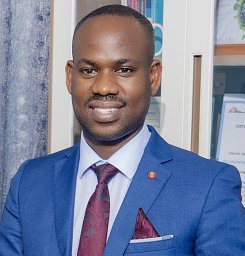 Atwine Daniel合作论文数Epicentre Mbarara1Akinniyi Osuntoki合作论文数1
 Jurgen Prestin合作论文数Institute of Mathematics, University of Lübeck1
 Mohamed Nagy Saad合作论文数Dept. of Electr. & Comput. Eng., Toronto Univ.1
 Tania S. Douglas合作论文数Department of Human Biology, University of Cape Town MRC/UCT Medical Imaging Research Unit1
 Arti Ahluwalia合作论文数departmental Research Centre, Inter;Faculty of Engineering;University of Pisa;Faculty of Engineering, University of Pisa1




