In clinical practice, treatment cycles are routinely started without the intention of undertaking fresh embryo transfer, as the freeze-all strategy is suitable for a multitude of alternative stimulation protocols as well as preimplantation genetic testing of embryos. Based on strengths, weaknesses, opportunities and threats (SWOT) analysis, it was evaluated whether a freeze-all strategy may be advised for all patients regardless of their (predicted) ovarian response. Therefore, the latest literature including the outcome of all randomized controlled trials (RCT) and their combined analysis were reviewed. Only two large trials, both from China, were considered conclusive and showed (following first embryo transfer) a higher chance of live birth following elective vitrification in comparison with fresh embryo transfer in women with polycystic ovary syndrome and in ovulatory women with a good prognosis. The other six trials showed either no difference in clinical outcome or were inconclusive by design. It is concluded that elective vitrification is likely favourable for high responders as it results in an improved chance of live birth following single embryo transfer, and is likely equally successful for normal responders. One trial in low responders indicated that the live birth rate may be compromised, which needs further confirmation in the future. This SWOT analysis of embryo transfer deferral also concluded that this approach allows the number of oocytes per stimulation cycle to be maximized safely in order to improve the chance of pregnancy. To minimize the risk for obstetric and perinatal complications in frozen-thawed embryo transfer cycles, (modified) natural cycles should be considered as the preferred approach for regular cycling women.
OBJECTIVE:To study whether the method of donor sperm cryopreservation, vitrification or slow freezing, affects embryological and clinical outcomes in in vitro fertilization (IVF) cycles. DESIGN:Retrospective cohort study. SUBJECTS:A total of 604 IVF cycles conducted between January 2023 and November 2024 using donor sperm cryopreserved by vitrification (n = 396) or slow freezing (n = 208). EXPOSURE:Cryopreservation method of donor sperm (vitrification vs. slow freezing). MAIN OUTCOME MEASURES:Fertilization rate, usable blastocyst rate, and clinical pregnancy rate after the first embryo transfer. RESULTS:Overall fertilization rates were similar between groups; however, stratified analysis by insemination technique revealed a significantly higher fertilization rate with vitrified sperm in conventional IVF cycles (76.4% vs. 65.9%; adjusted odds ratio [aOR] 1.95 [1.11-3.40]), whereas no difference was observed in intracytoplasmic sperm injection (ICSI) cycles (76.8% vs. 79.7%; aOR 0.84 [0.65-1.09]). Usable blastocyst and clinical pregnancy rates after the first embryo transfer were comparable between vitrification and slow freezing groups. Multivariable analyses adjusting for confounders confirmed these findings. CONCLUSION:Donor sperm vitrification is a safe alternative to conventional slow freezing and yields comparable blastocyst and clinical outcomes. Notably, in conventional IVF cycles, vitrification was associated with a significantly higher fertilization rate. This advantage was not observed in ICSI cycles, suggesting that vitrification better preserves sperm membrane functionality required for natural gamete interaction. Although clinical outcomes remain similar between both methods, reduced fertilization with slow-frozen sperm may decrease the number of embryos available, with potential implications for overall treatment efficiency.
BACKGROUND: Uterine artery pulsatility index is a key biomarker for preeclampsia screening and the most reliable indicator of uterine perfusion across all pregnancy trimesters. Although recent findings reveal a significant decrease in uterine artery pulsatility index during first trimester in artificial cycle frozen embryo transfer pregnancies, no previous study evaluated whether this decrease persists throughout the second and third trimesters, when hormonal treatment is discontinued. Considering the crucial role of uterine artery pulsatility index during the second half of pregnancy risk assessment, recommended by international guidelines to ensure early preeclampsia detection and proper pregnancy monitoring, we set out to perform a large retrospective study to evaluate the impact of endometrial preparation on second and third trimesters uterine artery pulsatility index. OBJECTIVE: The study aims to evaluate the possible impact of endometrial preparation for frozen embryo transfer on uterine vascular resistance during the second and third trimesters. STUDY DESIGN: This retrospective single-center study analyzed 27,495 singleton pregnancies that underwent Uterine Artery Pulsatility Index evaluation during the second trimester (20-22 weeks) of pregnancy at our University Hospital between January 2010 and November 2024. Among them, 23,547 were naturally conceived and 3948 resulted from Assisted Reproductive Technology (385 ovulation induction and intrauterine insemination, 864 in vitro fertilization frozen embryo transfer, and 2699 after frozen embryo transfer) (356 natural cycle frozen embryo transfer and 2343 artificial cycle frozen embryo transfer). Additionally, third trimester (35-37 weeks) Uterine Artery Pulsatility Index evaluations were available for 11,096 pregnancies. Pregnancies with fetal congenital abnormalities, aneuploidies, and twin pregnancies were excluded. The primary aim of the study was to investigate Uterine Artery Pulsatility Index values throughout pregnancy based on different types of conception. Analysis of covariance and linear mixed model (including potential confounders such as smoking, diabetes, race, chronic hypertension, aspirin administration, thrombophilia, age, weight, and oocyte donation) were used to analyze the association between mode of conception and log10-transformed multiple of the median values of Uterine Artery Pulsatility Index. RESULTS: The use of hormonal treatment in artificial cycle frozen embryo transfer cycles was associated with a significantly lower second-trimester Uterine Artery Pulsatility Index values 0.73 (artificial cycle frozen embryo transfer) as compared with all other modes of conception vs 0.89 (naturally conceived), 0.92 (ovulation induction and intrauterine insemination), 0.94 (fresh embryo transfer), and 0.89 (natural cycle frozen embryo transfer) (P<.001). Differences persisted during the third trimester with Uterine Artery Pulsatility Index values 0.95 for artificial cycle frozen embryo transfer vs 1.00 (naturally conceived), 1.03 (ovulation induction and intrauterine insemination), 1.00 (in vitro fertilization frozen embryo transfer), and 1.02 (natural cycle frozen embryo transfer) (P<.001). The results were confirmed after applying the multivariable regression analysis. Despite the improved uterine perfusion, artificial cycle frozen embryo transfer was associated with a 4-fold higher incidence of preeclampsia (5.2%) compared to natural cycle frozen embryo transfer (1.1%), naturally conceived (1. 4%), ovulation induction and intrauterine insemination (1%), and fresh embryo transfer (2.2%) (P<.001). CONCLUSION: The present study demonstrates that artificial cycle frozen embryo transfer is associated with reduced uterine vascular resistance across all pregnancy trimesters. This finding strongly supports the urgent need to revise the current second and third trimester preeclampsia risk assessment algorithm to ensure accurate early detection and proper management of high-risk pregnancies.
Sex-related differences in the QT interval become evident from puberty to menopause. Sex steroid hormones have been suggested to play a role in cardiac repolarization, though their impact remains a matter of debate. These effects are especially pronounced in women with inherited long QT syndrome (LQTS) and may face a higher risk of QT variability associated with endogenous or exogenous hormonal fluctuations. This narrative review aimed to summarize the impact of physiological hormonal fluctuations on QT interval in women with LQTS across the female lifespan, including menstrual cycle, pregnancy and postpartum, and menopause, concluding with exogenous hormonal administration such as oral contraceptives and ovarian stimulation in assisted reproductive technologies, highlighting potential genotype-specific differences. Insights gained from this review may inform more precise risk stratification, optimize individualized management, and improve safety for women with LQTS across their reproductive lifespan.
Research questionDoes luteinizing hormone (LH)-activity supplementation during ovarian stimulation (OS) influence embryo-ploidy rate compared with follicle-stimulating hormone (FSH)-only stimulation in in vitro fertilization (IVF) cycles with preimplantation genetic testing for aneuploidy (PGT-A)?MethodsRetrospective single-center cohort study (January 2018–January 2024) including 4,417 IVF/PGT-A cycles using gonadotropin-releasing hormone antagonist or progesterone-primed protocols. After exclusions, 952 cycles were selected via 1:1 propensity score-matching for female age and oocyte yield: 476 OS with recombinant FSH alone and 476 with added LH activity (recombinant LH or human menopausal gonadotropin). As the primary outcome, euploidy was assessed by trophectoderm biopsy and next-generation sequencing. Generalized estimating equations accounted for within-patient clustering. Secondary endpoints were clinical pregnancy, live birth, and miscarriage after frozen euploid embryo transfer (FEET).ResultsThere were no significant differences between FSH-only and FSH + LH activity groups with regard to oocyte yield, maturation, fertilization rates, or embryo euploidy rates (45.8% vs. 45.2%; p=0.749). Female age was identified as an independent negative predictor of euploidy (OR = 0.899; p< 0.001), while good-quality embryos had significantly higher odds of being euploid compared with poor-quality embryos (OR = 2.053; p< 0.001). Secondary outcomes showed no significant differences in clinical pregnancy rate, live birth rate, or miscarriage rate following FEET between groups. The addition of LH activity during ovarian stimulation was not independently associated with embryo ploidy status or live birth outcomes.ConclusionsIn a large matched IVF/PGT-A cohort, LH activity supplementation during OS did not improve blastocyst euploidy or reproductive outcomes. Gonadotropin regimens should be individualized rather than routinely including LH in unselected patients.
OBJECTIVE:To examine intrapatient variability in retrieved oocyte numbers across consecutive in vitro fertilization (IVF) ovarian stimulation (OS) cycles with an identical OS protocol. DESIGN:Cross-continental, multicenter retrospective cohort study. SUBJECTS:Patients undergoing OS for IVF (2014-2024) with ≥2 OS cycles within 6 months using the same OS protocol, gonadotropin type, and initial and daily dose; all underwent freeze-all-cycles and had ≥1 oocyte retrieved in each of the two consecutive cycles. For each patient, the earliest consecutive pair meeting criteria was analyzed. EXPOSURE:Oocyte yield in the first vs. consecutive OS cycle. MAIN OUTCOME MEASURES:Primary outcomes included: (i) average percentage change in oocyte yield between cycles (higher divided by lower oocyte yield); and (ii) 25th, 50th (median), and 75th percentiles of oocyte-yield percentage change, overall and by age groups (≤30, 31-35, 36-39, ≥40 years). Secondary outcomes included: (i) coefficient of determination (R2) between each cycle's oocyte count and the patient's average oocyte count across both cycles, representing the extent of variation explained by the patient's baseline profile; (ii) shifts between ovarian response categories, poor (1-3 oocytes), suboptimal (4-9), normal (10-14), and hyper-response (≥15); (iii) average and median percentage change in mature-oocyte yield. RESULTS:Overall, 801 cycle pairs met the inclusion criteria. Mean daily gonadotropin dosage was 361.5 ± 112.6 IU; with comparable demographic and cycle characteristics between cycles. Overall, the average percentage change in oocyte yield was 62.7%; the 25th, 50th (median), and 75th percentiles were 16.7%, 40%, and 80%, respectively. Fifty-percent of patients showed >33% difference in retrieved oocytes, and 381/801 (47.57%) shifted ovarian response categories, with 29/381 (7.61%) shifting across two categories. Median oocyte yield percentage change was 44.4% in women ≥40 vs. 33.3% in those ≤30 years. The coefficient of determination between each cycle and the average of the two cycles was 0.834, representing the optimal performance any prediction model could achieve when predicting oocyte yield given baseline characteristics alone. The average percentage change in mature oocyte yield was 74.5%, with a median of 50%. CONCLUSION:Cycle-to-cycle variations in retrieved oocyte yield exist despite the same cycle conditions, across all age groups, reflecting fluctuations in ovarian follicular readiness and response, challenging ovarian response categorization based on oocyte yield and stressing the importance of key performance indicators in IVF OS cycles.
ObjectivesThis study evaluated the efficacy of individualized follitropin delta dosing in patients with potential low response, defined by anti-Müllerian hormone (AMH) level <9 pmol/L, undergoing ovarian stimulation for assisted reproductive technology.MethodsA pooled analysis including 1844 patients from five randomized controlled trials was undertaken. The trials selected for the analysis used individualized dosing of follitropin delta, based on AMH level and body weight. Patients with potential low response (AMH screening level <9 pmol/L [~1.26 ng/mL]; n=329) were compared to patients with potential normal/high response (AMH level ≥9 pmol/L; n=1515). Patients with AMH <9 pmol/L all received the maximum daily dose of follitropin delta (12 µg).ResultsThe mean age was 34.6 years for patients with AMH <9 pmol/L and 32.2 years for patients with AMH ≥9 pmol/L, and the median AMH was 6 pmol/L and 20 pmol/L, respectively. In patients with AMH <9 pmol/L, the mean number of oocytes retrieved (6.3 oocytes) was significantly lower (p<0.0001) than in patients with AMH ≥9 pmol/L (10.8 oocytes), while no significant differences were observed for ongoing pregnancy rate (29.2% versus 31.8% in patients with AMH <9 pmol/L versus ≥9 pmol/L [p=0.32]) or live birth rate (28.6% versus 31.4% [p=0.32]). Seventeen patients (5.2%) with AMH <9 pmol/L and 33 patients (2.2%) with AMH ≥9 pmol/L had cycle cancellations due to poor response. Ovarian hyperstimulation syndrome (OHSS) occurred in 5 patients (1.5%) with AMH <9 pmol/L and 112 patients (7.4%) with AMH ≥9 pmol/L.ConclusionsFollitropin delta dosing in young patients with moderately reduced ovarian reserve, using 12 µg/day for ovarian stimulation, demonstrated clinically relevant fresh-cycle pregnancy outcomes as compared to patients with normal or high ovarian reserve. This pooled analysis adds evidence for the efficacy of follitropin delta across all ovarian reserve subgroups of the infertile population. The study was limited to fresh cycle outcomes; further studies are warranted focusing on cumulative outcomes and older patient populations with low ovarian reserve.
RESEARCH QUESTION:To investigate the potential interaction between endometrial thickness before Frozen Embryo Transfer and Placental Growth Factor levels during the first trimester screening of pregnancy. DESIGN:This single-centre retrospective observational study was conducted at the Reproductive Medicine Department of Dexeus University Hospital between January 2021 and June 2025. The analysis included singleton pregnancies resulting from both Artificial Cycle Frozen Embryo Transfer and Natural Cycle Frozen Embryo Transfer. All participants attended first-trimester pregnancy screening and delivered at Dexeus University Hospital in Barcelona, Spain. RESULTS:Overall, 1,757 singleton pregnancies after Frozen Embryo Transfer were analysed, including 1,080 autologous and 677 heterologous cycles. Higher endometrial thickness (>12 mm) was associated with lower first-trimester Placental Growth Factor levels (24.9 ± 16.3 vs. 29.6 ± 16.3 pg/mL; p = 0.04), a finding that remained significant after Multiples of the Median adjustment (0.80 ± 0.29 vs. 0.90 ± 0.35; p = 0.013) and Log10 transformation (-0.12 ± 0.16 vs. - 0.08 ± 0.18; p = 0.029). Linear regression confirmed a negative correlation between endometrial thickness and log10 MoM Placental Growth Factor (β = - 0.01; 95% CI - 0.01 to 0.00; p = 0.020). Obstetric and perinatal outcomes, including gestational age at delivery, birthweight, and the incidence of Hypertensive Disorders of Pregnancy, did not differ between groups. CONCLUSION:Our findings suggest a negative association between endometrial thickness prior to frozen embryo transfer and first-trimester placental growth factor levels. Further studies are warranted to confirm these results and to better elucidate the mechanisms underlying the potential impact of endometrial thickness on early placental angiogenesis.
Diminished ovarian reserve (DOR) is common in women with infertility and is associated with poorer in vitro fertilization (IVF) outcomes. Testosterone is widely used off-label in this patient group, although evidence for its efficacy and safety is limited. To address this, we conducted a triple-blind, placebo-controlled, randomized clinical trial evaluating whether transdermal testosterone gel prior to IVF improves clinical pregnancy rates in women with DOR. Females aged 18-43 with infertility and DOR according to the Bologna criteria were recruited at 10 fertility clinics in Europe between April 2015 and November 2022. Of 316 assessed for eligibility, 290 were enrolled and randomized. Two were excluded from the primary analysis as their treatment coincided with the onset of the COVID-19 pandemic, and they did not start ovarian stimulation, leaving 288 participants. Participants were randomized to 5.5 mg of transdermal testosterone or matching placebo once daily for ~9 weeks prior to ovarian stimulation. All participants received ovarian stimulation in a long GnRH-agonist cycle with 300IU/day of highly purified human menopausal gonadotropin; fresh embryo transfer was performed if an embryo was available. The primary outcome was clinical pregnancy rate following fresh embryo transfer, defined as an intrauterine gestational sac with an embryo demonstrating cardiac activity at ≥7 weeks' gestation. Of the 288 participants, 134 were randomized to testosterone and 154 to placebo. Clinical pregnancy rates did not differ significantly, occurring in 21 women (15.7%) in the testosterone group and 23 (14.9%) in the placebo group (risk ratio (RR), 1.05; 95% confidence interval (CI) 0.61 to 1.81, p = 0.86). The study was terminated for futility at the prespecified interim analysis based on a conditional power calculation once 70% of the target sample size were randomized. In this study, transdermal testosterone did not improve clinical pregnancy rates compared with placebo in patients with infertility and DOR. Trial Registration: ClinicalTrials.gov: NCT02418572, EudraCT: 2014-001835-35.
STUDY QUESTION:Does transdermal testosterone treatment improve fertility-related quality of life (QOL) in women with diminished ovarian reserve (DOR)? SUMMARY ANSWER:Transdermal testosterone for 9 weeks at a dose of 5.5 mg per day did not result in improved fertility-related QOL compared with placebo in women with DOR. WHAT IS KNOWN ALREADY:Reduced QOL is prevalent in women with infertility, many of whom have DOR. Several studies have shown a correlation between DOR and lower testosterone levels, and testosterone is frequently prescribed to women with DOR undergoing fertility treatment. Some studies have reported that testosterone therapy may improve wellbeing in pre- and post-menopausal women, though others have found no benefit. There are no studies evaluating the effect of testosterone on QOL in women undergoing fertility treatment. STUDY DESIGN, SIZE, DURATION:Pre-planned secondary analysis of a double-blind placebo-controlled randomized controlled trial that included 288 participants recruited between April 2015 and August 2022. Of these, 213 completed QOL surveys both before and after treatment and were eligible for inclusion in this analysis. PARTICIPANTS/MATERIALS, SETTINGS, METHODS:Participants were women aged 18-43 years with DOR according to the Bologna criteria and planning to undergo IVF treatment at one of eight fertility clinics in Spain, Belgium, and Denmark. Participants were randomized to 5.5 mg of transdermal testosterone per day as 1% gel (n = 106) or an identical placebo (n = 107), applied for a median of 60 days prior to commencing ovarian stimulation. QOL was assessed using the FertiQoL instrument prior to commencing the intervention, and at the completion of the intervention but prior to commencing ovarian stimulation. QOL scores were compared using a one-way ANCOVA adjusted for age, BMI, parity, history of IVF treatment, and baseline FertiQoL scores. MAIN RESULTS AND THE ROLE OF CHANCE:There were no significant differences in baseline characteristics between the testosterone (n = 106) and placebo (n = 107) groups. After adjustment, testosterone showed no benefit over placebo for the Total FertiQoL score (F(1,204)=0.07, P = 0.79), the Core and Treatment scores, nor for any of the included FertiQoL subscales. Total testosterone levels were higher in the testosterone group than the placebo group at the end of the treatment (3.2 ± 2.7 nmol/l vs 0.6 ± 0.4 nmol/l, P < 0.001). LIMITATION, REASONS FOR CAUTION:QOL was a secondary outcome in this trial, and participants were not recruited based on a low QOL. WIDER IMPLICATIONS OF THE FINDINGS:Considering the available evidence, including the current study, premenopausal women are unlikely to benefit from testosterone treatment with regard to wellbeing and QOL. This study provides further evidence that testosterone should not be seen as a treatment for low wellbeing or QOL. STUDY FUNDING/COMPETING INTEREST(S):The study was supported by unrestricted grants and support from Besins Healthcare, Roche Diagnostics, and Ferring Pharmaceuticals. The study medication and placebo were provided by Besins Healthcare. Funders had no access to patient data and had no role in the interpretation of the data, nor in the writing or approval of the final manuscript. The researchers were independent of the funders and had full access to all the data in the study. S.J.L. has received honoraria from Merck, Organon, and Hologic, consulting fees from Merck, and travel support from Merck, Organon, Besins Healthcare, and Ferring Pharmaceuticals. S.R.D. has received grants from NHMRC Australia, MS Australia, MRFF Australia, the Australian Heart Foundation, and Lawley Pharmaceuticals, consulting fees from Besins Healthcare, Astellas, and Abbott, honoraria from Theramex, Astellas, and Bayer, travel support from Astellas, and drugs/placebo from Lawley Pharmaceuticals for clinical trials; she is an Executive Board Member of the Australian Academy of Health and Medical Sciences. C.B. has received honoraria from Ferring Pharmaceuticals, IBSA, Organon, Merck A/S, and Abbott. A.G. has received honoraria from Lab Seid and travel support from Merck Serono. P.H. has received honoraria from Merck, IBSA, Gedeon Richter, and Besins Healthcare. L.D.L.F. has received consulting fees from Gedeon Richter, Ferring Pharmaceuticals, and Organon, travel support (personal and to institution) from Gedeon Richter, Ferring Pharmaceuticals, IBSA, Merck, Organon, and Theramex, and educational support (to institution) from Gedeon Richter and Merck. A.P. has received grants from Gedeon Richter, Ferring Pharmaceuticals, and Merck A/S, consulting fees from Gedeon Richter and Ferring Pharmaceuticals, honoraria from Ferring Pharmaceuticals, Gedeon Richter, Merck A/S, Abbott, and Organon, and travel support from Gedeon Richter. D.S. has received grants from Organon, Ferring Pharmaceuticals, Besins Healthcare, Gedeon Richter, and Vitrolife, honoraria from Organon, Ferring Pharmaceuticals, Besins Healthcare, Gedeon Richter, and Merck, travel support from Organon, Ferring Pharmaceuticals, Besins Healthcare, Gedeon Richter, and Merck, and is President of the Belgian Society for Reproductive Medicine. N.P.P. has received grants from Merck Serono, Ferring Pharmaceuticals, Theramex, Organon, Besins Healthcare, and Gedeon Richter, consulting fees from Merck Serono, Besins Healthcare, Organon, IBSA, FertilAI, and Alife, and honoraria from Merck Serono, Theramex, IBSA, Ferring Pharmaceuticals, Organon, Roche Diagnostics, and Besins Healthcare. S.G.M., F.M., and F.F. have no interests to declare. TRIAL REGISTRATION NUMBER:NCT02418572 (ClinicalTrials.gov).
What is the performance of a machine-learning) ML( model in predicting the number of oocytes retrieved during ovarian stimulation, compared to prediction by fertility specialists? The ML model consistently outperformed fertility specialists in predicting the number of oocytes in real-world IVF patients treated with r-hFSH-α originator. Fertility specialists often encounter challenges in accurately predicting oocyte retrieval outcomes before commencing the ovarian stimulation cycle, with frequent under- or overestimations. This variability stems from the subjective interpretation of patient-specific factors, such as age and ovarian reserve markers, during clinical decision-making. While existing research highlights the potential of AI tools to enhance clinical practice, further studies are needed to systematically compare the predictive performance of AI models with the expertise of fertility specialists. Twelve real-world IVF patient cases were provided in a web-based survey to 29 experienced fertility specialists from 29 clinics worldwide (Oct–Dec 2024). Patient profile data (age, height, weight, indication for ART treatment, AMH, AFC, freeze-all/fresh transfer) were provided. The r-hFSH-α starting dose was shared with the specialists (assuming no dose adjustment), who were requested to estimate the number of oocytes retrieved. Specialists completed a tutorial and quiz before the first case evaluation. A previously developed XGBoost (advanced tree-based that excels in speed and performance, particularly for structured data) machine learning model prediction of the number of oocytes retrieved according to patient profile and r-hFSH-α starting dose was compared to fertility specialists’ predictions. The predictions were evaluated against the ground truth number of oocytes retrieved. The accuracy of the machine learning model was then compared to specialist predictions. Performance was assessed using mean absolute error (MAE) and mean error (ME) to compare the predictions of the model and fertility specialists against the ground truth number of retrieved oocytes for each case. Physicians achieved an MAE of 3.77 oocytes, compared to an MAE of 1.94 oocytes achieved by the ML model, highlighting the model’s greater accuracy. Physicians achieved an ME of 1.97±4.43 (mean±standard deviation), showing a consistent tendency to underestimate the number of retrieved oocytes. In contrast, the ML model displayed a far more balanced ME of 0.16±2.64, demonstrating significantly lower bias and variability in predictions. To further compare performance, we ranked the physicians and the ML model for each case based on the accuracy of their predictions. A rank of 1 was assigned to the most accurate prediction (closest estimation to the ground-truth per case), while a rank of 30 was assigned to the least accurate. Across all cases, the ML model achieved the best average rank of 8.08, outperforming even the most accurate physicians. The three top-performing physicians had average ranks of 8.17, 10.79, and 10.96. The median physician rank was 15.62, highlighting that the ML model consistently delivered more accurate predictions than the majority of physicians. This survey involved a small cohort of reproductive specialists, assessing only 12 clinical cases. Additionally, the machine learning model’s predictions were based exclusively on r-hFSH-α originator-treated cycles. AI models may enhance prediction accuracy and consistency, enabling personalized ovarian stimulation protocols and supporting informed decision-making during patient consultation. Such tools can also provide AI-based second opinion in challenging cases and potentially improve consistency in r-hFSH-α dosing strategy among reproductive specialists. No
Frozen embryo transfer (FET) cycles have increased exponentially in the last decade. For many years, the most widely accepted protocol for endometrial preparation for FET cycles has been the artificial cycle (AC-FET), mainly due to the ease of coordinating the timing of embryo transfer with the operational needs of the IVF lab, the medical team and the patient. Accumulating data support that, due to the presence of corpus luteum, natural cycle frozen embryo transfer (NC-FET) is associated with better maternal and perinatal outcomes, especially lower preeclampsia risk, as compared with AC-FET. In this context, novel protocols for endometrial preparation in NC-FET are being tested, to allow better planning of NC-FET either through a flexible ovulation trigger or via initiation of progesterone administration independent of ovulation. Although several clinicians recommend a complete shift to NC-FET for all normo-ovulatory women to prevent pregnancy complications, reverting to a "back to nature" approach is not a comprehensive solution to the problem. Abandoning AC-FET, without any other action, will not solve the problem, simply because not all patients have the same risk. Preeclampsia is a multifactorial disease, and several factors, aside from AC-FET, may present a much higher risk of developing the condition. Therefore, a thorough assessment of preeclampsia risk before selecting a FET protocol, optimizing first-trimester screening algorithms and implementing primary prevention measures for truly at-risk patients-rather than stigmatizing and abandoning AC-FET-should be prioritized.
OBJECTIVE:The study aimed to compare the efficacy, in terms of mature oocytes, of dual trigger vs. agonist alone in good-prognosis patients undergoing elective fertility preservation. DESIGN:Randomized, controlled, single-center, superiority clinical trial. SUBJECTS:A total of 109 women were enrolled in this study between October 2021 and April 2023 with a 1:1 allocation. Eligible patients were ≤40 years old, with an antral follicular count of <20 and antimüllerian hormone level of ≤3 ng/mL undergoing elective fertility preservation cycles. INTERVENTION:Controlled ovarian stimulation was performed using 225-300 IU/d of follitropin α or β or 15-20 μg of follitropin δ, tailored to ovarian reserve and weight. Luteinizing hormone surge was suppressed through a progestin-primed ovarian stimulation protocol, with oral administration of micronized progesterone (200 mg daily) from the beginning of ovarian stimulation until the trigger day. As soon as at least three follicles measuring ≥18 mm were observed by ultrasound, patients were randomization to the intervention group (triptorelin 0.2 mg + recombinant human chorionic gonadotropin 250 mcg) or the control group trigger with gonadotropin-releasing hormone agonist (GnRH-a) alone (triptorelin 0.2 mg). MAIN OUTCOME MEASURES:The primary endpoint was the number of metaphase II (MII) oocytes retrieved after final oocyte maturation with dual trigger and GnRH-a trigger in patients undergoing elective fertility preservation. RESULTS:Overall, 109 patients were analyzed, 55 in the dual trigger group and 54 in the control arm (GnRH-a). No statistically significant differences were found regarding the total number of oocytes nor MII oocytes retrieved between the dual trigger and GnRH-a groups (9.22 ± 5.11 vs. 9.56 ± 5.16 [estimated mean difference, -0.34 {95% confidence interval, -2.29 to 1.61}] and 7.31 ± 4.63 vs. 7.94 ± 4.39 group [estimated mean difference, -0.64 {95% confidence interval, -2.07 to 0.80}], respectively). Likewise, no statistically significant differences were found regarding estradiol, progesterone, luteinizing hormone, and follicle-stimulating hormone levels on the day after the trigger. Notably, neither group exhibited any case of ovarian hyperstimulation syndrome. CONCLUSION:In patients undergoing fertility preservation, adding human chorionic gonadotropin to GnRH-a for triggering final oocyte maturation is not superior to the administration of GnRH-a alone in terms of MII oocytes. Therefore, the selection of the trigger method should be based on both patients' and clinicians' preferences, with a focus on patients' safety and convenience.
RESEARCH QUESTION:Can generative artificial intelligence (AI) models provide reliable counselling to fertility patients regarding real-world clinical questions? DESIGN:In this cross-sectional study, 12 clinical questions were developed to reflect common, real-life dilemmas encountered during fertility workup and treatment. Responses to each question were generated by two experienced fertility specialists, and two AI models - ChatGPT and Gemini. Eight leading internationally recognized fertility experts, blinded to the source of each reply, independently rated all the responses on a scale from 1 (strongly disagree) to 10 (strongly agree). Ratings were compared across all four repliers using non-parametric statistical tests. RESULTS:The replies authored by physicians received significantly higher overall scores than those generated by AI models (P < 0.001). The median scores were highest for Doctor A (9.0), followed by Doctor B (8.0), then ChatGPT (7.0) and finally Gemini, which received the lowest score (4.5). The proportion of high-scoring responses (≥8) was greatest for Doctor A (70.8%), followed by Doctor B (56.3%), then ChatGPT (47.9%) and finally Gemini (35.4%) (P < 0.001). CONCLUSIONS:Experienced fertility specialists outperformed generative AI models in providing accurate responses to complex clinical questions. Despite the growing accessibility and sophistication of AI tools, their use for individualized fertility counselling remains limited. Continued refinement and clinical validation of AI tools are essential before they can be considered reliable for patient-specific guidance. At present, AI should be viewed as a complementary resource rather than a substitute for expert clinical judgement.
RESEARCH QUESTION:Are there differences between patients with and without endometriosis in progesterone concentrations the day before artificial-cycle frozen embryo transfer (AC-FET) and in live birth rate (LBR) following subcutaneous progesterone when indicated? DESIGN:This retrospective cohort included 985 AC-FET cycles with vaginal progesterone (600 mg/day) from January 2019 to December 2022 at a university-affiliated fertility centre. Among them, 168 cycles (17.05%) were from patients with endometriosis (n = 128) and 817 from controls (n = 649). LBR was evaluated based on progesterone the day before transfer. Subcutaneous progesterone (25 mg/day) was given when <10.6 ng/ml. Cycles were divided into four groups: 1 (endometriosis, reference, 51 cycles) and 3 (controls, 274 cycles) with progesterone <10.6 ng/ml plus supplementation; 2 (endometriosis, 117 cycles) and 4 (controls, 543 cycles) with progesterone ≥10.6 ng/ml. RESULTS:The likelihood of progesterone <10.6 ng/ml before transfer was similar between groups (adjusted odds ratio [aOR] 0.98, 95% CI 0.65-1.47). After adjustment for age, BMI and embryo quality, LBR were comparable between group 1 and groups 2 (aOR 0.79, 95% CI 0.36-1.74), 3 (aOR 0.91, 95% CI 0.45-1.84) and 4 (aOR 1.39, 95% CI 0.71-2.74). Higher progesterone >90th percentile (23.31 ng/ml) on pregnancy test day was associated with higher clinical pregnancy (controls: 73% vs 50.5%, P <0.001; endometriosis: 83.3% vs 46.1%, P = 0.016) and LBR (controls: 63.5% vs 38.2%, P <0.001; endometriosis: 66.7% vs 32.8%, P = 0.027). CONCLUSIONS:Progesterone concentrations before transfer are comparable between endometriosis and control cycles. Vaginal progesterone with subcutaneous supplementation leads to comparable LBR, regardless of endometriosis status.
Oocytes are among the longest-lived cells in the body. Recent studies on mouse oocytes highlight unique adaptations in protein homeostasis (proteostasis) within these cells. However, the mechanisms of proteostasis in human oocytes remain virtually unstudied. We present the first large-scale study of proteostatic activity in human oocytes using over 100 freshly-donated oocytes from 21 healthy women aged 19–34. We analyzed the activity and distribution of lysosomes, proteasomes, and mitochondria in both immature and mature oocytes. In contrast to mice, where degradative activity increases during oocyte maturation, human oocytes exhibit nearly 2-fold lower degradative activity than surrounding somatic cells, which decreases further as oocytes mature. Oocyte maturation is also coupled with a decrease in mitochondrial membrane potential. We propose that reduced organelle activity preserves cellular components during the prolonged maturation in human oocytes. Our findings highlight the need to directly investigate human oocyte biology to address challenges in female fertility. ### Competing Interest Statement The authors have declared no competing interest.
What factors influence fertility-associated quality of life (QOL) in women with diminished ovarian reserve (DOR), and does testosterone improve QOL in this population? Several factors are associated with QOL in women with DOR. Transdermal testosterone treatment for ∼9 weeks did not improve fertility-related QOL in women with DOR. Reduced QOL is common in women with infertility, including women with DOR. Previous studies have linked low serum androgen levels to reduced QOL, and lower testosterone levels are associated with poorer ovarian response, suggesting DOR may be a state of relative androgen-deficiency. Interventional studies of testosterone in various populations of women with low androgen levels report variable results; some show improvements in QOL, whilst others find no benefit. To date, there are no studies evaluating the effect of testosterone treatment on QOL in infertile women with DOR. Therefore, we sought to investigate whether testosterone treatment impacts QOL in this population. Pre-planned secondary analysis of a RCT of transdermal testosterone in women with DOR. Of the 288 participants in the main RCT who were randomised between April 2015 and August 2022, 264 (91.7%) completed the FertiQoL survey at the first visit, whilst 214 (74.3%) completed it both before and after testosterone treatment. Participants included in this analysis were recruited from eight tertiary fertility clinics in Spain, Belgium and Denmark. Eligible participants were women aged 18-43 with DOR according to the Bologna criteria who were planning to undergo IVF. Participants were randomised to ∼9 weeks of 5.5mg of transdermal testosterone daily or placebo prior to commencing ovarian stimulation. Participants completed the psychometrically validated FertiQoL survey prior to commencing the study drug and at the conclusion of the study drug but prior to commencing ovarian stimulation. Testosterone levels were measured at study entry and after treatment. At baseline, prior to commencing the study medication, several factors were associated with QOL scores in the multiple linear regression analysis. There was a positive association between QOL scores and age (β = 0.66; 95% CI 0.16 to 1.16), previous birth (β = 7.38; 95% CI 1.94 to 12.82) and country (β = 4.81; 95% CI 1.21 to 8.42), and a negative association between QOL and previous IVF treatment (β=-8.45; 95% CI -13.54 to -3.36). Regarding the effects of testosterone treatment, the two study groups were similar at baseline. Following ∼9 weeks of treatment with testosterone or placebo, and after adjustment for previous birth, number of IVF cycles, age, BMI and the relevant baseline score, testosterone treatment was not associated with improvements in total FertiQoL scores (marginal means 73.0 (95% CI 70.6 to 75.4) vs 73.2 (95% CI 70.7 to 75.6)). There was also no significant treatment effect observed on Core or Treatment FertiQoL scores, nor any of the FertiQoL subscale scores. The well-defined inclusion criteria enhance the internal validity, however limit extrapolation to other populations. The duration of treatment was 9 weeks, which may be too brief to observe differences. The testosterone dose achieved serum levels at the upper end of the reference range, whilst some studies have targeted supraphysiological levels. This study identifies predisposing and protective factors for poor QOL in women with DOR, which could help to identify at-risk patient and offer targeted interventions. The inter-country differences in QOL merit further research. In this study, 9 weeks of transdermal testosterone treatment did not improve QOL in women with DOR. Yes
RESEARCH QUESTION:Is the age of oocyte donors associated with usable and top-quality blastocyst rates? DESIGN:Retrospective cross-sectional study (January 2016-October 2022) of 1738 oocyte recipient cycles with culture to blastocyst stage. Cycles were categorized according to the age of oocyte donors (Group I, <20 years; Group II, 20-25 years; Group III, ≥26 years). Usable and top-quality blastocyst rates were compared using chi-squared test. For the multivariable analysis, a generalized logistic mixed model was applied to estimate the odds for every endpoint. The probabilities of obtaining one usable blastocyst and one top-quality blastocyst based on the number of inseminated oocytes were estimated. RESULTS:The usable blastocyst rate was 42.8%, and this differed significantly with the age of the oocyte donor: 33.2% for Group I, 42.9% for Group II, and 43.5% for Group III (P < 0.001). When adjusting for covariates, the usable blastocyst rate remained significantly lower for Group I compared with Group III (OR 0.65, 95% CI 0.49-0.86). The top-quality blastocyst rate was 56.7%, and this did not differ significantly with the age of the oocyte donor (P = 0.565). In order to have the same 90% likelihood of obtaining at least one usable blastocyst as the reference group (Group III), two more oocytes (six versus four) are required when the oocyte donor is aged <20 years. Similarly, three more oocytes (12 versus nine) are needed to have the same 90% chance of obtaining at least one top-quality blastocyst. CONCLUSIONS:In comparison with oocyte donors aged ≥26 years, those aged <20 years have a significantly lower usable blastocyst rate but a comparable top-quality blastocyst rate. These findings allow for adjustment of the number of oocytes assigned to recipients based on the age of the donor to ensure comparable outcomes across age groups while preventing the creation of too many supernumerary embryos.
 Christophe Blockeel合作论文数72
 Herman Tournaye合作论文数68
 Davide Mauri合作论文数Klebsiella oxytoca Working Group, Thessaloníki, Greece59
 Panagiotis Drakopoulos合作论文数Division of Obstetrics and Gynecology, University Hospital of Geneva43
 Ignacio Rodriguez合作论文数Department of Obstetrics, Gynaecology and Reproductive Medicine, Institut Universitari Dexeus42
 Dominic Stoop合作论文数Centre for Reproductive Medicine, Free University of Brussels42
 Michel De Vos合作论文数35
 Francisca Martinez合作论文数35Peter Humaidan合作论文数32Buenaventura Coroleu合作论文数31
 Paul Devroey合作论文数Vrije Universiteit Brussel27Valachis Antonis合作论文数25
 Santos-Ribeiro Samuel合作论文数Universitair Ziekenhuis Brussel, Vrije Universiteit Brussel23
 Michel Camus合作论文数22
 Santiago Espinosa Garcia合作论文数Dept Anaesthesiol, Hosp Gen Univ Gregorio Maranon20
 Racca Annalisa合作论文数IRCCS San Martino Hospital and National Institute for Cancer Research, University of Genoa20Antonis Valachis合作论文数20
 Sandra Garcia合作论文数18Greta Verheyen合作论文数17Monica Parriego合作论文数16Pinborg Anja合作论文数15Spyridon Tzioras合作论文数15
 Sesh Kamal Sunkara合作论文数14
 Lisbet Van Landuyt合作论文数14Ana Raquel Neves合作论文数13Giovanni Casazza合作论文数Istituto di Statistica Medica e Biometria, University of Milan13
 Arne van de Vijver合作论文数12Konstantinos Kamposioras合作论文数PACMeR: Medical Oncology12
 Pedro N. Barri合作论文数Service of Reproductive Medicine;Department of Obstetrics and Gynaecology;Institut Universitari Dexeus;Institut Clinic of Obstetrics and Gynaecology;Faculty of Medicine, University of Barcelona;Hospital Clinic-Institut d'Investigacions Biomediques August;University of Barcelona, Institut Universitari Dexeus12
 Ellen Anckaert合作论文数12Lluc Coll合作论文数11
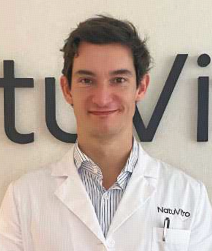 Iñaki González-Foruria合作论文数NatuVitro11
 Elisabet Clua合作论文数Department of Obstetrics, Gynaecology and Reproductive Medicine, Institut Universitari Dexeus11
 Ioannis E. Messinis合作论文数Department of Obstetrics and Gynaecology;Medical School;University of Thessalia11S. Santos Ribeiro合作论文数10
 Vaiarelli Alberto合作论文数Universitair Ziekenhuis Brussel, Vrije Universiteit Brussel10Basil C. Tarlatzis合作论文数10E.G. Papanikolaou合作论文数10Patrick Haentjens合作论文数Universitair Ziekenhuis Brussel, Vrije Universiteit Brussel10Maria Tsappi合作论文数10
 Evangelos G. Papanikolaou合作论文数University Hospital;Free University;Centre for Reproductive Medicine;University Hospital, Free University10Pehr Lind合作论文数10
 Pedro Montoya-Botero合作论文数Conceptum Unidad Fertil Country9Tsali Lamprini合作论文数9La Marca Antonio合作论文数9
 Shari Mackens合作论文数9Ivan Cortinovis合作论文数9Zavos Apostolos合作论文数Medical School, University of Thessalia8




