RATIONAL AND OBJECTIVE:Formative feedback is an important strategy to improve resident learning. The purpose of our study is to evaluate the impact on frequency, quality and perceptions of resident formative feedback after implementation of a diagnostic breast imaging specific entrustable professional activity based mid-rotation checklist. MATERIAL AND METHODS:In this IRB-approved multicenter study, a six-step methodology was used to develop the validated EPA based checklist, participant pre- and post-implementation surveys, and analyze the collected data. RESULTS:26 out of 32 (81%) residents and 7 out of 9 (78%) teaching attendings found the structured feedback checklist helpful in evaluating residents' performance on the diagnostic breast imaging rotation. 9 of 9 (100%) attending stated it improved their ability to give specific, timely, actionable and thoughtful feedback and 6 of 9 (67%) agreed that their feedback was more structured. 27 of 32 (84%) residents found that the feedback they received allowed them to tailor their studying to areas that needed improvement during the remainder of their rotation. CONCLUSION:Mid-rotation feedback using a structured EPA-based checklist improves the frequency and quality of resident formative feedback from both a residents' and attendings' perspective.
Purpose: To characterize how the adoption of virtual residency interviews (2020-2021 cycle) has impacted the geographic distribution of radiology resident matches. Methods: University -based interventional (IR) and diagnostic radiology (DR) residency programs from 2017 to 2021 were identified using a national residency database (FRIEDA). Public applicant data were obtained from official residency program websites. Medical schools and residency programs were categorized by US census regions. Geographic applicant distribution before and after the initiation of virtual interviews was statistically assessed using Chi-square tests. The effect of virtual interviews on the probability of matching within the same geographic region as one's medical school was evaluated with multivariate logistic regression. Results: 4358 radiology residents (88% diagnostic, 12% interventional) matched at 102 radiology programs during the study period. 71% (n = 3115 residents) had data available for analysis. 56.3% of DR and 49.3% of IR residents matched in the same geographic region as their medical school. The geographic distribution of applicants who matched at Southern IR residency programs significantly changed after implementation of virtual interviews (p < 0.0001). Virtual interviews did not increase the odds of matching in the same region as one's medical school for IR (OR 1.11, p = 0.08) or DR (OR 1.01, p = 0.58) applicants. Top -20 ranked DR programs had lower odds of in -region matches (OR 0.87, p < 0.001). Conclusion: With few exceptions, shifting to virtual residency interviews did not significantly affect the geographic distribution of IR or DR residency matches. Top -ranked DR programs match more regionally diverse applicants. (c) 2024 The Association of University Radiologists. Published by Elsevier Inc. All rights reserved.
Despite the importance of diagnostic imaging across specialties, radiology is often given little emphasis in medical school education. resulting in medical students having little, if any, exposure to radiology. This may negatively impact students’ appreciation of the role of radiology in the workup of a variety of chief complaints or evaluation of treatment response.
RATIONALE AND OBJECTIVES:While the ACGME requires Resident as Teacher (RAT) training, curricula in radiology remain limited. Our study was performed to examine radiology residents (RR) and teaching faculty (TF) perceptions about RAT training.MATERIALS AND METHODS:In 2021, anonymous online surveys were administered to all RR (53-item) and to all TF (24-item) of a radiology residency program. Content domains included attitudes about RAT training and learning topics.RESULTS:Response rates were 97% (38/39) for RR and 54% (58/107) for TF. Most RR desired training to become better educators to medical students (MS) (81%) and other residents (83%). Seventy-seven percent of RR reported the importance regarding how to give feedback to other learners, while 94% desired formal training on delivering case presentations. While 94% of RR reported that resident feedback was valuable, only 6% reported always giving feedback to MS. Seventy-two percent of RR did not apply at least some best-practices in their reading room teaching. Fifty-nine percent of RR wanted TF to observe their own teaching skills and provide feedback although 70% reported rarely or never receiving TF feedback. Ninety-three percent of TF reported RR should receive RAT training, while 88% reported that feedback of RR to MS was important.CONCLUSION:RR and TF strongly endorsed the need for RAT training. RR anticipate teaching to be an important part of their careers. We identified learning topics and possible gaps regarding how TF are meeting RR needs, which could inform the development of RAT curricula.
To fulfill ACGME requirements, radiology residency programs are required to provide an educational experience that includes a core didactic curriculum for each subspecialty. Although developing and delivering such a core curriculum may not present a problem for large academic programs, it can present a significant challenge for smaller programs with limited faculty in each subspecialty area. Success of the core curriculum lectures series developed for cardiothoracic radiology by the Society of Thoracic Radiology and for musculoskeletal radiology by the International Skeletal Society in collaboration with the Society for Skeletal Radiology prompted the idea of creating a comprehensive core curriculum lecture series encompassing all subspecialties. This paper aims to describe the multi-society collaborative effort entailed in building a curated, on line resident focused core curriculum lecture series detailing the barriers encountered, effects of the COVID-19 pandemic and impact of the finished project.
Rationale and Objectives: Meeting the Accreditation Council for Graduate Medical Education scholarly activity requirement can be chal-lenging for residents. Time to engage in research is one of the commonly perceived barriers. To address this barrier, our residency pro-gram implemented a focused academic time initiative of a half day per week that can be taken while on rotation. At the end of the third year of implementation, we assessed the effectiveness of this initiative on the productivity of resident scholarly activity.Materials and Methods: Radiology resident scholarly activity submitted to the Accreditation Council for Graduate Medical Education web-based Accreditation Data System were reviewed and compared to the three academic years before (July 1, 2012-June 30, 2015) and three academic years after (July 1, 2015-June 30, 2018) implementing the focused research time. The types of scholarly activity, which consisted of peer-reviewed journal publications, national conference presentations, and textbook chapters were captured. PubMed-Indexed for MEDLINE (PMID) number was used to confirm publications. Descriptive statistics were used to analyze the data.Results: The total number of residents per year, ranging between 37-40, was similar between the academic years 2012-2015 (116 resi-dents total) and 2015-2018 (117 residents total). After initiating focused academic time, the number of publications increased from 45 to 75 (67%), presentations at conferences increased from 112 to 128 (14%), the number of textbook chapters increased from 4 to 15 (275%), and total number of first author publications by residents increased from 21 to 28 (33% increase).Conclusion: Longitudinal focused academic time of half a day per week increased productivity of scholarly activity among our radiology residents.
Background Previous studies suggest that use of artificial intelligence (AI) algorithms as diagnostic aids may improve the quality of skeletal age assessment, though these studies lack evidence from clinical practice. Purpose To compare the accuracy and interpretation time of skeletal age assessment on hand radiograph examinations with and without the use of an AI algorithm as a diagnostic aid. Materials and Methods In this prospective randomized controlled trial, the accuracy of skeletal age assessment on hand radiograph examinations was performed with (n = 792) and without (n = 739) the AI algorithm as a diagnostic aid. For examinations with the AI algorithm, the radiologist was shown the AI interpretation as part of their routine clinical work and was permitted to accept or modify it. Hand radiographs were interpreted by 93 radiologists from six centers. The primary efficacy outcome was the mean absolute difference between the skeletal age dictated into the radiologists’ signed report and the average interpretation of a panel of four radiologists not using a diagnostic aid. The secondary outcome was the interpretation time. A linear mixed-effects regression model with random center- and radiologist-level effects was used to compare the two experimental groups. Results Overall mean absolute difference was lower when radiologists used the AI algorithm compared with when they did not (5.36 months vs 5.95 months; P = .04). The proportions at which the absolute difference exceeded 12 months (9.3% vs 13.0%, P = .02) and 24 months (0.5% vs 1.8%, P = .02) were lower with the AI algorithm than without it. Median radiologist interpretation time was lower with the AI algorithm than without it (102 seconds vs 142 seconds, P = .001). Conclusion Use of an artificial intelligence algorithm improved skeletal age assessment accuracy and reduced interpretation times for radiologists, although differences were observed between centers. Clinical trial registration no. NCT03530098 © RSNA, 2021 Online supplemental material is available for this article. See also the editorial by Rubin in this issue.
RATIONALE AND OBJECTIVES:The Covid-19 pandemic ushered a sudden need for residency programs to develop innovative socially distant and remote approaches to effectively promote their program. Here we describe our experience using the social virtual reality (VR) platform Mozilla Hubs for the pre-interview social during the 2020-2021 radiology residency virtual recruitment season, provide results of a survey sent to assess applicants' attitudes towards the VR pre-interview social, and outline additional use-cases for the emerging technology.MATERIALS AND METHODS:A VR Meeting Hall dedicated to the pre-interview social was designed in Mozilla Hubs. To assess applicants' impressions of the Mozilla Hubs pre-interview social, applicants were sent an optional web-based survey. Survey respondents were asked to respond to a series of eleven statements using a five-point Likert scale of perceived agreement: Strongly Agree, Agree, Neutral, Disagree, Strongly Disagree. Statements were designed to gauge applicants' attitudes towards the Mozilla Hubs pre-interview social and its usefulness in helping them learn about the residency program, particularly in comparison with pre-interview socials held on conventional video conferencing software (CVCS).RESULTS:Of the 120 residency applicants invited to the Mozilla Hubs pre-interview social, 111 (93%) attended. Of these, 68 (61%) participated in the anonymous survey. Most applicants reported a better overall experience with Mozilla Hubs compared to CVCS (47/68, 69%), with 10% (7/68) reporting a worse overall experience, and 21% (14/68) neutral. Most applicants reported the Mozilla Hubs pre-interview social allowed them to better assess residency culture than did pre-interview socials using CVCS (41/68, 60%). Seventy-two percent of applicants reported that the Mozilla Hubs pre-interview social positively impacted their decision to strongly consider the residency program (49/68).CONCLUSION:Radiology residency applicants overall preferred a pre-interview social hosted on a social VR platform, Mozilla Hubs, compared to those hosted on CVCS. Applicants reported the use of a social VR platform reflected positively on the residency and positively impacted their decision to strongly consider the program.
Rationale and Objectives: The educational value of the daily resident readout, a vital component of resident training, has been markedly diminished due to a significant decrease in imaging volume and case mix diversity. The goal of this study was to create a "simulated" daily readout (SDR) to restore the educational value of the daily readout. Materials and Methods: To create the SDR the following tasks were performed; selection of cases for a daily worklist for each resident rotation, comprising a combination of normal and abnormal cases; determination of the correct number of cases and the appropriate mix of imaging modalities for each worklist; development of an ''educational'' environment consisting of separate ''instances'' of both our Picture Archive Communication System and reporting systems; and the anonymization of all of the cases on the worklists. Surveys of both residents and faculty involved in the SDR were performed to assess its effectiveness. Results: Thirty-two residents participated in the SDR. The daily worklists for the first 20 days of the SDR included 3682 cases. An average of 480 cases per day was dictated by the residents. Surveys of the residents and the faculty involved in the SDR demonstrated that both agreed that the SDR effectively mimics a resident's daily work on rotations and preserves resident education during the Coronavirus Disease 2019 crisis. Conclusion: The development of the SDR provided an effective method of preserving the educational value of the daily readout experience of radiology residents, despite severe decreases in imaging exam volume and case mix diversity during the Coronavirus Disease 2019 pandemic. (c) 2020 The Association of University Radiologists. Published by Elsevier Inc. All rights reserved.
Models for diagnostic reasoning in radiology have been based on the observed behaviors of experienced radiologists but have not directly focused on the thought processes of novices as they improve their accuracy of image interpretation. By collecting think-aloud verbal reports, the current study was designed to investigate differences in specific thought processes between medical students (novices) as they learn and radiologists (experts), so that we can better design future instructional environments. Seven medical students and four physicians with radiology training were asked to interpret and diagnose pediatric elbow radiographs where fracture is suspected. After reporting their diagnosis of a case, they were given immediate feedback. Participants were asked to verbalize their thoughts while completing the diagnosis and while they reflected on the provided feedback. The protocol analysis of their verbalizations showed that participants used some combination of four processes to interpret the case:gestalt interpretation, purposeful search, rule application,andreasoning from a prior case.All types of processes except reasoning from a prior case were applied significantly more frequently by experts. Further, gestalt interpretation was used with higher frequency in abnormal cases while purposeful search was used more often for normal cases. Our assessment of processes could help guide the design of instructional environments with well-curated image banks and analytics to facilitate the novice's journey to expertise in image interpretation.
Purpose: Acclimating residents to radiology residency requires attention to new responsibilities, educational material, and social cohesion. To this end, we instituted a structured orientation week for incoming residents and assessed its impact. Procedures: During the first weeks of July 2016 and 2017, first year residents attended a five day orientation free of clinical duties, consisting of didactics, hands-on training sessions, and social events. After two orientation cohorts, residents who completed orientation week, and two cohorts who had not, were given a voluntary, anonymous survey using Likert scale questions (1 [worst] to 5 [best]) regarding preparedness for responsibilities, learning, and social cohesion. Residents were asked which components were or would have been helpful. Independent samples t-tests were performed to evaluate differences between the two groups (two-tailed p < 0.05). Findings: 21/37 (57%) residents participated. Higher percentages of residents who participated in the orientation week gave scores >= 4 when asked about preparedness for rotations (70% vs. 36%), learning new material (80% vs. 36%), and class cohesiveness (90% vs. 70%). Mean scores on these questions were also higher for these residents with regards to: preparedness for new responsibilities (3.7 vs. 2.9), learning new material (3.8 vs. 2.9), and class cohesiveness (4.5 vs. 3.8), with differences approaching significance (p = 0.09-0.15). Individual components receiving most votes of >= 4 were social outings, resident lunches, didactic lectures, and PACS training. Conclusion: A weeklong orientation program free of clinical duties was valued by residents and contributed to acclimation to new responsibilities, education, and social cohesion.
RATIONAL AND OBJECTIVES:Radiology has one of the lowest female representation rates in medicine and recruiting female residents is a challenge for some residency programs. There is limited understanding of gender differences among residency applicants during program selection. The study objective is to investigate which program factors were considered most important by radiology residency applicants and to assess for differences by gender, race and ethnicity.MATERIALS AND METHODS:An anonymous survey was distributed electronically to diagnostic and interventional radiology residency programs in the US and Canada via the Association of Program Coordinators in Radiology (APCR). Residents were asked to evaluate the importance of 30 factors during evaluation of residency programs using a 5-point Likert scale (1 = not important, 5 = extremely important).RESULTS:370 residents and 1 fellow completed the survey. Of the respondents, 269 were male (72.5%) and 101 were female (27.2%). The most important factors to respondents during program selection were program culture (4.42), geographic location (4.17), fellowship placement (4.14), and imaging and/or procedure volume (3.98). There was a significant difference between male and female respondents in the importance of program culture (p = 0.002), composition of current residents (p = 0.007), percentage of current female residents (p < 0.0001), program size (p = 0.047), call schedule (p = 0.025), percentage of female faculty (p < 0.0001), faculty ethnic and racial diversity (p < 0.0001), resident ethnic and racial diversity (p < 0.0001), which female respondents ranked more highly.CONCLUSION:Applicants consider many factors during residency program selection. Program culture, geographic location, fellowship placement and imaging and/or procedural volume were most important. There were significant differences by gender, race and ethnicity in importance of several factors.
RATIONALE AND OBJECTIVES:The Alliance of Directors and Vice Chairs in Education group identified the need to develop an education budget template as resource for our community. Having a framework and working knowledge of budgetary considerations is crucial to those with general oversight and executive managerial responsibility for departmental educational programs.METHODS:An online survey was sent to all the Alliance of Directors and Vice Chairs in Education members. Survey questions included education funding sources, presence of vice chair of finance, expectation of revenue generation, existing education budget, funding decision-makers, education budget formulation and approval, vice chair of education's role in budget, education budget line items, and income statement review.RESULTS:The survey response rate was 41/81 (51%). A majority 26/41 (63%) of respondents had an education budget that typically included funding for all medical students, residents, and fellows but only a minority of respondents report they developed 10/22 (45%), approved 6/22 (27%), or regularly reviewed 6/21 (29%) this budget. In sharp contrast was the role of department chairs and administrators, who presumably all participated in this process. To assist in education budget development and review, as well as meet the need to improve participants' financial accounting knowledge as a key tenet of faculty professional development, the authors developed sample budget templates and an income statement primer.CONCLUSION:Our survey results suggested the need for an educational budget framework and financial accounting resources for those in radiology education posts, and resources have been provided.
Radiologists are facing increasing workplace pressures that can lead to decreased job satisfaction and burnout. The increasing complexity and volumes of cases and increasing numbers of noninterpretive tasks, compounded by decreasing reimbursements and visibility in this digital age, have created a critical need to develop innovations that optimize workflow, increase radiologist engagement, and enhance patient care. During their workday, radiologists often must navigate through multiple software programs, including picture archiving and communication systems, electronic health records, and dictation software. Furthermore, additional noninterpretive duties can interrupt image review. Fragmented data and frequent task switching can create frustration and potentially affect patient care. Despite the current successful technological advancements across industries, radiology software systems often remain nonintegrated and not leveraged to their full potential. Each step of the imaging process can be enhanced with use of information technology (IT). Successful implementation of IT innovations requires a collaborative team of radiologists, IT professionals, and software programmers to develop customized solutions. This article includes a discussion of how IT tools are used to improve many steps of the imaging process, including examination protocoling, image interpretation, reporting, communication, and radiologist feedback. ©RSNA, 2018.
Purpose: The purpose of this study was to reduce radiation exposure during pediatric central venous line (CVL) placement by implementing a radiation safety process including a radiation safety briefing and a job instruction model with a preradiation time-out. Methods: We reviewed records of all patients under 21 who underwent CVL placement in the operating room covering 22 months before the intervention through 10 months after 2013-2016. The intervention consisted of a radiation safety briefing by the surgeon to the intraoperative staff before each case and a radiation safety time-out. We measured and analyzed the dose area product (DAP), total radiation time pre- and postintervention, and the use of postprocedural chest radiograph. Results: 100 patients with valid DAP measurements were identified for analysis (59 preintervention, 41 postintervention). Following implementation of the radiation safety process, there was a 79% decrease in median DAP (61.4 vs 13.1 rad*cm(2), P < 0.001) and a 73% decrease in the median radiation time (28 vs 7.6 s, P < 0.001). Additionally, there was a significant reduction in use of confirmatory CXR (95% vs 15%, P < 0.01). Conclusion: A preoperative radiation safety briefing and a radiation safety time-out supported by a job instruction model were effective in significantly lowering, the absorbed doses of radiation in children undergoing CVL insertion. Type of study: Case-control study. (C) 2018 Elsevier Inc. All rights reserved.
To preserve radiology rounds in the changing health care environment, we have introduced virtual radiology rounds, an initiative enabling clinicians to remotely review imaging studies with the radiologist.
Rapid and accurate diagnosis of appendicitis, particularly with respect to the presence or absence of perforation, is essential in guiding appropriate management. Although many studies have explored sonographic findings associated with acute appendicitis, few investigations discuss specific signs that can reliably differentiate perforated appendicitis from acute appendicitis prior to abscess formation.
Shielding of the gonads is a dose-saving strategy in pediatric radiography and its use is the law in New York and the majority of other states. However, routine use of gonadal shields is controversial because of concerns that important diagnostic information can be obscured.
Objective: To prospectively evaluate the diagnostic utility of interlobar renal arterial resistive index values, in predicting the presence of vesicoureteral reflux in children with sonographically normal appearing kidneys. Methods: We investigated 35 children with a history of urinary tract infection with grayscale and color Doppler ultrasound (US) fol-lowed by a standard voiding cystouretherogram to assess the presence of vesicoureteral reflux. All renal units (individ-ual kidneys) were morphologically normal without evidence of hydronephrosis. Resistive index (RI) was measured at the level of the interlobar arteries. Results: Of a total of 67 kidney units, 3 kidney units yielded no diagnostic results due to technical factors (crying, agitation). 46 kidney units (69%) were not associated with any degree of reflux. 17 kidney units (25%) were associated with low-mid grade reflux (grades I - III). 4 kidney units (6%) were associated with high grade reflux (grades IV - V). No statistically significant correlation was found between the interlobar resistive indices of sonographically normal appearing kidneys and the presence, absence, or degree of vesicoureteral reflux. Conclusions: The results of this study suggest that resistive index measurements taken in the interlobar arteries of sonographically normal appearing kidneys cannot be used to predict the presence or absence of vesicoureteral reflux.
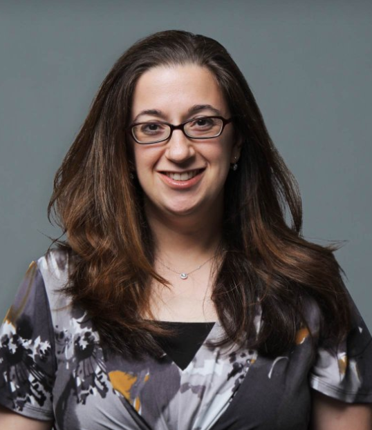 Naomi A. Strubel合作论文数Division of Pediatric Radiology, Department of Radiology, New York University8
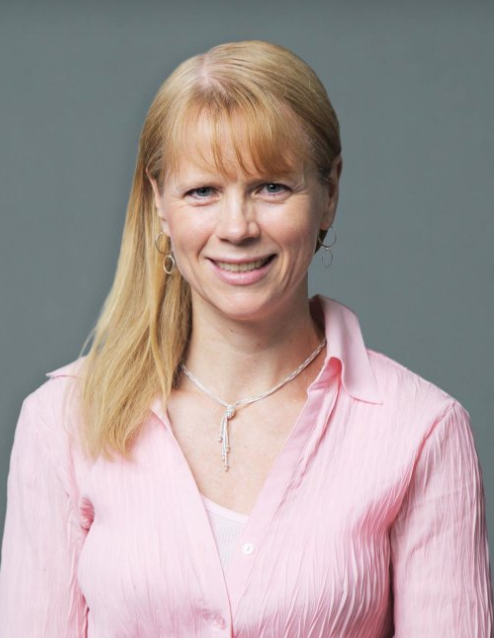 Lynne P. Pinkney合作论文数Division of Pediatric Radiology, Department of Radiology, New York University7
 Amodio John合作论文数5
 Milla Sarah S合作论文数5Rafael Rivera合作论文数5
 Howard B. Ginsburg合作论文数Department of Surgery, Grossman School of Medicine, New York University;Department of Pediatrics, Grossman School of Medicine, New York University4
 Michael P. Recht合作论文数Division of Musculoskeletal Imaging, Department of Radiology, New York University4
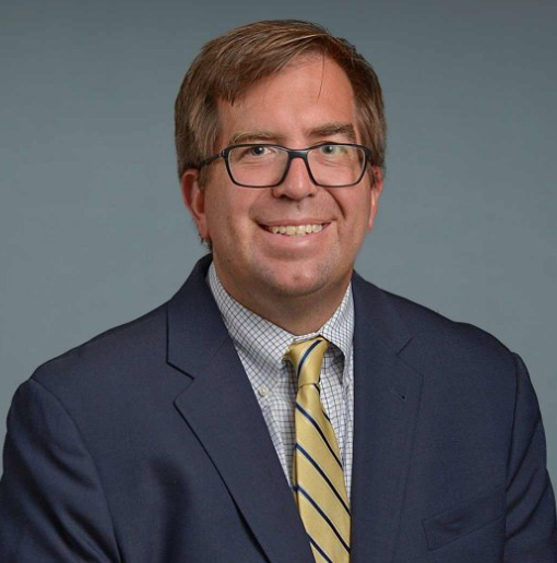 Jason C. Hoffmann合作论文数Department of Radiology, New York University3
 Shailee V. Lala合作论文数Division of Pediatric Radiology, Department of Radiology, New York University3Sandra Tomita合作论文数3James S. Babb合作论文数NYU Langone Medical Center3Mary W Chang合作论文数2Kopal Kulkarni合作论文数2
 Monica Sheth合作论文数2Sohail Contractor合作论文数2Beatrix Hyemin Choi合作论文数2Michael Macari合作论文数2
 Jason C. Fisher合作论文数Division of Pediatric Surgery, Department of Surgery, Grossman School of Medicine, New York University2
 Georgeann McGuinness合作论文数Division of Thoracic Imaging, Department of Radiology, New York University2
 Cecilia L. Mercado合作论文数Division of Breast Imaging, Department of Radiology, New York University2Vinay Prabhu合作论文数Division of Abdominal Imaging, Department of Radiology, New York University2
 Danny C. Kim合作论文数Division of Abdominal Imaging, Department of Radiology, New York University2
 Sharon L. Gardner合作论文数Division of Pediatric Hematology-Oncology, Department of Pediatrics, Grossman School of Medicine, New York University2
 Genevieve L. Bennett合作论文数Department of Radiology, Grossman School of Medicine, New York University;New York University Langone Hospitals2Pinkney Lynne P合作论文数2
 Slanetz Priscilla J合作论文数Beth Israel Deaconess Medical Center, Harvard Medical School2
 Monnier Chloe合作论文数2
 Jeffrey B Alpert合作论文数2
 Nathan Kuppermann合作论文数Department of Emergency Medicine and Pediatrics, School of Medicine, University of Caifornia, Davis;Radiology Outcomes Research Laboratory, University of California San Francisco2
 Mark Bittman合作论文数Department of Radiology, Boston Children's Hospital and Harvard Medical School2M. Alba Greco合作论文数2George C. Nomikos合作论文数2Lawrence Bodenstein合作论文数2
 Scott W. Atlas合作论文数Hoover Institution, Stanford University2Sandra L. Moore合作论文数2David P. Naidich合作论文数2Maciek Bobinski合作论文数2
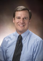 Joseph J. Zorc合作论文数Department of Pediatrics, University of Pennsylvania School of Medicine2Barry S. Leitman合作论文数2
 Charles G Macias合作论文数Emergency Medicine, Baylor College of Medicine and Texas Children's Hospital2
 Kathy N. Shaw合作论文数Department of Pediatrics, The Children's Hospital of Philadelphia2Jennifer M. Martino合作论文数2John Amodio合作论文数2Keith A. Kuenzler合作论文数2Chloe Chhor合作论文数1Susan E. Sharp合作论文数1P S Dayan合作论文数1
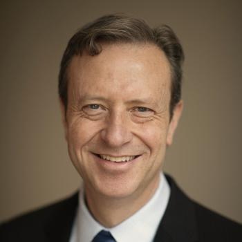 David Larson合作论文数Department of Radiology, Stanford University School of Medicine1




