Cultures of primary mouse bile duct epithelial cells are a valuable tool to study cholangiocyte secretion and bile formation. However, freshly isolated cells have a limited ability to expand in culture. Here we report a novel isolation and culture technique for normal mouse cholangiocytes (NMC) that enables long-term growth without compromising function. Mouse cholangiocytes were isolated and cultured in conditioned medium (CM) that was subsequently supplemented with ROCK inhibitor Y-27632. Expression of cholangiocyte markers was assessed by qPCR, immunofluorescence, and Western blotting. Patch clamp techniques were used to measure cAMP-activated Cl- current, Ca2+-activated Cl- current, and volume-stimulated Cl- current. We obtained NMC cultures that were polarized and maintained a cholangiocyte phenotype for over 50 passages. Functional studies show that ion channel activity is maintained in NMC regardless of the number of passages and despite removal of CM. NMC also perform other physiological functions such as ATP release and intracellular Ca2+ changes in response to stimulation with bile acids. Thus, our isolation procedure produces viable NMC that maintain biophysical properties in long-term culture. We also demonstrate the utility of NMC in studies investigating the cellular mechanisms responsible for cholangiocyte secretion and bile formation.
The Wnt/β-catenin signaling pathway is critical for liver homeostasis. We have previously shown that hepatocyte β-catenin plays a pleiotropic role in cholestatic injury. However, the role of cholangiocyte β-catenin signaling during cholestasis remains unclear. Inducible-Osteopontin (OPN)-Cre-β-catenin-floxed C57BL/6 mice were used in two cholestasis models. Mdr2 knockout (KO)-β-catenin-floxed:OPN-Cre mice were administered tamoxifen to delete β-catenin from cholangiocytes. Wild-type and cholangiocyte β-catenin KO mice were also administered a 3,5‐diethoxycarbonyl‐1,4‐dihydrocollidine (DDC) diet to induce cholestasis. Serum was collected to evaluate liver enzymes. qRT-PCR and immunohistochemistry/immunofluorescence assays were performed on whole livers to assess injury, vascular remodeling, and hepatocyte reprogramming. Livers were isolated for transmission electron microscopy. Isolated cholangiocytes were analyzed by RNA-seq. Cholangiocytes were treated with β-catenin siRNA and lipopolysaccharide in vitro to determine changes in angiogenic factors and NF-κB activation. Conditioned media from cholangiocytes were used to evaluate endothelial cell proliferation in vitro. Mice lacking cholangiocyte β-catenin showed similar levels of hepatobiliary injury compared to controls. We observed more hepatocytes expressing cholangiocyte markers and ductular cells expressing β-catenin in β-catenin KO animals, indicating enhanced hepatocyte reprogramming. Interestingly, cholangiocyte β-catenin KO also had fibrotic hepatic arteries and increased angiogenesis versus controls. Histology and transmission electron microscopy revealed increased basement membrane formation and loss of fenestrations in the sinusoids of β-catenin KO animals. RNA-seq of isolated β-catenin KO cholangiocytes revealed increased expression of angiogenesis pathways that was associated with NF-κB activation. In vitro studies silencing β-catenin in cholangiocytes induced Vegf and Pdgfb expression. Lipopolysaccharide stimulation increased NF-κB nuclear localization in β-catenin-silenced cholangiocytes. Stimulated media from these cells promoted endothelial cell proliferation, recapitulating the angiogenic phenotype found in vivo. β-catenin signaling in cholangiocytes is a novel mediator of cell–cell communication, and its loss induces a pro-angiogenic phenotype and supports hepatocyte reprogramming during cholestasis, both of which may prevent accelerated liver injury.
The gut-liver axis is the bidirectional relationship between the gut microbiota and the liver. Dysbiosis in the gut-liver axis and disrupted bile acid homeostasis contribute to cholestatic liver disease pathogenesis. Patients afflicted with cholestasis have accelerated bone loss, a higher incidence of fractures, and are at risk of developing osteoporosis. However, the mechanisms underlying bone loss are largely unknown. The study purpose was to investigate the role of the gut-liver axis and bile acid signaling on skeletal homeostasis during cholestasis. Male C57BL/6J specific-pathogen-free mice were administered 3,5-diethoxycarbonyl-1,4-dihydrocollidine from age 11 to 15 weeks to induce cholestasis. 16s rDNA sequencing was performed on colonic contents. Livers were processed for qRT-pCR. Trabecular and cortical bone were analyzed by micro-CT. Osteoclast/osteoblast outcomes were determined by histomorphometry. Bile acid concentrations in serum and bone marrow were assessed by mass spectrometry. MC3T3-E1 and RAW 264.7 cells were stimulated with bile acids at concentrations found in the bone marrow to determine their effects on osteoblasts and osteoclasts. Cholestatic mice had less bone mass than controls, attributed to increases in osteoclasts and decreases in osteoblasts. Following cholestatic injury, mice show dysbiotic shifts in their colonic bacteriome, increased expression of hepatic bile acid efflux transporters, and elevated bone marrow bile acids. In vitro, bile acids from the bone marrow of cholestatic mice suppressed osteoblastogenesis and promoted osteoclastogenesis, which was rescued by stimulating cells with a farnesoid X receptor agonist. This study introduces the gut-liver axis as a novel regulator of skeletal homeostasis during cholestatic liver disease through dysregulated bile acid signaling.
Porphyrias are rare metabolic disorders arising from defects in heme biosynthesis, leading to accumulation of toxic porphyrin intermediates, mitochondrial dysfunction, and liver injury. Current therapies are limited in efficacy, emphasizing the need for novel treatments. Prior studies showed hepatocyte-specific β-catenin deletion attenuates porphyrin accumulation and liver injury in 3,5-diethoxycarbonyl-1,4-dihydrocollidine (DDC)-induced porphyria. We hypothesized that inhibiting components of the Wnt-β-catenin-glutamine synthesis (GS) pathway reduces heme synthesis and also enhances porphyrin clearance by activating autophagy and improving mitochondrial quality control. We combined pharmacologic Wnt inhibition and hepatocyte-specific GS deletion in murine models of porphyria. Readouts included spatial transcriptomics, targeted metabolomics, immunohistochemistry, confocal mt-Keima imaging, high-resolution respirometry, and transmission electron microscopy. Human liver biopsies and explants from porphyria patients were also examined by dual-label immunohistochemistry. Wnt inhibition during DDC suppressed upregulation of heme biosynthesis genes, reduced porphyrin intermediate accumulation, and enhanced autophagic flux. GS deletion attenuated porphyrin biosynthesis by limiting intracellular glutamine. Wnt and GS deletion produced additive increases in autophagy, restored zonation, and further reduced porphyrin accumulation. Wnt inhibition restored mitophagy, whereas GS deletion primarily improved mitochondrial coupling efficiency. Wnt inhibition also decreased fibrosis in a genetic mouse model of porphyria. Patient samples mirrored murine findings, with heme enzymes and autophagy inversely correlated with β-catenin expression in porphyria cutanea tarda. By disrupting Wnt-GS signaling, we establish a link between increased autophagy, reduced porphyrin formation, and heme pathway regulation in mouse and human liver. These findings identify the Wnt signaling pathway as a potential therapeutic target in porphyria.Abbreviations: ALA: δ-Aminolevulinic acid; AIP: acute intermittent porphyria; ALAS: aminolevulinic acid synthase; ALAD: aminolevulinic acid dehydratase; ALP: alkaline phosphatase: AST: aspartate aminotransferase; ALT: alanine aminotransferase; DAB: 3,3'-diaminobenzidine; DDC: 3,5-diethoxycarbonyl-1,4-dihydrocollidine; EPP: erythropoietic protoporphyria; Fech: ferrochelatase; GAPDH: glyceraldehyde 3-phosphate dehydrogenase; GS: glutamine synthesis; H&E: hematoxylin and eosin: HO-1: heme oxygenase 1; IHC: immunohistochemistry; KO: knockout; LAMP1: lysosomal associated membrane protein 1; LC3: microtubule-associated protein 1 A/1B-light chain 3; mTOR: mechanistic target of rapamycin; PBG: porphobilinogen; PBS: phosphate-buffered saline; PP-IX: protoporphyrin-IX; PCT: porphyria cutanea tarda; RCR: respiratory control ratio; SQSTM1/p62: sequestosome 1; TEM: transmission electron microscopy; Wnt-I: Wnt-C59 (inhibitor).
The liver has a critical role in regulating host metabolism, immunity, detoxification, and homeostasis. Proper liver function is essential for host health, and dysregulation of hepatic signaling pathways can lead to the onset of disease. The Wnt/β-catenin signaling pathway is an important regulator of liver homeostasis and function. Throughout life, hepatic Wnt/β-catenin signaling contributes to liver development and growth, metabolic zonation, and regeneration. Extensive research has demonstrated that aberrant Wnt/β-catenin signaling drives liver pathologies, including cancers, steatohepatitis, and cholestasis. In this review, we discuss the Wnt/β-catenin pathway as it pertains to liver function and how disruptions in this pathway contribute to the onset and progression of liver diseases. Further, we discuss ongoing research that targets the Wnt/β-catenin pathway for the treatment of liver pathologies.
The porphyrias are a group of metabolic disorders that are caused by defects in one of the eight enzymes that synthesize heme. A common feature of all porphyrias is accumulation of porphyrin precursors or porphyrins, which are intermediates of the heme biosynthesis pathway. Approximately 15% of heme biosynthesis occurs in the liver, and excessive hepatic production of porphyrin precursors caused by heme enzyme deficiencies can lead to neurovisceral manifestations. Additionally, in erythropoietic protoporphyria, porphyrins accumulate in the liver, leading to hepatic injury. These rare diseases have few effective medical therapies, and disease mechanisms are not always well understood. Animal models have provided a platform to study the pathophysiology of disease and test emerging therapies. In this review, the last of a three-part series, we describe the animal models that have been generated to study porphyrias with hepatic involvement. For each model, we discuss mechanisms of injury, phenotypic features, and the similarities and contrasts to human porphyria. We also describe preclinical studies that have utilized the model for therapeutic interventions. Overall, animal-based studies have made significant contributions to our understanding of porphyria and may lead to innovative therapies in the future.
Hepatic porphyrias are a group of metabolic disorders that are characterized by overproduction and accumulation of porphyrin precursors in the liver. These porphyrins cause neurologic symptoms as well as cutaneous photosensitivity, and in some cases patients can experience life-threatening acute neurovisceral attacks. This review describes the acute hepatic porphyrias in detail, including acute intermittent porphyria, hereditary coproporphyria, and variegate porphyria, as well as the hepatic porphyrias with cutaneous manifestations such as porphyria cutanea tarda and hepatoerythropoietic porphyria. Each section will cover disease prevalence, clinical manifestations, and current therapies, including strategies to manage symptoms. Finally, we review new and emerging treatment modalities, including gene therapy through use of adeno-associated vectors and chaperone therapies such as lipid nanoparticle and small interfering RNA-based therapeutics.
BACKGROUND:Cholestasis is an intractable liver disorder that results from impaired bile flow. We have previously shown that the Wnt/β-catenin signaling pathway regulates the progression of cholestatic liver disease through multiple mechanisms, including bile acid metabolism and hepatocyte proliferation. To further explore the impact of these functions during intrahepatic cholestasis, we exposed mice to a xenobiotic that causes selective biliary injury. METHODS:α-naphthylisothiocyanate (ANIT) was administered to liver-specific knockout (KO) of β-catenin and wild-type mice in the diet. Mice were killed at 6 or 14 days to assess the severity of cholestatic liver disease, measure the expression of target genes, and perform biochemical analyses. RESULTS:We found that the presence of β-catenin was protective against ANIT, as KO mice had a significantly lower survival rate than wild-type mice. Although serum markers of liver damage and total bile acid levels were similar between KO and wild-type mice, the KO had minor histological abnormalities, such as sinusoidal dilatation, concentric fibrosis around ducts, and decreased inflammation. Notably, both total glutathione levels and expression of glutathione-S-transferases, which catalyze the conjugation of ANIT to glutathione, were significantly decreased in KO after ANIT. Nuclear factor erythroid-derived 2-like 2, a master regulator of the antioxidant response, was activated in KO after ANIT as well as in a subset of patients with primary sclerosing cholangitis lacking activated β-catenin. Despite the activation of nuclear factor erythroid-derived 2-like 2, KO livers had increased lipid peroxidation and cell death, which likely contributed to mortality. CONCLUSIONS:Loss of β-catenin leads to increased cellular injury and cell death during cholestasis through failure to neutralize oxidative stress, which may contribute to the pathology of this disease.
 Satdarshan Paul Singh Monga合作论文数Department of Pathology, School of Medicine, University of Pittsburgh42
 Satdarshan P. Monga合作论文数26Sucha Singh合作论文数26
 Pamela K Cornuet合作论文数24
 Liu Silvia合作论文数Department of Biostatistics, University of Pittsburgh School of Medicine19Karis Kosar合作论文数17Minakshi Poddar合作论文数14
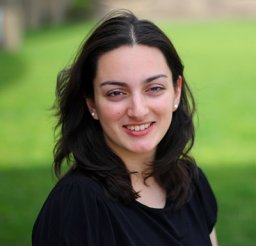 Laura Molina合作论文数Sch Med, Univ Pittsburgh13Goel C合作论文数Allergy and Immunology Section, CSIR-Institute of Genomics and Integrative Biology;Academy of Scientific and Innovative Research (AcSIR),, CSIR;CSIR-Institute of Genomics and Integrative Biology, Academy of Scientific and Innovative Research (AcSIR),11Rong Zhang合作论文数School of Biological and Health Systems Engineering, Arizona State University10
 Tirthadipa Pradhan Sundd合作论文数Sch Med, Univ Pittsburgh10
 George K. Michalopoulos合作论文数Department of Pathology, School of Medicine, University of Pittsburgh10
 Aaron Bell合作论文数10William C. Bowen合作论文数10Michael Oertel合作论文数9Xiaochao Ma合作论文数9Sungjin Ko合作论文数Univ Pittsburgh, Pittsburgh, PA 15260 USA8
 Michael D Thompson合作论文数8Oluwashanu Balogun合作论文数7Shikai Hu合作论文数7Junyan Tao合作论文数7
 Aatur D. Singhi合作论文数Division of Anatomic Pathology, Department of Pathology, School of Medicine, University of Pittsburgh;Developmental Laboratory, University of Pittsburgh Medical Center7
 Ravi Vats合作论文数Pittsburgh Heart Liver & Blood Vasc Med Inst, Univ Pittsburgh6
 Reben Raeman合作论文数University of Pittsburgh6Russell Jacquelyn合作论文数6
 Jianhua Luo (骆建华)合作论文数Laboratory of Aerospace Intelligent Photoelectric Technology, Shanghai Jiao Tong University6Qin Li合作论文数5Orr Anne合作论文数5Yang Jing合作论文数5
 Lili Zhou (周丽丽)合作论文数School of Basic Medical Sciences, Southern Medical University;Department of Nephrology, Nanfang Hospital, Southern Medical University;National Clinical Research Center for Kidney and Urological Diseases5
 Hirohisa Okabe合作论文数Department of Gastroenterological Surgery, Kumamoto University5
 Gang Zeng (曾钢)合作论文数School of Intelligence Science and Technology, Peking University5
 Prithu Sundd合作论文数5Shirish Paranjpe合作论文数5
 An Jiang (蒋安)合作论文数The Second Affiliated Hospital, Xi'an Jiaotong University4
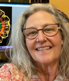 Donna Beer Stolz合作论文数McGowan Institute for Regenerative Medicine, University of Pittsburgh;Center for Biologic Imaging, School of Medicine, University of Pittsburgh;Departments of Cell Biology and Pathology, University of Pittsburgh4
 Marc Abrams合作论文数4
 Morgan Preziosi合作论文数4
 Simon C. Watkins合作论文数Department of Cell Biology, School of Medicine, University of Pittsburgh;Pittsburgh Center for Interdisciplinary, University of Pittsburgh;Department of Immunology, McGowan Institute for Regenerative Medicine, University of Pittsburgh4Mohammed Hassan合作论文数4Sarangarajan Ranganathan合作论文数4
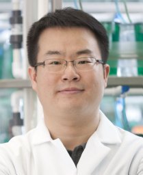 Donghun Shin合作论文数Department of Preventive Medicine and Institute for Medical Science, Keimyung University4Satdarshan Pal Singh Monga合作论文数3Prince Awuah合作论文数3Anu Balogun合作论文数3Harvinder Saggi合作论文数3
 Elizabeth Lee合作论文数Johns Hopkins Bloomberg Sch Publ Hlth, Dept Epidemiol, Baltimore, MD 21205 USA3
 Dean Yimlamai合作论文数Molecular Medicine, Pharmacology, and Physiology, Biological & Biomedical Sciences, Yale University3Antonella Cusimano合作论文数Policlinico P. Giaccone, Università di Palermo3




