Abstract Small cell lung cancer (SCLC) presents as a highly chemosensitive malignancy but acquires cross-resistance after relapse. This transformation is nearly inevitable in patients but has been difficult to capture in laboratory models. Here, we present a preclinical system that recapitulates acquired cross-resistance, developed from 51 patient-derived xenograft (PDX) models. Each model was tested in vivo against three clinical regimens: cisplatin plus etoposide, olaparib plus temozolomide, and topotecan. These drug-response profiles captured hallmark clinical features of SCLC, such as the emergence of treatment-refractory disease after early relapse. For one patient, serial PDX models revealed that cross-resistance was acquired through MYC amplification on extrachromosomal DNA (ecDNA). Genomic and transcriptional profiles of the full PDX panel revealed that MYC paralog amplifications on ecDNAs were recurrent in relapsed cross-resistant SCLC, and this was corroborated in tumor biopsies from relapsed patients. We conclude that ecDNAs with MYC paralogs are recurrent drivers of cross-resistance in SCLC. Significance: SCLC is initially chemosensitive, but acquired cross-resistance renders this disease refractory to further treatment and ultimately fatal. The genomic drivers of this transformation are unknown. We use a population of PDX models to discover that amplifications of MYC paralogs on ecDNA are recurrent drivers of acquired cross-resistance in SCLC. This article is featured in Selected Articles from This Issue, p. 695
Acquired ecMYCN amplification in MGH1578 serial models and lineage oncogene expression compared with ecDNA status across the PDX panel.
Reconstruction of ecDNAs containing MYC paralogs and MYCL FISH in MGH1501-1A metaphase chromosomes.
Abstract The retinoblastoma protein (RB) suppresses the activity of the E2F transcription factor family, controlling cellular proliferation. Recent studies, however, indicate that RB's role in chromatin organization, which is not yet fully understood, might be distinct from this E2F-dependent regulation. Employing chromosome conformation capture at a single nucleosome resolution, we identified RB as a cell cycle-regulated repressor of cohesin-dependent loop formation at topologically associating domain (TAD) boundaries. RB depletion increased the number and size of cohesin-dependent loops and strengthened topologically associating domains (TADs). This phenomenon was specific to the G1 phase and was not observed in the S phase, indicating that this novel RB function is regulated by the cell cycle. Mechanistically, RB showed extensive colocalization with cohesin in the human genome, and it impacted cohesin’s distribution on the chromatin. Active RB reduced cohesin from RB-bound TAD boundaries and decreased cohesin activity therein, as assessed by the increased K105/106 acetylation of the cohesin subunit SMC3. This led to reduced insulation in chromosome conformation capture assays. Importantly, by weakening the insulation activity of the adjacent insulators, RB non-canonically enhanced the expression of non-E2F target genes salient for cell adhesion and extracellular matrix organization. When RB was lost, cells showed a more rapid cellular detachment rate and an elevated migration rate, indicating that this novel RB function controls specific transcriptional programs rather than arbitrary genes to regulate the process of cell adhesion and migration. Overall, we conclude that RB has a central role in the interplay between cell cycle and chromatin organization, by repressing the cohesin-dependent loop formation at TAD boundaries. This RB function safeguards E2F-independent transcriptional programs driven by active enhancers and helps maintain cellular adhesion to the extracellular matrix. Citation Format: Hanjun Lee, Ioanna-Maria Gkotinakou, Badri Krishnan, Nicholas J. Dyson, Michael S. Lawrence, Ioannis Sanidas. RB represses cohesin-dependent loop formation and activates E2F-independent transcription [abstract]. In: Proceedings of the American Association for Cancer Research Annual Meeting 2024; Part 1 (Regular Abstracts); 2024 Apr 5-10; San Diego, CA. Philadelphia (PA): AACR; Cancer Res 2024;84(6_Suppl):Abstract nr 1648.
Translesion synthesis (TLS) is a cellular mechanism through which actively replicating cells recruit specialized, low-fidelity DNA polymerases to damaged DNA to allow for replication past these lesions. REV1 is one of these TLS DNA polymerases that functions primarily as a scaffolding protein to organize the TLS heteroprotein complex and ensure replication occurs in the presence of DNA lesions. The C-Terminal domain of REV1 (REV1-CT) forms many protein-protein interactions (PPIs) with other TLS polymerases, making it essential for TLS function and a promising drug target for anti-cancer drug development. We utilized several lead identification strategies to identify various small molecules capable of disrupting the PPI between REV1-CT and the REV1 Interacting Regions (RIR) present in several other TLS polymerases. These lead compounds were profiled in several in vitro potency and PK assays to identify two scaffolds (1 and 6) as the most promising for further development. Both 1 and 6 synergized with cisplatin in a REV1-dependent fashion and demonstrated promising in vivo PK and toxicity profiles.
MYC paralog copy number and ecDNA status in SCLC cell lines, PDX models and biopsy samples derived before treatment or after relapse.
MYC paralog amplifications that were detected or omitted by segmented copy number analysis, and patient survival compared with ecDNA status of PDX models.
Genomic analysis of chromosome 17 focal amplification in MGH1531-5BX and analysis of ecDNA junction mutations in MGH1518-3A.
Despite the great significance of the tumor suppressor Rb in cancer biology, the full spectrum of its binding pattern in human cells was yet to be established. Here, we report that rather than exclusively associating with E2F-bound promoters, chromatin-bound Rb associates with three distinct types of loci—E2F1-bound promoters, AP-1-bound enhancers, and CTCF-bound insulators. The binding pattern of Rb correlated with cohesin binding and chromatin accessibility in all three types of Rb-bound regions. When cells entered the S phase after the G1/S checkpoint, Rb redistributed from promoters to enhancers, leading to distinct transcriptional changes. These Rb-bound enhancers were cell-type-specific, in contrast to Rb-bound promoters that were constitutively present throughout most human cell types. Overall, our study provides a comprehensive view on the role of the tumor suppressor Rb, which has a well-preserved role in the G1 phase of regulating transcription at E2F1-bound promoters, while having a cell-type-specific role in the S phase when cells are determined to replicate. Citation Format: Hanjun Lee, Ioanna M. Gkotinakou, Ioannis Sanidas, Michael S. Lawrence, Nicholas J. Dyson. The non-canonical role of Rb in enhancers and insulators [abstract]. In: Proceedings of the American Association for Cancer Research Annual Meeting 2023; Part 1 (Regular and Invited Abstracts); 2023 Apr 14-19; Orlando, FL. Philadelphia (PA): AACR; Cancer Res 2023;83(7_Suppl):Abstract nr 1420.
Computational pipelines for chromatin immunoprecipitation sequencing analysis can neglect colocalization events that occur in a mere subset of the genome. Here, we detail a streamlined approach for assessing colocalization of chromatinbound proteins using the bedGraph2Cluster and PanChIP algorithms. Using histone modifications as an example, bedGraph2Cluster performs clustering analysis on chromatin binding patterns of target proteins. PanChIP then compares these clusters with a reference library of chromatin binding patterns and measures the overlap in peaks, capturing the heterogeneity in chromatin binding and colocalization patterns.For complete details on the use and execution of this protocol, please refer to Sanidas et al. (2022).1
The retinoblastoma tumor suppressor (RB) prevents G1 to S cell cycle transition by inhibiting E2F activity. This function requires that RB remains un- or underphosphorylated (the so-called active forms of RB). Recently, we showed that active forms of RB cause widespread changes in nuclear architecture that are visible under a microscope. These phenotypes did not correlate with cell cycle arrest or repression of the E2F transcriptional program, but appeared later, and were associated with the appearance of autophagy or in IMR-90 cells with senescence markers. In this perspective, we describe the relative timing of these RB-induced events and discuss the mechanisms that may underlie RB-induced chromatin dispersion. We consider the relationship between RB-induced dispersion, autophagy, and senescence and the potential connection between dispersion and cell cycle exit.
Supp Table 1: APA effected PUM target in breast tumors Supp Table 2: primers and siRNAs used Supp Fig 1: TNBC tumor 3'UTR RT-PCR, FOXO1, E2F4 and PTEN Supp Fig 2: TNBC tumors 3'UTR RT-PCR NRAS Supp Fig 3: TNBC tumors 3'UTR RT-PCR c-JUN Supp Fig 4: NRAS, PTEN and FOXO1 regulation in multiple cell lines Supp Fig 5: NRAS and c-JUN stability assays Supp Fig 6: Regulation of 3'UTR length
 Benjamin J Drapkin合作论文数30
 Anna F. Farago合作论文数Massachusetts General Hospital Cancer Center and Harvard Medical School,Boston26
 Marcello Stanzione合作论文数Tech Univ Dresden, Fac Med, Inst Physiol Chem, Fetscherstr 74, D-01307 Dresden, Germany25David T. Myers合作论文数24Sarah Phat合作论文数24
 Michael S. Lawrence合作论文数Harvard Medical School;Lawrence Laboratory, Mass General Cancer Center, Massachusetts General Hospital24Jun Zhong合作论文数23
 Brandon Nicolay合作论文数Massachusetts General Hospital Cancer Center, Harvard Medical School20E Harlow合作论文数19
 Miles Wayne O合作论文数Massachusetts General Hospital Cancer Center, Harvard Medical School16Christensen Camilla L合作论文数16
 Rex Hung合作论文数Department of Pathology, Massachusetts General Hospital16
 Kwok-Kin Wong合作论文数Department of Medicine, Grossman School of Medicine, New York University15
 Jun-Yuan Ji合作论文数15Michael Korenjak合作论文数14Edmond Wong合作论文数14
 Wise Jillian F合作论文数Univ Texas MD Anderson Canc Ctr14Wilhelm Haas合作论文数Department of Cell Biology, Harvard Medical School14Hanieh Mazloom-Farsibaf合作论文数13Victor D. Chien合作论文数13Seth Hamilton合作论文数13Braeden Freitas合作论文数13
 Sihan Wu合作论文数13Shreoshi Pal Choudhuri合作论文数University of Texas Southwestern Medical Center13Collin Gilbreath合作论文数13Di Yang合作论文数13Jun Yi Stanley Lim合作论文数13
 Nam Sung Moon合作论文数Department of Biology, Faculty of Science, McGill University13Meador Catherine B合作论文数13
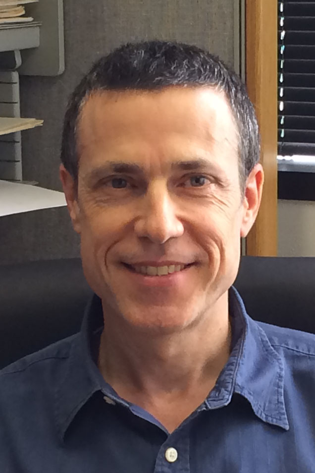 Luc Girard合作论文数Department of Hamon Center for Therapeutic Oncology Research, Medical School, University of Texas Southwestern Medical Center;Department of Pharmacology, University of Texas Southwestern Medical Center13
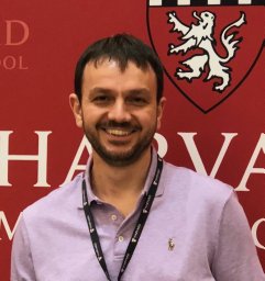 Ioannis Sanidas合作论文数Laboratory of Biochemistry, The Aristotle University13
 Jung-Whan Kim合作论文数Department of Toxicology, National Institute of Toxicological Research13
 Maxim V Frolov合作论文数11
 Luisa Di Stefano合作论文数10Amity L Manning合作论文数Cancer Center and Harvard Medical School, Massachusetts General Hospital10
 Daniel Arie Haber合作论文数Harvard Medical School;Medicine-MGH Cancer Center, Massachusetts General Hospital10
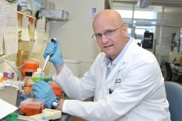 Frederick A. Dick合作论文数London Regional Cancer Program;Children's Health Research Institute;Department of Biochemistry, University of Western Ontario;Children's Health Research Institute, University of Western Ontario10Katrin Tschöp合作论文数9Boukhali Myriam合作论文数9
 Lecia Sequist合作论文数Cancer Early Detection and Diagnostics Clinic, Massachusetts General Hospital Cancer Center;Harvard Medical School;Dana-Farber Cancer Center8
 Nabeel M. Bardeesy合作论文数Center for Cancer Research, Massachusetts General Hospital;Harvard Medical School;KFCCR Faculty, Mass General Research Institute;Harvard Stem Cell Institute;OncoRev, LLC8Aaron N. Hata合作论文数Massachusetts General Hospital Cancer Center/Department of Medicine, Harvard Medical School8
 Shyamala Maheswaran合作论文数Harvard Medical School, Harvard University;Mass General Research Institute, Massachusetts General Hospital8
 Alice Shaw合作论文数Harvard Medical School;Department of Medical Oncology, Dana-Farber Cancer Institute7O. Stevaux合作论文数7Michelle S. Longworth合作论文数7
 Beow Yeap合作论文数Cancer Center, Massachusetts General Hospital;Massachusetts General Hospital Biostatistics;Harvard Medical School7
 Jacqueline A. Lees合作论文数MIT Department of Biology7
 Anabel Herr合作论文数6




