Enrichment analysis is frequently used in combination with differential expression data to investigate potential commonalities amongst lists of genes and generate hypotheses for further experiments. However, current enrichment analysis approaches on pathways ignore the functional relationships between genes in a pathway, particularly OR logic that occurs when a set of proteins can each individually perform the same step in a pathway. As a result, these approaches miss pathways with large or multiple sets because of an inflation of pathway size (when measured as the total gene count) relative to the number of steps. We address this problem by enriching on step-enabling entities in pathways. We treat sets of protein-coding genes as single entities, and we also weight sets to account for the number of genes in them using the multivariate Fisher’s noncentral hypergeometric distribution. We then show three examples of pathways that are recovered with this method and find that the results have significant proportions of pathways not found in gene list enrichment analysis.
OBJECTIVE While the utilization of machine learning (ML) for data analysis typically requires significant technical expertise, novel platforms can deploy ML methods without requiring the user to have any coding experience (termed AutoML). The potential for these methods to be applied to neurosurgical video and surgical data science is unknown. METHODS AutoML, a code-free ML (CFML) system, was used to identify surgical instruments contained within each frame of endoscopic, endonasal intraoperative video obtained from a previously validated internal carotid injury training exercise performed on a high-fidelity cadaver model. Instrument-detection performances using CFML were compared with two state-of-the-art ML models built using the Python coding language on the same intraoperative video data set. RESULTS The CFML system successfully ingested surgical video without the use of any code. A total of 31,443 images were used to develop this model; 27,223 images were uploaded for training, 2292 images for validation, and 1928 images for testing. The mean average precision on the test set across all instruments was 0.708. The CFML model outperformed two standard object detection networks, RetinaNet and YOLOv3, which had mean average precisions of 0.669 and 0.527, respectively, in analyzing the same data set. Significant advantages to the CFML system included ease of use, relatively low cost, displays of true/false positives and negatives in a user-friendly interface, and the ability to deploy models for further analysis with ease. Significant drawbacks of the CFML model included an inability to view the structure of the trained model, an inability to update the ML model once trained with new examples, and the inability for robust downstream analysis of model performance and error modes. CONCLUSIONS This first report describes the baseline performance of CFML in an object detection task using a publicly available surgical video data set as a test bed. Compared with standard, code-based object detection networks, CFML exceeded performance standards. This finding is encouraging for surgeon-scientists seeking to perform object detection tasks to answer clinical questions, perform quality improvement, and develop novel research ideas. The limited interpretability and customization of CFML models remain ongoing challenges. With the further development of code-free platforms, CFML will become increasingly important across biomedical research. Using CFML, surgeons without significant coding experience can perform exploratory ML analyses rapidly and efficiently.
Importance:Surgical data scientists lack video data sets that depict adverse events, which may affect model generalizability and introduce bias. Hemorrhage may be particularly challenging for computer vision-based models because blood obscures the scene. Objective:To assess the utility of the Simulated Outcomes Following Carotid Artery Laceration (SOCAL)-a publicly available surgical video data set of hemorrhage complication management with instrument annotations and task outcomes-to provide benchmarks for surgical data science techniques, including computer vision instrument detection, instrument use metrics and outcome associations, and validation of a SOCAL-trained neural network using real operative video. Design, Setting, and Participants:For this quailty improvement study, a total of 75 surgeons with 1 to 30 years' experience (mean, 7 years) were filmed from January 1, 2017, to December 31, 2020, managing catastrophic surgical hemorrhage in a high-fidelity cadaveric training exercise at nationwide training courses. Videos were annotated from January 1 to June 30, 2021. Interventions:Surgeons received expert coaching between 2 trials. Main Outcomes and Measures:Hemostasis within 5 minutes (task success, dichotomous), time to hemostasis (in seconds), and blood loss (in milliliters) were recorded. Deep neural networks (DNNs) were trained to detect surgical instruments in view. Model performance was measured using mean average precision (mAP), sensitivity, and positive predictive value. Results:SOCAL contains 31 443 frames with 65 071 surgical instrument annotations from 147 trials with associated surgeon demographic characteristics, time to hemostasis, and recorded blood loss for each trial. Computer vision-based instrument detection methods using DNNs trained on SOCAL achieved a mAP of 0.67 overall and 0.91 for the most common surgical instrument (suction). Hemorrhage control challenges standard object detectors: detection of some surgical instruments remained poor (mAP, 0.25). On real intraoperative video, the model achieved a sensitivity of 0.77 and a positive predictive value of 0.96. Instrument use metrics derived from the SOCAL video were significantly associated with performance (blood loss). Conclusions and Relevance:Hemorrhage control is a high-stakes adverse event that poses unique challenges for video analysis, but no data sets of hemorrhage control exist. The use of SOCAL, the first data set to depict hemorrhage control, allows the benchmarking of data science applications, including object detection, performance metric development, and identification of metrics associated with outcomes. In the future, SOCAL may be used to build and validate surgical data science models.
OBJECTIVE Virtual reality (VR) and augmented reality (AR) systems are increasingly available to neurosurgeons. These systems may provide opportunities for technical rehearsal and assessments of surgeon performance. The assessment of neurosurgeon skill in VR and AR environments and the validity of VR and AR feedback has not been systematically reviewed. METHODS A systematic review following the Preferred Reporting Items for Systematic Reviews and Meta-Analyses (PRISMA) guidelines was conducted through MEDLINE and PubMed. Studies published in English between January 1990 and February 2021 describing the use of VR or AR to quantify surgical technical performance of neurosurgeons without the use of human raters were included. The types and categories of automated performance metrics (APMs) from each of these studies were recorded. RESULTS Thirty-three VR studies were included in the review; no AR studies met inclusion criteria. VR APMs were categorized as either distance to target, force, kinematics, time, blood loss, or volume of resection. Distance and time were the most well-studied APM domains, although all domains were effective at differentiating surgeon experience levels. Distance was successfully used to track improvements with practice. Examining volume of resection demonstrated that attending surgeons removed less simulated tumor but preserved more normal tissue than trainees. More recently, APMs have been used in machine learning algorithms to predict level of training with a high degree of accuracy. Key limitations to enhanced-reality systems include limited AR usage for automated surgical assessment and lack of external and longitudinal validation of VR systems. CONCLUSIONS VR has been used to assess surgeon performance across a wide spectrum of domains. The VR environment can be used to quantify surgeon performance, assess surgeon proficiency, and track training progression. AR systems have not yet been used to provide metrics for surgeon performance assessment despite potential for intraoperative integration. VR-based APMs may be especially useful for metrics that are difficult to assess intraoperatively, including blood loss and extent of resection.
DNAproDB (https://dnaprodb.usc.edu) is a web-based database and structural analysis tool that offers a combination of data visualization, data processing and search functionality that improves the speed and ease with which researchers can analyze, access and visualize structural data of DNA-protein complexes. In this paper, we report significant improvements made to DNAproDB since its initial release. DNAproDB now supports any DNA secondary structure from typical B-form DNA to single-stranded DNA to G-quadruplexes. We have updated the structure of our data files to support complex DNA conformations, multiple DNA-protein complexes within a DNAproDB entry and model indexing for analysis of ensemble data. Support for chemically modified residues and nucleotides has been significantly improved along with the addition of new structural features, improved structural moiety assignment and use of more sequence-based annotations. We have redesigned our report pages and search forms to support these enhancements, and the DNAproDB website has been improved to be more responsive and user-friendly. DNAproDB is now integrated with the Nucleic Acid Database, and we have increased our coverage of available Protein Data Bank entries. Our database now contains 95% of all available DNA-protein complexes, making our tools for analysis of these structures accessible to a broad community.
TFBSshape (https://tfbsshape.usc.edu) is a motif database for analyzing structural profiles of transcription factor binding sites (TFBSs). The main rationale for this database is to be able to derive mechanistic insights in protein-DNA readout modes from sequencing data without available structures. We extended the quantity and dimensionality of TFBSshape, from mostly in vitro to in vivo binding and from unmethylated to methylated DNA. This new release of TFBSshape improves its functionality and launches a responsive and user-friendly web interface for easy access to the data. The current expansion includes new entries from the most recent collections of transcription factors (TFs) from the JASPAR and UniPROBE databases, methylated TFBSs derived from in vitro high-throughput EpiSELEX-seq binding assays and in vivo methylated TFBSs from the MeDReaders database. TFBSshape content has increased to 2428 structural profiles for 1900 TFs from 39 different species. The structural profiles for each TFBS entry now include 13 shape features and minor groove electrostatic potential for standard DNA and four shape features for methylated DNA. We improved the flexibility and accuracy for the shape-based alignment of TFBSs and designed new tools to compare methylated and unmethylated structural profiles of TFs and methods to derive DNA shape-preserving nucleotide mutations in TFBSs.
 Arman Roshannai合作论文数4Guillaume Kugener合作论文数4Yichao Zhu合作论文数4
 Dhiraj J. Pangal合作论文数Dept Neurosurg, Univ Southern Calif4
 Daniel Donoho合作论文数Department of Neurosurgery, Brigham and Women’s Hospital, BTM 4, 60 Fenwood Road, Boston, MA 02115, USA4
 Gabriel Zada合作论文数Keck School of Medicine, University of Southern California4Justin Chan合作论文数3Tyler Cardinal合作论文数3
 Elizabeth Lechtholz-Zey合作论文数2Aditya Sinha合作论文数2Andrew J. Hung合作论文数2
 Vardan Papyan合作论文数Department of Mathematics, University of Toronto2
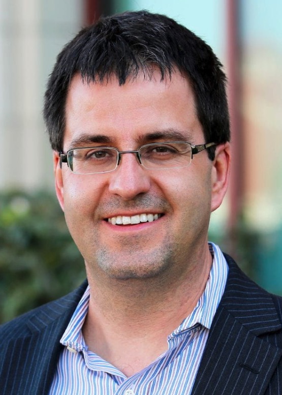 Remo Rohs合作论文数Michelson Center for Convergent Bioscience, University of Southern California;Department of Computer Science, Viterbi School of Engineering, University of Southern California;Department of Physics and Astronomy, University of Southern California;Norris Comprehensive Cancer Center, Keck School of Medicine, University of Southern California;Department of Chemistry, University of Southern California;Department of Quantitative and Computational Biology, University of Southern California2
 Anima Anandkumar合作论文数Anima AI + Science Lab, California Institute of Technology;Division of Engineering and Applied Science, California Institute of Technology;Department of Computing and Mathematical Sciences, California Institute of Technology2Jared M Sagendorf合作论文数1Vyom Unadkat合作论文数1Dustin Ebert合作论文数1
 Casey Collet合作论文数1Shivani Sundaram合作论文数1Sasha Lasky合作论文数1Yingfei Wang合作论文数Quantitative and Computational Biology, Departments of Biological Sciences, Chemistry, Physics & Astronomy, and Computer Science, University of Southern California, Los Angeles, CA 90089, USA.1
 Xiaoyan Han合作论文数Operations Research and Information Engineering, Cornell University1
 Tsu-Pei Chiu合作论文数1
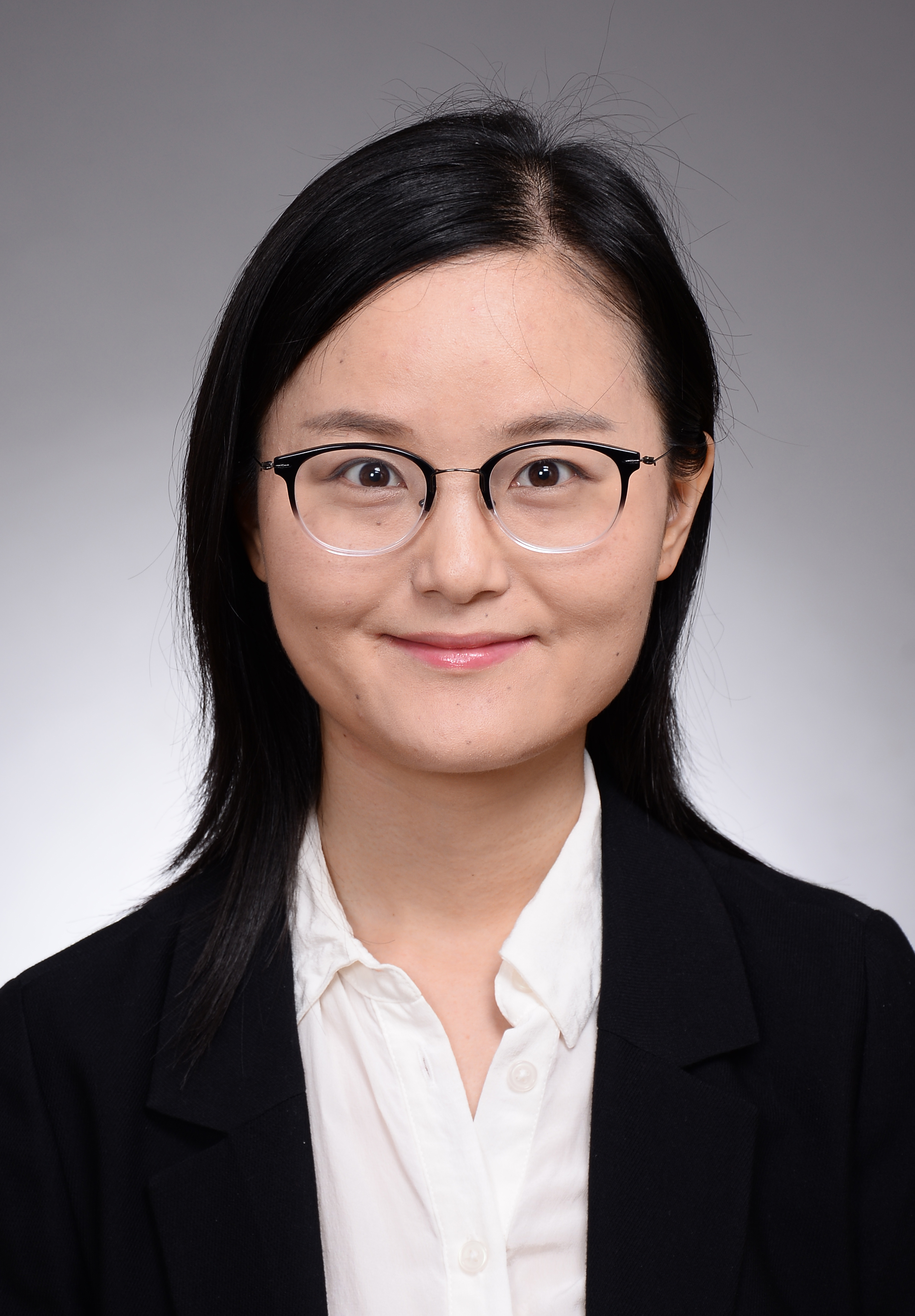 Beibei Xin (辛蓓蓓)合作论文数College of Agronomy and Biotechnology, China Agricultural University1
 Helen Berman合作论文数Department of Chemistry and Chemical Biology, School of Arts and Sciences, Rutgers, The State University of New Jersey1Kimberly Van Auken合作论文数1
 Bozena Wrobel合作论文数1
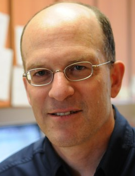 Paul W. Sternberg合作论文数Sternberg Lab, Division of Biology and Biological Engineering, California Institute of Technology1




