Coronary artery anomalies (CAAs) are a rare but clinically significant group of congenital abnormalities that are associated with myocardial ischaemia, malignant arrhythmias and sudden cardiac death, particularly in young people and athletes. Despite increasing recognition of these conditions and advances in cardiovascular imaging, there are still significant challenges regarding their classification, risk stratification and management, particularly with respect to surgical indications. This review provides a comprehensive overview of the current evidence on the classification, pathophysiology, diagnosis and management of CAAs, with a particular focus on surgical decision-making and contemporary guideline recommendations. A systematic literature search was conducted up to February 2025 using PubMed and Google Scholar. Priority was given to international guidelines, consensus statements, systematic reviews, meta-analyses and large observational studies. CAAs encompass a broad spectrum of anatomical variants and clinical presentations. Among these, anomalies of coronary origin are the most extensively studied. Surgical management is well established for coronary arteries arising from the pulmonary artery, particularly for anomalous left coronary artery from the pulmonary artery (ALCAPA). Substantial advances have also been made in the diagnosis, risk stratification, and treatment of anomalous aortic origin of a coronary artery (AAOCA), which has become a major focus of contemporary guideline recommendations. For patients with AAOCA, surgical correction, including unroofing, coronary reimplantation or coronary artery bypass grafting, is recommended for individuals with symptoms and/or high-risk anatomical features. In contrast, the diagnosis and management of myocardial bridging, coronary artery fistulas, and coronary artery ectasia remain controversial, with considerable variability in the indications for medical, percutaneous, and surgical treatment. The management of CAAs is an evolving field. While there is consensus for a limited number of anomalies, most cases still require individualised decision-making. It is essential to develop standardised diagnostic frameworks, improved risk stratification tools and outcome-based management criteria. A multidisciplinary, evidence-based approach involving cardiologists, cardiac imagers, interventional cardiologists and cardiac surgeons is crucial in order to optimise patient outcomes and reduce the risk of adverse cardiovascular events, including sudden cardiac death.
Donation after circulatory death (DCD) is increasingly implemented worldwide and represents a growing source of transplantable organs. Abdominal normothermic regional perfusion (A-NRP), typically performed using a modified venoarterial extracorporeal perfusion circuit, improves outcomes in liver and kidney transplantation but requires reliable exclusion of thoracic, coronary, and cerebral reperfusion. This is commonly achieved by supraceliac aortic occlusion. In donors with severe iliofemoral or aortoiliac disease, femoral access for balloon placement may not be feasible, limiting the applicability of A-NRP. We describe a novel percutaneous subclavian approach for aortic occlusion balloon placement and report limited initial institutional experience in two controlled DCD donors using aggregated procedural feasibility data. This planned alternative route may be considered in selected anatomically challenging cases when standard femoral balloon placement is not feasible.
The aorta is a distinct organ that requires a comprehensive and multidisciplinary approach and close collaboration between specialized aortic teams and primary care physicians. This article highlights the importance of this collaboration by linking a clinical case to the key principles of aortic disease elective management in accordance with recent clinical practice guidelines.
The aorta, once viewed as a passive conduit, is now recognized as an active organ crucial for hemodynamic regulation and vascular homeostasis. Thoracic aortic aneurysms (TAAs), particularly those involving the ascending aorta, often remain silent until life-threatening complications such as dissection or rupture occur. Current management primarily relies on aortic diameter criteria, yet up to 60% of type A dissections occur at sizes below the 5.5 cm surgical threshold, revealing the limitations of this approach. This narrative review summarizes recent advances in understanding ascending aortic aneurysms, including insights into their genetic and degenerative mechanisms, the role of novel morphological and hemodynamic markers, and the potential of advanced imaging techniques. It also explores evolving surgical strategies, from conventional open repair, still the gold standard, to minimally invasive and investigational endovascular approaches. By integrating biological, morphological, and clinical factors, emerging strategies aim to move beyond diameter alone toward more personalized risk assessment. This paradigm shift may improve early detection, optimize surgical timing, and ultimately enhance outcomes for patients with ascending aortic aneurysms.
(1) Background: There is a need for a novel surrogate marker to ease decision making when facing ascending aortic dilatation. In this article, we study the ratio between ascending and descending aorta diameters as a potential one. (2) Methods: Retrospective observational cohort study, including all the patients who underwent surgery for acute type A aorta dissection (aTAAD) between January 2014 and September 2020 at our center. A total of 50 patients were included. Clinical and demographic data were collected. The anatomical measurements were made including orthogonal maximal diameters of the ascending and descending aorta, post-dissection whole circumference length (post-wCL), post-dissection true lumen circumference length (post-tCL), and surface and sphericity indices of the ascending and descending aorta. Pre-dissection ascending aorta diameter (pre-AAD) and pre-dissection descending aorta diameter (pre-DAD) were calculated as well as the ratio between them and compared with reference values. (3) Results: Of the pre-AAD patients, 96% had smaller than the recommended 55 mm. The ratio between the descending and ascending aorta pre-dissection diameters was significantly smaller compared to the reference value (0.657 ± 0.125 versus 0.745 ± 0.016 with a mean difference of −0.088 and a p < 0.001). (4) Conclusions: The 55 mm threshold for aorta maximal diameter is an insufficient criterion when assessing the risk of dissection. The ratio between DAD and AAD is a parameter worthy of analysis as a tool to stratify the risk of dissection.
Stingray punctures are relatively common in several coastal regions and may result in chronic wounds and disability due to injury and envenomation. Vascular complications are rare but can be severe. We describe a case of high-flow arteriovenous fistula resulting from a stingray injury that required a venous bypass.
COVID19 altered and impacted medical and surgical practice around the world. Standard of care and routine procedures are disrupted. Majors shift in personnel, and ad hoc new team as well as delocalization and working with new infrastructures are further challenges to be dealt with. This review of three very unusual scenarios illustrates pitfalls and dangers harbored in the re-shaped landscape of COVID19 exemplifying the narrow path bridging from the medical and surgical comfort zone to uncharted territory and eventually leading to collateral damage.Le Covid-19 a profondément modifié et sévèrement impacté les pratiques médicales et chirurgicales à long terme. Les standards de prise en charge et les procédures de routine sont altérés, voire perturbés. Des mutations majeures au niveau du personnel et des équipes de même que la délocalisation ou le travail avec de nouvelles infrastructures sont autant de défis à relever, encore aujourd’hui. Trois scénarios inhabituels illustrent les pièges et les dangers qui se cachent dans le paysage marqué par le Covid-19. Ces exemples démontrent la marge étroite entre la zone de confort médicale et chirurgicale classique et l’appréhension d’une situation inhabituelle qui risque d’entraîner des dommages collatéraux pour les patients.
Extrinsic compression is a potential cause of stent failure. We have described the case of a 65-year-old paraplegic patient with acute right leg ischemia. His medical history was relevant for aortobifemoral bypass, followed by kissing covered stent reconstruction of a proximal anastomotic false aneurysm. The computed tomography scan showed collapse of the right iliac covered stent with ipsilateral iliofemoral graft thrombosis and partial collapse of the left iliac covered stent. He underwent emergent right iliac limb open thrombectomy and redo covered stent relining. The cause of compression was found to be daily deep abdominal massages for intestinal evacuation. The endovascular device should be tailored to the patient's particularities.
Acute lower-limb ischemia after abdominal aortic surgery is rare but potentially limb- threatening. Thus, prompt diagnosis and management are paramount. Only early identification and rapid correction might prevent ischemia-related complications.
COVID19 altered and impacted medical and surgical practice around the world. Standard of care and routine procedures are disrupted. Majors shift in personnel, and ad hoc new team as well as delocalization and working with new infrastructures are further challenges to be dealt with. This review of three very unusual scenarios illustrates pitfalls and dangers harbored in the re-shaped landscape of COVID19 exemplifying the narrow path bridging from the medical and surgical comfort zone to uncharted territory and eventually leading to collateral damage.
Driveline infection is one of the most frequent complications following left ventricular assist device (LVAD) treatment and there is no consensus for its management. The standard approach to treat foreign-body infection is complete device ablation, which is not always feasible and therefore not an elected method for LVAD driveline infections. Here we share the results from a series of cases successfully treated for driveline infection by negative pressure wound therapy (NPWT) therapy. Between 2016 and 2020, five male patients were hospitalized in our unit with a driveline infection of HeartMate III-LVAD®. Ultrasonography and/or thoraco-abdominal CT confirmed the diagnosis, infection localization, and abscess formation. Following an antibiotic treatment, an urgent surgical abscess drainage and debridement of the infected tissues were performed. At the end of the procedure, NPWT was applied. NPWT re-dressing and debridement of wound was performed every 3-4 days. The wound was closed surgically after obtaining negative culture results and good healing. The patients were discharged in good condition, without signs of infection. Two patients underwent successful heart transplantation after 1 and 13 months. Other patients did not show any residual or recurrent infection during the follow-up within 25 months. Driveline infection following LVAD implantation is a significant complication and a challenging in terms of management for both; the surgical team and the patient. These results from our case series report a successful and less invasive approach by using NPWT for the treatment of LVAD driveline infections.
Objectives: Bioprosthetic valve deterioration remains a major limitation following aortic valve replacement. Favorable results have been reported with an autologous pericardium aortic valve neocuspidization. Methods: Seventy patients (31 women and 39 men) (mean age, 62 +/- 12 years) with aortic stenosis (n = 52 [74%]) or aortic regurgitation (n = 18 [26%]) underwent the aortic valve neocuspidization procedure. Thirty-four patients (49%) had a tricuspid valve, 35 (50%) had a bicuspid valve, and 1 (1%) had a monocuspid valve. European System for Cardiac Operative Risk Evaluation and Society of Thoracic Surgeons scores were, respectively, 2.2% +/- 2% and 2.0% +/- 1.8%. Four patients (6%) had active endocarditis and 2 (3%) had endocarditis sequelae. One patient (1%) had fibroelastoma. A combined procedure was performed in 33 patients (46%). Results: The follow-up period was 24 +/- 12 months. One patient (1%) died in hospital and 1 patient (1%) underwent conventional valve replacement for significant aortic regurgitation. Postoperative peak and mean pressure gradients were respectively 14 +/- 5 and 8 +/- 3 mm Hg. Aortic valve area was 2.5 +/- 0.6 cm(2). During follow-up, no patients died. Reintervention occurred in 2 patients (3%). At last follow-up, peak pressure gradient was 13 +/- 7 mm Hg, mean pressure gradient was 7 +/- 4 mm Hg, and aortic valve area was 2.3 +/- 0.7 cm(2). There was 1 recurrence of moderate aortic stenosis (1%). All patients were in New York Heart Association functional class I (90%) or II (10%). Freedom from major valve-related events was 92.1%, (98.5% for death, 95.2% for reintervention, and 95.2% for endocarditis). Conclusions: In our experience, the midterm outcomes of the aortic valve neocuspidization procedure with autologous glutaraldehyde fixed pericardium were acceptable for survival, operative risk and valve-related complications, for our all-comer patient population with various aortic valve diseases.
ECMO is the most frequently used mechanical support for patients suffering from low cardiac output syndrome. Combining IABP with ECMO is believed to increase coronary artery blood flow, decrease high afterload, and restore systemic pulsatile flow conditions. This study evaluates that combined effect on coronary artery flow during various load conditions using an in vitro circuit. In doing so, different clinical scenarios were simulated, such as normal cardiac output and moderate-to-severe heart failure. In the heart failure scenarios, we used peripheral ECMO support to compensate for the lowered cardiac output value and reach a default normal value. The increase in coronary blood flow using the combined IABP-ECMO setup was more noticeable in low heart rate conditions. At baseline, intermediate and severe LV failure levels, adding IABP increased coronary mean flow by 16%, 7.5%, and 3.4% (HR 60 bpm) and by 6%, 4.5%, and 2.5% (HR 100 bpm) respectively. Based on our in vitro study results, combining ECMO and IABP in a heart failure setup further improves coronary blood flow. This effect was more pronounced at a lower heart rate and decreased with heart failure, which might positively impact recovery from cardiac failure.
Outflow tract abnormalities are the most frequent congenital heart defects. These are due to the absence or dysfunction of the two main cell types, i.e., neural crest cells and secondary heart field cells that migrate in opposite directions at the same stage of development. These cells directly govern aortic arch patterning and development, ascending aorta dilatation, semi-valvular and coronary artery development, aortopulmonary septation abnormalities, persistence of the ductus arteriosus, trunk and proximal pulmonary arteries, sub-valvular conal ventricular septal/rotational defects, and non-compaction of the left ventricle. In some cases, depending on the functional defects of these cells, additional malformations are found in the expected spatial migratory area of the cells, namely in the pharyngeal arch derivatives and cervico-facial structures. Associated non-cardiovascular anomalies are often underestimated, since the multipotency and functional alteration of these cells can result in the modification of multiple neural, epidermal, and cervical structures at different levels. In most cases, patients do not display the full phenotype of abnormalities, but congenital cardiac defects involving the ventricular outflow tract, ascending aorta, aortic arch and supra-aortic trunks should be considered as markers for possible impaired function of these cells. Neural crest cells should not be considered as a unique cell population but on the basis of their cervical rhombomere origins R3-R5 or R6-R7-R8 and specific migration patterns: R3-R4 towards arch II, R5-R6 arch III and R7-R8 arch IV and VI. A better understanding of their development may lead to the discovery of unknown associated abnormalities, thereby enabling potential improvements to be made to the therapeutic approach.
Brachiocephalic vein aneurysm is an extremely rare clinical entity, with a limited number of case reports in the literature. We report the case of a 74-year-old woman with a left brachiocephalic vein aneurysm (3 × 3 × 6.5 cm) that was discovered fortuitously by contrast-enhanced chest computed tomography. Surgical intervention was performed simultaneously during tricuspid valve surgery with coronary artery bypass grafting. This paper discusses the details of our case, the surgical treatment of brachiocephalic vein aneurysms, and the different treatment options.
Introduction - We report the case of a patient treated by an endovascular aortic repair (EVAR) for an infrarenal aneurysm that developed a type Ia endoleak. His aneurysms enlarged to 10 cm mandating a rapid repair. We successfully used an off-the-shelf thoracic branched graft with four branches to the visceral arteries by a bilateral femoral and left axillary artery approach. Methods - An 85-year-old male patient presented to our clinic for a 10 cm abdominal artic aneurysm (AAA). He was previously treated by EVAR for a 6 cm diameter aneurysms two years prior to his presentation using a Talent bifurcated device (Medtronic, Santa Rosa, Calif) with embolisation of the right hypogastric artery. He was lost to follow-up and presented one year later with an aortic rupture because of a distal migration of the main body of the device and a proximal type I endoleak. He was treated with a proximal cuff extension using an Endurant device (Medtronic, Santa Rosa, Calif). A year later, he presented again with a ruptured aneurysm due to a type III endoleak after disconnection of the main body and the proximal extension. He was managed urgently with a new Endurant bifurcated endograft extending from the most distal renal artery towards the previous bilateral iliac branches. During his Ct scan follow-up a proximal aortic dilation was noted reaching 35 mm in diameter at the level of the visceral aorta with a large posterior type Ia endoleak and a progression of his aneurysm to 10 cm (figure 1A). Since we could not wait for a custom made device, the off-the-shelf t-Branch (Cook, Bloomington, Ind) was used. It consists of a thoracic graft with four branches for each of the visceral arteries. It was introduced by the femoral artery and positioned in a way that the branches are 3 centimetres above their corresponding target vessels. Then through the left axillary approach each vessel was catheterised and stented one after the other (figure 1B). Results - The completion angiogram showed a successfully excluded aneurysm with absence of type I or III endoleak (figure 1C). All target vessels were patent. The patient had a smooth recovery; mainly he did not develop any signs of spinal cord ischemia and was discharged 10 days later. On the one-month follow-up Ct scan, the aneurysm was still excluded, no signs of endoleaks were noted and the visceral branches were still patent. Conclusion - The t-Branch is a ready to use endograft that is helpful in treated urgent cases of type Ia endoleaks following previous EVAR that could not wait for a custom made device. References1Sobocinski J, d'Utra G, O'Brien N, et al. Off-the-shelf fenestrated endografts: a realistic option for more than 70% of patients with juxtarenal aneurysms. J Endovasc Ther. 2012;19:165–172.2Bisdas T, Donas KP, Bosiers M, et al. Anatomical suitability of the t-branch stent-graft in patients with thoracoabdominal aortic aneurysms treated using custom-made multibranched endografts. J Endovasc Ther. 2013;20:672–677.3Adib Khanafer, Manar Khashram, Carmen-Maria Ruiz, Dana Mann, Andrew Laing. Use of the Off-the-Shelf t-Branch Device to Treat an Acute Type Ia Endoleak in a Symptomatic Juxtarenal Abdominal Aortic Aneurysm. J Endovasc Ther. 2016:23:212–215.
External iliac artery endofibrosis is a rare disease described mainly in male endurance athletes. It presents as claudication of the lower limb during near-maximum effort. The patients lack the usual risk factors for atherosclerosis, which makes diagnosis challenging. We present a case of external iliac artery endofibrosis in a female competitive cyclist. The initial surgical management was complicated by early recurrence due to intimal hyperplasia. After secondary drug-eluting balloon angioplasty, the patient was able to resume competition. As such, it is important to maintain a high index of suspicion when evaluating a patient presenting with claudication symptoms in this setting. Primary treatment is surgical, and in cases of early recurrence angioplasty may be indicated. Most patients can return to full activity after healing is complete.
Background: Proximal aortic dilation following open and endovascular aortic repair (EVAR) is a well-known phenomenon. If severe enough, it may lead to late onset of type Ia endoleak that jeopardizes the proximal seal. Methods: We report the case of a patient previously treated by EVAR for an infrarenal aneurysm who developed a type Ia endoleak after proximal aortic dilation. His aneurysms enlarged to 10 cm mandating a rapid repair without the delay for a custom-made device. Results: We successfully used the off-the-shelf thoracic t-Branch graft (Cook, Bloomington, IN) with its 4 branches to treat the dilated aorta and seal the endoleak. Conclusion: The off-the-shelf t-Branch is a useful option in patient with previous EVAR presenting with proximal aortic dilation and endoleak who cannot wait for a custom-made device.
Carotid artery atherosclerosis remains one of the major causes of stroke. The efficacy of carotid artery recanalization has already been established and is considered significant for symptomatic stenosis >70% and moderate for stenosis between 50-69 %. Regarding asymptomatic stenosis, carotid artery recanalization should be reserved for carefully selected patients with a stenosis between 70-90 % and an estimated procedural risk at less than 3 %. Two therapeutic options are currently available : angioplasty with stent placement and endarterectomy. While the endovascular approach may be associated with a higher periprocedural complication rate of stroke, there remains a significant decrease in the rate of myocardial infarction and peripheral nerve palsy compared to surgery. As a result, each case should be reviewed with a multidisciplinary approach in order to propose the best therapeutic strategy.
L’athérosclérose des artères carotides est une des causes majeures d’infarctus cérébral. Le bénéfice de la revascularisation carotidienne est très important pour les sténoses symptomatiques de 70 % ou plus, et modéré pour celles de 50 à 69 %. Pour les sténoses asymptomatiques, la revascularisation carotidienne devrait être proposée chez des patients bien sélectionnés, avec une sténose entre 70 et 90 %, si le risque de la procédure peut être estimé à moins de 3 %. Deux approches sont possibles : le stenting et la chirurgie. Le stenting semble être associé à un nombre plus important d’AVC périprocéduraux, mais à un risque moindre d’infarctus du myocarde et de paralysie des nerfs crâniens par rapport à la chirurgie. Chaque cas devrait être discuté dans le cadre d’un colloque multidisciplinaire pour proposer la meilleure stratégie thérapeutique au patient.
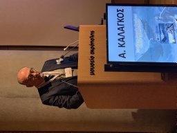 Afksendiyos Kalangos合作论文数Department of Pediatric Cardiac Surgery, Mitera Children’s Hospital26
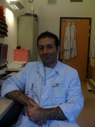 Gemayel Gino合作论文数University Hospital of Geneva, University of Geneva14
 Christoph Huber合作论文数Divison of Cardiac and Vascular Surgery, University Hospital Geneva14
 Mugnai Damiano合作论文数13Bernard Faidutti合作论文数13
 Mustafa Cikirikcioglu合作论文数Cardiac Surgeon;University Hospital of Geneva;Cardiac Surgeon, University Hospital of Geneva10René Prêtre合作论文数8Marc Licker合作论文数8
 Aristotelis Panos合作论文数Cardiac Surg, Univ Hosp Geneva7Anastase Spiliopoulos合作论文数6
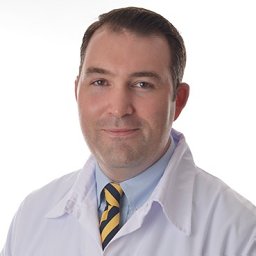 Patrick Myers合作论文数Service de Chirurgie Cardiaque, Centre Hospitalier Universitaire Vaudois6Khabiri Ebrahim合作论文数5John Robert合作论文数5
 Karim Bendjelid合作论文数Department of Anesthesiology, Pharmacology and Intensive Care, Geneva University Hospitals Intensive Care Unit5
 Raphael Giraud合作论文数5
 Christoph Ellenberger合作论文数Department of Anesthesiology;University Hospital of Geneva;Department of Anesthesiology, University Hospital of Geneva5D. Mugnai合作论文数4Roman Sztajzel合作论文数4John Diaper合作论文数4Marco Roffi合作论文数4Dominique Vala合作论文数4Afksendyios Kalangos合作论文数4Tornike Sologashvili合作论文数3Parmeseeven Mootoosamy合作论文数3
 Michel Montessuit合作论文数3D. Delay合作论文数3
 Paul Vaucher合作论文数Sch Hlth Sci Fribourg, Univ Appl Sci & Arts Western Switzerland HES SO3Lovblad Karl-Olof合作论文数3Marc Righini合作论文数Department de Medecine, Universite de Geneve3Francesco Puccinelli合作论文数3Marco Vannotti合作论文数3
 Bernard Favrat合作论文数Department of Ambulatory Care and Community Medicine, University of Lausanne3Ivan Bruschweiler合作论文数3Maurice Beghetti合作论文数3Patrick Bodenmann合作论文数3J Sierra合作论文数3A. Bissery合作论文数2A Baldovinos合作论文数2A.P. Narata合作论文数2Gregory Khatchatourian合作论文数2E Khabiri合作论文数2Gaia Pollorsi合作论文数2
 Andrea Carcaterra合作论文数2
 Jorge Sierra合作论文数2Lydia Wuarin合作论文数2Nurcan Yilmaz合作论文数2H Yilmaz合作论文数2Gregory Khatchatourov合作论文数2
 Philippe Reymond合作论文数Geneva University Hospital, University of Geneva2




