An 81 year old man underwent re-vascularization for symptomatic coronary disease, using a standard sternal approach. Postoperative mediastinitis was treated by sternal reopening and VAC therapy. Secondary sternal closure was realized after favorable clinical course and normalization of inflammatory parameters. Patient left the hospital, but was readmitted 2 weeks later for acute bleeding from the lower part of the sternal scar. Thoracic CT-scan revealed bleeding from the heart. Intra-operatively we found partial sternal dehiscence and a hole in the right ventricle. Hemostasis was achieved by direct suture. After VAC and antibiotic treatment the sternum was closed 7 weeks later with favorable clinical outcome.
Schon seit über fünf Jahrhunderten hat man sich mit der Aortenklappe befasst, wie die Zeichnungen von Leonardo da Vinci (1452–1519) und die Sammlung des Anatomen und Arztes Andreas Vesalius (1514–1564) in dessen Werk «De humani Corporis Fabrica» zeigen. Allerdings sollte noch viel Zeit verstreichen, bis am 13. Juli 1912 die erste Operation an der Aortenklappe bei einem lebenden Menschen vorgenommen wurde. Die beiden Chirurgen Théodore Tuffier und Alexis Carrel führten eine Aortenklappendilatation durch, indem sie die Aortenklappenöffnung mit dem Finger durch die Wand der Hauptschlagader erweiterten. Bereits 1947 eröffnete Sir Russel Claude Brock mit seinem Kardioskop alternative Zugangswege; er führte das Gerät durch die Arteria subclavia retrograd ein und dilatierte auf diesem Weg die Stenose. Schon 1952 ersetzte Hufnagel eine Aortenklappe durch eine «Lucite ball valve» in der Aorta descendens, also in nicht-anatomischer Position. Erst am 6. Mai 1953 wurde die erste Herz-Lungen-Maschine erfolgreich eingesetzt. Schliesslich konnte am 10. März 1960 Harken erstmals eine künstliche Klappe in der Aorta in anatomisch korrekter Position einsetzen.
tion de l’anatomiste et médecin Andreas Vesales (1514–1564) dans son ouvrage «De humani Corporis Fabrica». Il faudra pourtant attendre le début du siècle passé, soit le 13 juillet 1912 pour assister à la première opération sur la valve aortique in vivo. On doit cette première dilatation manuelle d’une sténose aortique par l’invagination de l’aorte ascendante aux chirurgiens Théodore Tuffier et Alexis Carrel. Déjà en 1947, le baron Russel Claude Brock ouvrait la voie d’accès alternatifs avec son cardioscope qu’il introduisait par l’artère sous-clavière de manière rétrograde pour pouvoir dilater une sténose aortique. En 1952 déjà, Hufnagel remplaçait une valve aortique par une «Lucite ball valve» dans l’aorte descendante, c’est-à-dire en position extra anatomique. Rappelons tout de même qu’il fallut attendre le 6 mai 1953 pour que la première machine cœur-poumons voie le jour avec succès. Finalement, le 10 mars 1960, Harken parvint à implanter la première valve artificielle en position aortique (= in situ).
The hybrid treatment of aortic aneurysms is indicated in patients having the ostia of supra aortic or visceral branches taken in to the aneurysm. Indeed, these lesions are not eligible for classic endovascular treatment because the existing endoprostheses cannot provide perfusion of the side branches without inducing major endoleaks. The surgical technique consists of 2 steps: firstly, a by-pass between normal aorta and the major aortic branches involved in the aneurysm is performed to guarantee the perfusion of the organs such as brain, bowel, and after endoprosthesis deployment. Secondly, the endoprosthesis is deployed using the classical technique to isolate the aneurysm. The hybrid approach provides safe and reliable treatment of complex aortic aneurysms with mortality and morbidity rate far below the classical open surgery.
The Admiral, a new microporous membrane oxygenator with a low surface area, decreased priming volume and two separate reservoirs, was tested in 30 adult patients. This study was undertaken to evaluate blood path resistance, gas exchange capabilities and blood trauma in clinical use, with and without shed blood separation. Patients were divided into 3 groups. Group 1 had valve surgery without separation of suction, Group 2 had coronary artery bypass grafting (CABG) with direct blood aspiration and Group 3 had coronary artery bypass grafting with shed blood separation. The suctioned, separated, cardiotomy blood in Group 3 was treated with an autotransfusion device at the end of bypass before being returned to the patient. Theoretical blood flow could be achieved in all cases without problem. The pressure drop through the oxygenator averaged 88 ± 13 mmHg at 4 l/min and 109 ± 12 mmHg at 5 l/min. O2 transfer was 163 ± 27 ml/min. Free plasma haemoglobin rose in all groups, but significantly less in group 3. Lactate dehydrogenase (LDH) rose significantly in Groups 1 and 2. Platelets decreased in all groups without significant differences. Clinical experience with this new oxygenator was safe, the reduced membrane surface did not impair gas exchange and blood trauma could be minimized easily by separating shed blood, using the second cardiotomy reservoir.
Following acute myocardial infarction, necrotic cardiac tissue is replaced by scar leading to ventricular remodeling and pump failure. Transplantation of autologous bone marrow-derived cells into the heart, early post-infarct, aims to prevent ventricular remodeling. This strategy has been evaluated in four controlled, randomized clinical trials, which provided mixed results. A transient improvement in ventricular function was observed in one trial, and a modest improvement (the duration of which remains to be determined) in an additional trial, whereas two trials showed negative results. A modest benefit of bone marrow cell transplantation was also observed in patients with chronic ischemic heart disease. Despite mixed results reported so far, cell therapy of heart disease still is in its infancy and has considerable room for improvement.
Zwischen 1995 und 2005 wuchs die Anzahl der jährlich von uns mit endovaskulären Techniken versorgten Aortenaneurysmen (EVAR) von 0 auf 50, und dies auf allen Stufen der Aorta. Zu unserer Organisation gehören ein breites Team von Chirurgen, ein Lager mit 3 kompletten Familien von Endoprothesen (gerade Endoprothesen, konische Endoprothesen, und Bifurkationen), ein mobiler Wagen mit Zubehör (Einführungsbestecke, Führungsdrähte, Katheter, Ballone etc.) und ein Apparat auf Rädern für die intravaskuläre Ultraschalluntersuchung (IVUS). Letzterer erlaubt es zusammen mit einer mobilen Durchleuchtungsanlage (C-Bogen), in jedem Operationssaal unserer Institution endovaskulär Aneurysmen zu analysieren, und dies in der Regel ohne Angiographie bzw. Kontrastmittel. Deshalb sind wir nicht mehr auf eine ausgiebige bildgebende präoperative Abklärung potenzieller Kandidaten für eine endovaskuläre Sanierung von Aneurysmen angewiesen und können rupturierte Aneurysmen der Bauchaorta oder der thorakalen Aorta ohne Verzug behandeln. Bei der endovaskulären Sanierung von Aortenaneurysmen unterscheiden wir zwischen Prozessschritten (Indikationsstellung, Darstellung der Zugangsgefäße, Ausmessen mittels IVUS und Roadmapping mittels Durchleuchtung, Implantatwahl, Implantatinsertion, Positionierung, Implantatabwurf, Erfolgsbeurteilung, Rekonstruktion der Zugangsgefäße und Nachkontrolle) und Kompetenzstufen (Assistent, Oberarzt, Leitender Arzt). Unsere ultraschallgestützte Technik zur endovaskulären Sanierung von Aneurysmen wurde mittels IVUS-Transporter und Telementoring erfolgreich auch anderen Institutionen zur Verfügung gestellt.
Verhinderung katastrophaler Blutungen anlässlich einer Resternotomie durch vorangehende kardiale Dekompression mittels EKZ, peripherer Smart-Kanülierung und schwerkraftgetriebener Drainage.
Objective Avoid catastrophic haemorrhage during sternal re-entry by prior cardiac decompression using CPB with gravity driven smart venous drainage. Methods Prospective study of trans-femoral cannulation of the inferior vena cava, the right atrium and/or superior vena cave respectively with the smartcanula (R) and cardiac decompression with CPB prior to sternal re-entry in 20 consecutive cardiac reoperations. Results Smart venous cannulation was successful in 20/20 patients who underwent the following operations: A) 9/20 patients (45%) a redo valve procedure, B) 5/20 (25%) a redo aortic procedure, C) 4/20 (20%) a redo congenital repair, and D) 2/20 (10%) other re-operations. For the entire series, target blood flow was 4.2 +/- 0.3 l/min whereas the flow achieved prior to sternal re-entry accounted for 4.3 +/- 0.6 l/min (NS). For patients requiring single venous cannulation (A, B, and D: 16/20), target flow was 4.3+0.3 l/min and achieved flow prior to sternal re-entry 4.6 +/- 0.5 l/min (= 106%) vs. 4.6+0.5 l/min (= 106%) after sternal re-entry (NS). For patients requiring dual venous cannulation (group C= congenitals) and a target pump flow of 4+0.1 l/min, the chest was opened after single remote venous cannulation at flow of 3.4 +/- 0.7 l/min (86%) which increased to 4.1 l/min with a second cannula (102%: NS). For 36F and 63 cm long smartcanula (R) (n=10) target flow was 4.4 +/- 0.2 l/min and achieved flow accounted for 4.9 +/- 0.2 l/min (111%; p <0.05), whereas for shorter 36F cannula (<53 cm) target flow was 4.0 +/- 0.1 l/min and achieved flow accounted for 4.0+0.2 l/min (100%: NS). Conclusion Smart femoral cannulation allows for full flow prior to sternal reentry and efficient cardiac decompression. Higher blood flows can be achieved with longer smartcannulas (R).
Objectives: To assess the performance of 45F vs. 36F smartcanula in CPB with gravity drainage alone. Methods: Twenty patients were randomly assigned to two groups receiving for venous drainage a smartcanula which is collapsed over a mandrel for trans-atrial insertion into the inferior vena cava and expanded in situ to either 45F or 36F. Results: Valve replacementyrepair was realized in 7y10 andyor CABG in 6y10 for 36F (69"13 years) vs. 5y10 and 5y10, respectively, for 45F (63"11 years: NS). Body weight and surface area (BSA) were 83"9 kg (1.9"0.2 m , max 2.2 m ) for 36F vs. 79"6 kg: NS (1.9"0.1 m (NS), max 2.1 m ) for 45F. Insertion and access orifice diameter 2 2 2 2 (area) was 6 mm and 10 mm (78.5 mm ) for the 36F vs. 6 mm and 13 mm (132 mm ) for the 45F (q69%). Calculated target pump flow 2 2 (2.4 lyminym ) was 4.7"0.4 lymin for 36F vs. 4.5"0.3 lymin for 45F. Achieved pump flow accounted for 5.0"0.3 lymin for 36F (8% above 2 target) vs. 4.8"0.3 lymin for 45F (8% above target): NS. The water balance during the pump run (clear volume added minus hemofilter and urine output) was 2.2"0.3 l for 36F vs. 2.0 l for 45F: NS. Conclusion: Due to its ‘open’ wall (the vena cava provides the seal), its reduced wall thickness (range: 0.0–0.4 mm), and its self-expanding design, the 36F smartcanula requiring a 30F access orifice has sufficient drainage capacity by gravity alone for full CPB in adults with a BSA up to 2.2 mm . 2 2006 Published by European Association for Cardio-Thoracic Surgery. All rights reserved.
Objective: Transthoracic echocardiography (TTE) has been used clinically to disobstruct venous drainage cannula and to optimise placement of venous cannulae in the vena cava but it has never been used to evaluate performance capabilities. Also, little progress has been made in venous cannula design in order to optimise venous return to the heart lung machine. We designed a self-expandable Smartcanula (R) (SC) and analysed its performance capability using echocardiography. Methods: An epicardial echocardiography probe was placed over the SC or control cannula (CTRL) and a Doppler image was obtained. Mean (V-m) and maximum (V-max) velocities, flow and diameter were obtained. Also, pressure drop (Delta P-CPB) was obtained between the central venous pressure and inlet to venous reservoir. LDH and Free Hb were also compared in 30 patients. Comparison was made between the two groups using the student's t-test with statistical significance established when p < 0.05. Results: Age for the SC and CC groups were 61.6 +/- 17.6 years and 64.6 +/- 13.1 years, respectively. Weight was 70.3 +/- 11.6 kg and 72.8 +/- 14.4 kg, respectively. BSA was 1 80 +/- 0.2 m(2) and 1.82 +/- 0.2 m(2), respectively. CPB times were 114 +/- 53 min and 108 +/- 44 min, respectively. Cross-clamp time was 59 15 min and 76 29 min, respectively (p = NS). Free-Hb was 568 +/- 142 U/l versus 549 +/- 271 U/l post-CPB for the SC and CC, respectively (p NS). LDH was 335 +/- 73 mg/l versus 354 +/- 116 mg/l for the SC and CC, respectively (p NS). V. was 89 10cm/s (SC) versus 63 +/- 3 cm/s (CC), V-mm was 139 +/- 23 cm/s (SC) versus 93 +/- 11 cm/s (CC) (both p < 0.01). Delta P-CPB was 30 +/- 10 mmHg (SC) versus 43 +/- 13 mmHg (CC) (p < 0.05). A Bland-Altman test showed good agreement between the two devices used concerning flow rate calculations between CPB and TTE (bias 300 ml +/- 700 ml standard deviation). Conclusions: This novel Smartcanula design, due to its self-expanding principle, provides superior flow characteristics compared to classic two stage venous cannula used for adult CPB surgery. No detrimental effects were observed concerning blood damage. Echocardiography was effective in analysing venous cannula performance and velocity patterns. (c) 2006 Elsevier B.V. All rights reserved.
Devices for venous cannulation have seen significant progress over time: the original, rigid steel cannulas have evolved toward flexible plastic cannulas with wire support that prevents kinking, very thin walled wire wound cannulas allowing for percutaneous application, and all sorts of combinations. In contrast to all these rectilinear venous cannula designs, which present the same cross-sectional area over their entire intravascular path, the smartcanula concept of "collapsed insertion and expansion in situ" is the logical next step for venous access. Automatically adjusting cross-sectional area up to a pre-determined diameter or the vessel lumen provides optimal flow and ease of use for both, insertion and removal. Smartcanula performance was assessed in a small series of patients (76 +/- 17 kg) undergoing redo procedures. The calculated target pump flow (2.4 L/min/m2) was 4.42 +/- 61 L/ min. Mean pump flow achieved during cardiopulmonary bypass was 4.84 +/- 87 L/min or 110% of the target. Reduced atrial chatter, kink resistance in situ, and improved blood drainage despite smaller access orifice size, are the most striking advantages of this new device. The benefits of smart cannulation are obvious in remote cannulation for limited access cardiac surgery, but there are many other cannula applications where space is an issue, and that is where smart cannulation is most effective.
Background. The objective of the present study was to compare current results of prosthetic valve replacement following acute infective native valve endocarditis (NVE) with that of prosthetic valve endocarditis (PVE). Prosthetic valve replacement is often necessary for acute infective endocarditis. Although valve repair and homografts have been associated with excellent outcome, homograft availability and the importance of valvular destruction often dictate prosthetic valve replacement in patients with acute bacterial endocarditis.Methods. A retrospective analysis of the experience with prosthetic valve replacement following acute NVE and PVE between 1988 and 1998 was performed at the Montreal Heart Institute.Results. Seventy-seven patients (57 men and 20 women, mean age 48 +/- 16 years) with acute infective endocarditis underwent valve replacement. Fifty patients had NVE and 27 had PVE. Four patients (8%) with NVE died within 30 days of operation and there were no hospital deaths in patients with PVE. Survival at 1, 5, and 7 years averaged 80% +/- 6%, 76% +/- 6%, and 76% +/- 6% for NVE and 70% +/- 9%, 59% +/- 10%, and 55% +/- 10% for PVE, respectively (p = 0.15). Reoperation-free survival at 1, 5, and 7 years averaged 80% +/- 6%, 76% +/- 6%, and 76% +/- 6% for NVE and 45% +/- 10%, 40% +/- 10%, and 36% +/- 9% for PVE (p = 0.003). Five-year survival for NVE averaged 75% +/- 9% following aortic valve replacement and 79% +/- 9% following mitral valve replacement. Five-year survival for PVE averaged 66% +/- 12% following aortic valve replacement and 43% +/- 19% following mitral valve replacement (p = 0.75). Nine patients underwent reoperation during follow-up: indications were prosthesis infection in 4 patients (3 mitral, 1 aortic), dehiscence of mitral prosthesis in 3, and dehiscence of aortic prosthesis in 2.Conclusions. Prosthetic valve replacement for NVE resulted in good long-term patient survival with a minimal risk of reoperation compared with patients who underwent valve replacement for PVE. In patients with PVE, those who needed reoperation had recurrent endocarditis or noninfectious periprosthetic dehiscence. (C) 2000 by The Society of Thoracic Surgeons.
A 74-year-old woman (43 kg, 139 cm) was admitted to the Montreal Heart Institute for unstable angina. She had had left carotid endarterectomy 4 years earlier and was awaiting surgery for right carotid occlusive disease in another hospital. An unstable angina syndrome developed for which she was referred to our center. A coronary angiogram revealed a proximal occlusion of the left anterior descending artery, severe stenosis of the left circumflex artery with poor runoff, and a 70% stenosis of the dominant right coronary artery. Left ventricular contraction was within normal limits at echocardiography. She was scheduled for a double coronary artery bypass of the left anterior descending and right coronary arteries without extracorporeal bypass to minimize the risk of stroke. Anesthesia was induced with sufentanil, midazolam, and pancuronium. The blood pressure was 180/75 mm Hg and the central venous pressure, 5 mm Hg. Catheterization (Swan-Ganz catheter, Baxter Healthcare Corp, Edwards Division, Santa Ana, Calif) showed a normal pulmonary artery pressure (19/9 mm Hg) and cardiac output was 2.4 L/min (cardiac index: 1.9 L · min–1 · m–2). The initial blood gas values were within normal limits. Concomitantly to sternotomy and the beginning of the internal thoracic artery dissection, a right lower thigh incision was performed for endoscopic saphenectomy (Vasoview, Uniport Endoscopic Vessel Harvesting System, Origin Medsystems, Inc, a subsidiary of Eli Lilly and Company, Indianapolis, Ind). After identification of the internal saphenous vein and insertion of a port, a seal was achieved at the level of the knee incision and carbon dioxide insufflation was initiated at a flow of 2 L/min to obtain a pressure of 15 mm Hg. The saphenous vein was dissected up to the level of the femoral crux. During isolation of branches, 1 small collateral vein was torn off near the crux, 8 minutes after the start of the endoscopic dissection. Simultaneously, systemic blood pressure suddenly fell to 55 mm Hg systolic (Fig 1, A ), followed by ischemic changes (depression of the ST segment in leads DII and V5 , Fig 1, B ). Capnography showed an immediate rise of end-tidal carbon dioxide to 48mmHg (Fig 1, D ). Carbon dioxide insufflation was immediately discontinued and the internal thoracic artery retractor was taken down to rule out any cardiac compression, but there was no improvement in the hemodynamic status. A sternal retractor was put in and cardiac dissection was begun as the extracorporeal circuit was primed. One hundred micrograms of phenylephrine hydrochloride (Neo-Synephrine) was administered and a perfusion of norepinephrine was started. The blood gas sample drawn during the hypotensive episode showed respiratory acidosis with a pH of 7.27 and a PCO 2 level of 64 mm Hg. There were no changes in ventilation pressures and the arterial oxygen saturation was 100%. Pulmonary artery pressure rose to a level of 60mmHg systolic with a central venous pressure of 19 mm Hg (Fig 1, C ). Ventilatory rate and tidal volume were increased with an inspired oxygen fraction of 100%. Ischemic changes regressed as the blood pressure was restored. The total duration of hypotension was 4 minutes. After hemodynamic stabilization without further need for inotropic support, the harvesting of the left internal thoracic artery was resumed and the saphenous vein was taken out by the bridging technique. The 2 bypasses were completed uneventfully off-pump with mechanical stabilization. The patient was extubated 12 hours after the operation with no evidence of neurologic deficit or elevation of serum creatine kinase MB levels. The patient was discharged on postoperative day 6 without complications. The internal saphenous vein used for coronary artery bypass is usually harvested by the open technique, allowing direct visualization. Many potential complications may occur with the traditional technique, including dehiscence, skin necrosis, or wound infection. These complications are increased in the case of obesity, diabetes mellitus, and peripheral vascular disease. To reduce these complications and to improve the functional result and, to a lesser degree, the cosmetic appearance, new techniques are now available with the current trend of minimally invasive coronary surgery. These include the classic bridging technique, the tunneling technique, and endoscopic harvesting with or without gas insufflation. Serious venous gas embolism, a rare complication of endoscopic procedures with an incidence of about 1 in 7500 cases in laparoscopic surgery,1Herron DM Vernon JK Gryska PV Reines HD Venous gas embolism during endoscopy.Surg Endosc. 1999; 13: 276-279Crossref PubMed Scopus (32) Google Scholar has not yet been reported in endoscopic saphenectomy. The mechanisms of carbon dioxide embolism usually involve absorption into the circulation, carbon dioxide being highly soluble in blood, or, more seriously, direct entry of gas into the vascular bed generally via an injury to a vessel. This is dependent on the gradient between the central venous pressure and the pressure of insufflation, as demonstrated by Bazin and colleagues2Bazin JE Gillart T Rasson P Conio N Aigouy L Schoeffler P Haemodynamic conditions enhancing gas embolism after venous injury during laparoscopy: a study in pigs.Br J Anaesth. 1997; 78: 570-575Crossref PubMed Scopus (50) Google Scholar during laparoscopy in a porcine model. By extrapolation, one can assume that the mechanism is similar in case of injury to the saphenous vein and in the presence of relative hypovolemia, as in our patient. Similarly, gas embolism has been reported during hysteroscopy with patients in the Trendelenburg position.3Brooks PG Venous air embolism during operative hysteroscopy.J Am Assoc Gynecol Laparosc. 1997; 4: 399-402Abstract Full Text PDF PubMed Scopus (41) Google Scholar This important yet simple factor should be taken into consideration and the operating table should be set in the Fowler position, which facilitates the harvesting of the internal thoracic artery pedicle. Manifestations of carbon dioxide embolism are “gas lock” in the right atrium, pulmonary gas embolism, paradoxic embolism with or without patent foramen ovale with coronary embolism or neurologic disturbances, bradycardia or arrhythmia, cardiovascular collapse resulting from acute reduction in the peripheral resistance, and cardiopulmonary arrest.4Joris JL Anesthetic management of laparoscopy.in: Anesthesia. : Churchill Livingstone, New York1994: 2011-2029Google Scholar The detection of carbon dioxide embolism is based on the widely established end-tidal carbon dioxide monitoring. Indeed, carbon dioxide embolization causes a biphasic change in end-tidal carbon dioxide.4Joris JL Anesthetic management of laparoscopy.in: Anesthesia. : Churchill Livingstone, New York1994: 2011-2029Google Scholar The decrease in end-tidal carbon dioxide is usually encountered in case of embolism, being preceded by an initial increase resulting from pulmonary excretion of carbon dioxide, as in our patient (Fig 1, D ). Transesophageal echocardiography, increasingly used in modern cardiac surgery, is a more sensitive tool for detecting gas bubbles during laparoscopy5Mann C Boccara G Fabre JM Grevy V Colson P The detection of carbon dioxide embolism during laparoscopy in pigs: a comparison of transesophageal Doppler and end-tidal carbon dioxide monitoring.Acta Anaesthesiol Scand. 1997; 41: 281-286Crossref PubMed Scopus (21) Google Scholar and could be used routinely in this clinical setting, allowing in addition the evaluation of regional contraction. Furthermore, clinical evaluation using transesophageal echocardiography is in progress in our center to assess the precise incidence of this phenomenon. We are aware that the majority of episodes are not clinically significant, as demonstrated in a series of laparoscopic cholecystectomies.6Derouin M Couture P Boudreault D Girard D Gravel D Detection of gas embolism by transesophageal echocardiography during laparoscopic cholecystectomy.Anesth Analg. 1996; 82: 119-124PubMed Google Scholar If hypercapnia occurs, the endoscopic procedure insufflation should be immediately suspended and simultaneous compression of the proximal venous axis at the groin should be instituted. Trendelenburg position with tilting of the patient to the left, pharmacologic manipulations, and central venous pressure elevation by volume repletion to restore the hemodynamic status should be implemented promptly and the operation resumed with conversion to the open or bridging technique. In case of a major embolism, aspiration of gas from the right ventricle via the thermodilution catheter or via a direct puncture using a needle may relieve a gas lock in the right ventricle.1Herron DM Vernon JK Gryska PV Reines HD Venous gas embolism during endoscopy.Surg Endosc. 1999; 13: 276-279Crossref PubMed Scopus (32) Google Scholar In refractory cases, cardiopulmonary bypass may be required. In the case reported here, the conjunction of multiple factors, that is, injury to the vein in an elderly woman with low weight and friable tissues, with concomitant hypovolemia in the supine position, enabled gas embolism to occur. No untoward consequences occurred because of vigilant intraoperative monitoring and prompt intervention. Knowledge of the predisposing factors and preventive measures should keep the rate of this complication low and enable patients to have the full benefits of this advance in coronary artery bypass surgery.
AIM OF THE STUDY:To analyse the course of upper limb edema in patients with an arteriovenous fistula used for dialysis and to analyse the available therapeutic options.STUDY DESIGN:Retrospective study of patients with this type of edema, who were treated in our institution from 1992 to 1996.PATIENTS AND METHODS:Seven consecutive patients with an arterioveinous fistula treated for edema of the upper extremity, were reviewed. The fistula was created at the elbow in 6 patients and at the forearm in 1. The edema appeared immediately after operation in 4 patients and after a delay in 3 patients. Stenosis (3 patients) or occlusion (2 patients) of the subclavian vein was documented in 5 patients who were investigated by angiography.RESULTS:The edema regressed spontaneously in 4 patients because collaterals developed in 3 patients, and the fistula thrombosed in 1 patient. Surgical intervention allowed regression of the edema in the other 3 patients: excessive output of the fistula was reduced in 2 patients and an axillojugular bypass was performed in 1 patient. The fistula remained effective in 6 patients. Another fistula was performed on the contralateral arm in 1 patient.CONCLUSION:Non-operative management is recommended in patients who develop edema immediately after creation of the fistula, because spontaneous regression is likely. Measures aimed at reducing the output of the fistula or enhancing the venous capacities of the arm are required when edema appears at a later stage. The fistula can be saved in the majority of cases.
Background. Control of hemorrhage in patients with active bleeding from rupture of the aortic arch is difficult, because of the location of the bleeding and the impossibility of cross-clamping the aorta without interfering with cerebral perfusion. A precise and swift plan of management helped us salvage some patients and prompted us to review our experience.Methods. Six patients with active bleeding of the aortic arch in the mediastinum and pericardial cavity (5 patients) or left pleural cavity (1 patient), treated between 1992 and 1996, were reviewed. Bleeding was reduced by keeping the mediastinum under local tension (3 patients) or by applying compression on the bleeding site (2 patients), or both (1 patient) while circulatory support, retransfusion of aspirated blood, and hypothermia were established. The diseased aortic arch was replaced during deep hypothermic circulatory arrest, which ranged from 25 to 40 minutes. In 3 patients, the brain was further protected by retrograde (2 patients) or antegrade (1 patient) cerebral perfusion.Results. Hemorrhage from the aortic arch was controlled in all patients. Two patients died postoperatively, one of respiratory failure and the other of abdominal sepsis. Recovery of neurologic function was assessed and complete in all patients. The 4 survivors are well 8 to 49 months after operation.Conclusions. An approach relying on local tamponade to reduce bleeding, rapid establishment of circulatory support and hypothermia, retransfusion of aspirated blood, and swift repair of the aortic arch under circulatory arrest allows salvage of patients with active bleeding from an aortic arch rupture. (C) 1998 by The Society of Thoracic Surgeons.
We report a patient with Marfan's syndrome and pectus excavatum who underwent open heart surgery with simultaneous correction of the sternal malformation. Permanent internal stabilization, achieved by bilateral overlapping of the bevelled ends of the lowest ribs and reinforced with sternal closure wires offered a maintained postoperative chest wall stability, avoided the potential postoperative complications of cardiac compression, and improved the aesthetic appearance of the anterior chest wall. The increased risk of bleeding due to extensive dissection was minimized by postponing the repair of pectus excavatum to when protamin is administered after termination of cardiopulmonary bypass.
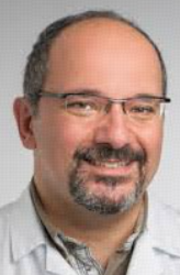 Piergiorgio Tozzi合作论文数Lausanne University Hospital10
 L. Von Segesser合作论文数8
 Enrico Ferrari合作论文数7J Horisberger合作论文数6Patrick Ruchat合作论文数5F Stumpe合作论文数4René Prêtre合作论文数3Bettina Marty合作论文数3David Jegger合作论文数3Giuseppe Siniscalchi合作论文数3Nicolas Murith合作论文数3P Page合作论文数2Y Hebert合作论文数2Pelletier L C合作论文数2Michel Pellerin合作论文数2Michel Carrier合作论文数2
 Louis Paul Perrault合作论文数Département de Pharmacologie et Physiologie, Faculté de Médecine, Université de Montréal;Département de Chirurgie, Faculté de Médecine, Université de Montréal;Centre de Recherche de l'Institut de Cardiologie de Montréal2
 Christoph Huber合作论文数Divison of Cardiac and Vascular Surgery, University Hospital Geneva2
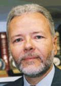 Ludwig K. Von Segesser合作论文数University Hospital CHUV2Ivan Bruschweiler合作论文数2P Gersbach合作论文数2S. Marinakis合作论文数1P-G Chassot合作论文数1M Bogen合作论文数1M Burki合作论文数1P Vogt合作论文数1M Hurni合作论文数1A Mucciolo合作论文数1R Marchand合作论文数1P Auger合作论文数1
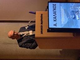 Afksendiyos Kalangos合作论文数Department of Pediatric Cardiac Surgery, Mitera Children’s Hospital1Niclauss Lars合作论文数1Berdajs Denis A合作论文数Department of Cardiovascular Surgery, University Hospital1Tremblay I合作论文数1
 Ludwig Karl von Segesser合作论文数Department of Cardiovascular Surgery, University Hospital Lausanne1
 Raymond Cartier合作论文数1Kalonji T合作论文数1De Stefano E合作论文数1
 Giuseppe Vassalli合作论文数1Augusto Gallino合作论文数1
 Tiziano Moccetti合作论文数Cardiocentro Ticino;Faculty of Medicine, University of Zurich1A. Bouveret合作论文数1
 Salah Dine Qanadli合作论文数1Bernard Faidutti合作论文数1Vassilios P Argitis合作论文数1I Mallabiabarrena合作论文数1R. Blain合作论文数1Olivier Chavanon合作论文数Institution: Grenoble University Hospital1G Mucciolo合作论文数1




