Supplementary Tables S1-S4 from Genome-Wide Screening of Genomic Alterations and Their Clinicopathologic Implications in Non–Small Cell Lung Cancers
FISH with whole chromosome or region-specific painting probes made from either flow-sorted or microdissected chromosomes has revolutionized cytogenetics. Generation of paints from flow-sorted chromosomes relies on the use of an expensive and sophisticated fluorescence-activated cell sorter and suspensions of freshly prepared chromosomes. Preparation of paints from microdissected materials requires an inverted microscope with appropriate micromanipulators and metaphase chromosome spreads on coverslips. Painting probes made from flow-sorted chromosomes generally have better chromosomal coverage and can be used in a wide range of applications between distantly related species, while region-specific probes from microdissection enable higher-resolution analyses restricted to comparative painting between closely related species. Here we provide detailed protocols on generation probes from both flow-sorted and microdissected chromosomes.
Chromosomal rearrangements are common in cancer. More than 50% occur in common fragile sites and disrupt tumor suppressors. However, such rearrangements are not known in gastric cancer. Here we report recurrent 18q2 breakpoints in 6 of 17 gastric cancer cell lines. The rearranged chromosome 18, t(9; 18), in MKN7 cells was flow sorted and identified by reverse chromosome painting. High-resolution tiling array hybridization mapped breakpoints to DOK6 (docking protein 6) intron 4 in FRA18C (18q22.2) and an intergenic region in 9q22.2. The same rearrangement was detected by FISH in 22% of 99 primary gastric cancers. Intron 4 truncation was associated with reduced DOK6 transcription. Analysis of The Cancer Genome Atlas stomach adenocarcinoma cohort showed significant correlation of DOK6 expression with histological and molecular phenotypes. Multiple oncogenic signaling pathways (gastrin-CREB, NGF-neurotrophin, PDGF, EGFR, ERK, ERBB4, FGFR1, RAS, VEGFR2 and RAF/MAP kinase) known to be active in aggressive gastric cancers were strikingly diminished in gastric cancers with low DOK6 expression. Median survival of patients with low DOK6-expressing tumors was 2100 days compared with 533 days in patients with high DOK6-expressing tumors (log-rank P = 0.0027). The level of DOK6 expression in tumors predicted patient survival independent of TNM stage. These findings point to new functions of human DOK6 as an adaptor that interacts with diverse molecular components of signaling pathways. Our data suggest that DOK6 expression is an integrated biomarker of multiple oncogenic signals in gastric cancer and identify FRA18C as a new cancer-associated fragile site.
The ability to identify the clinical nature of the recurrent duplication of chromosome 17q12 has been limited by its rarity and the diverse range of phenotypes associated with this genomic change. In order to further define the clinical features of affected patients, detailed clinical information was collected in the largest series to date (30 patients and 2 of their siblings) through a multi‐institutional collaborative effort. The majority of patients presented with developmental delays varying from mild to severe. Though dysmorphic features were commonly reported, patients do not have consistent and recognizable features. Cardiac, ophthalmologic, growth, behavioral, and other abnormalities were each present in a subset of patients. The newly associated features potentially resulting from 17q12 duplication include height and weight above the 95th percentile, cataracts, microphthalmia, coloboma, astigmatism, tracheomalacia, cutaneous mosaicism, pectus excavatum, scoliosis, hypermobility, hypospadias, diverticulum of Kommerell, pyloric stenosis, and pseudohypoparathryoidism. The majority of duplications were inherited with some carrier parents reporting learning disabilities or microcephaly. We identified additional, potentially contributory copy number changes in a subset of patients, including one patient each with 16p11.2 deletion and 15q13.3 deletion. Our data further define and expand the clinical spectrum associated with duplications of 17q12 and provide support for the role of genomic modifiers contributing to phenotypic variability. © 2015 Wiley Periodicals, Inc.
Introduction: Increasing the usage of hearts from marginal donors and from donors after circulatory death (DCD) has the potential to expand the donor pool. Such hearts may be resuscitated when subjected to ex vivo perfusion. In addition, this may provide an opportunity for viability testing prior to transplant. We describe a novel ex vivo perfusion system designed to reanimate porcine DCD hearts. The same system was subsequently tested using a human heart from a marginal brainstem-dead donor. Method: In the first phase of the study 23 porcine hearts were procured following circulatory death. All hearts were subjected to a period of primary warm ischemia followed by 120 minutes of hypothermic preservation. The period of hypothermic preservation was initially static cold storage (SCS); then oxygenated machine perfusion and finally a combination of static cold storage and oxygen persufflation via the coronary sinus. Ex vivo perfusion of the hearts was performed with a normothermic, oxygenated blood-based solution in our Langendorff system. In the second phase of the study the same system was used to perfuse a human heart from a marginal brainstem-dead donor. This donor had not met criteria for consideration of heart donation. Results: 15 of the 23 (65.2%) DCD porcine hearts reanimated following reperfusion on the ex vivo system. Reanimation was achieved with 63.6% (7/11) in the SCS group; 33.3% (2/6) in the machine perfusion group and 100% (6/6) in the persufflation group. The human heart was placed in the system after a cold ischemic period of 7 hours 4 minutes and perfused with a mid-thermic temperature solution for a further 2 hours 40 minutes to allow for correction of hyperkalemia before warming. The heart started to work after a further hour and then maintained until the experiment was terminated after a further two hours. Conclusion: The ex vivo perfusion system described can potentially resuscitate marginal hearts including those from DCD. The system devised in this study could also be used as a platform to functionally assess marginal human hearts. This mode of viability testing would be an essential step to determine suitability for transplant.
BACKGROUND:Human genome sequencing has transformed our understanding of genomic variation and its relevance to health and disease, and is now starting to enter clinical practice for the diagnosis of rare diseases. The question of whether and how some categories of genomic findings should be shared with individual research participants is currently a topic of international debate, and development of robust analytical workflows to identify and communicate clinically relevant variants is paramount.METHODS:The Deciphering Developmental Disorders (DDD) study has developed a UK-wide patient recruitment network involving over 180 clinicians across all 24 regional genetics services, and has performed genome-wide microarray and whole exome sequencing on children with undiagnosed developmental disorders and their parents. After data analysis, pertinent genomic variants were returned to individual research participants via their local clinical genetics team.FINDINGS:Around 80,000 genomic variants were identified from exome sequencing and microarray analysis in each individual, of which on average 400 were rare and predicted to be protein altering. By focusing only on de novo and segregating variants in known developmental disorder genes, we achieved a diagnostic yield of 27% among 1133 previously investigated yet undiagnosed children with developmental disorders, whilst minimising incidental findings. In families with developmentally normal parents, whole exome sequencing of the child and both parents resulted in a 10-fold reduction in the number of potential causal variants that needed clinical evaluation compared to sequencing only the child. Most diagnostic variants identified in known genes were novel and not present in current databases of known disease variation.INTERPRETATION:Implementation of a robust translational genomics workflow is achievable within a large-scale rare disease research study to allow feedback of potentially diagnostic findings to clinicians and research participants. Systematic recording of relevant clinical data, curation of a gene-phenotype knowledge base, and development of clinical decision support software are needed in addition to automated exclusion of almost all variants, which is crucial for scalable prioritisation and review of possible diagnostic variants. However, the resource requirements of development and maintenance of a clinical reporting system within a research setting are substantial.FUNDING:Health Innovation Challenge Fund, a parallel funding partnership between the Wellcome Trust and the UK Department of Health.
A single Mendelian trait has been mapped to the human Y chromosome: Y-linked hearing impairment. The molecular basis of this disorder is unknown. Here, we report the detailed characterization of the DFNY1 Y chromosome and its comparison with a closely related Y chromosome from an unaffected branch of the family. The DFNY1 chromosome carries a complex rearrangement, including duplication of several noncontiguous segments of the Y chromosome and insertion of similar to 160 kb of DNA from chromosome 1, in the per-centric region of Yp. This segment of chromosome 1 is derived entirely from within a known hearing impairment locus, DFNA49. We suggest that a third copy of one or more genes from the shared segment of chromosome 1 might be responsible for the hearing-loss phenotype.
Nature 496, 498–503 (2013); doi:10.1038/nature12111 In this Letter, five authors were inadvertently omitted: Sharmin Begum and Christine Lloyd from the Wellcome Trust Sanger Institute, and Christa Lanz, Günter Raddatz and Stephan C. Schuster from the Max Planck Institute for Developmental Biology. David Elliot was incorrectly listed as David Eliot, Beverley Mortimore was incorrectly listed as Beverly Mortimer, and James D.
P2289 | BENCH Exome sequencing of multiple affected individuals from an Irish family with Brugada Syndrome uncovers a novel locus for the disorder J.G. Barc1, R.F. Marsman1, S. Le Scouarnec2 , Y. Mizusawa1, A.V. Postma3, N. Carter2, R. Redon4, A.A.M. Wilde1, P. Mckeown5, C.R. Bezzina1. 1Academic Medical Center, Heart Failure Research Center, Department of Experimental Cardiology, Amsterdam, Netherlands; 2Wellcome Trust Sanger Institute, Cambridge, United Kingdom; 3Academic Medical Center, Heart Failure Research Center, Dept. of Anatomy, Embryology and Physiology, Amsterdam, Netherlands; 4INSERM UMR1087 CNRS UMR 6291, l’Institut du Thorax, University Hospital of Nantes, Nantes, France; 5Queen’s University Belfast, Dentistry and Biomedical Sciences, Belfast, United Kingdom
Down syndrome (DS) is caused by trisomy of chromosome 21 (Hsa21) and presents a complex phenotype that arises from abnormal dosage of genes on this chromosome. However, the individual dosage-sensitive genes underlying each phenotype remain largely unknown. To help dissect genotype - phenotype correlations in this complex syndrome, the first fully transchromosomic mouse model, the Tc1 mouse, which carries a copy of human chromosome 21 was produced in 2005. The Tc1 strain is trisomic for the majority of genes that cause phenotypes associated with DS, and this freely available mouse strain has become used widely to study DS, the effects of gene dosage abnormalities, and the effect on the basic biology of cells when a mouse carries a freely segregating human chromosome. Tc1 mice were created by a process that included irradiation microcell-mediated chromosome transfer of Hsa21 into recipient mouse embryonic stem cells. Here, the combination of next generation sequencing, array-CGH and fluorescence in situ hybridization technologies has enabled us to identify unsuspected rearrangements of Hsa21 in this mouse model; revealing one deletion, six duplications and more than 25 de novo structural rearrangements. Our study is not only essential for informing functional studies of the Tc1 mouse but also (1) presents for the first time a detailed sequence analysis of the effects of gamma radiation on an entire human chromosome, which gives some mechanistic insight into the effects of radiation damage on DNA, and (2) overcomes specific technical difficulties of assaying a human chromosome on a mouse background where highly conserved sequences may confound the analysis. Sequence data generated in this study is deposited in the ENA database, Study Accession number: ERP000439.
clicking here. colleagues, clients, or customers by , you can order high-quality copies for your If you wish to distribute this article to others here. following the guidelines can be obtained by Permission to republish or repurpose articles or portions of articles ): May 1, 2012 www.sciencemag.org (this information is current as of The following resources related to this article are available online at http://www.sciencemag.org/content/318/5849/420.full.html version of this article at: including high-resolution figures, can be found in the online Updated information and services, http://www.sciencemag.org/content/suppl/2007/09/27/1149504.DC1.html can be found at: Supporting Online Material 224 article(s) on the ISI Web of Science cited by This article has been http://www.sciencemag.org/content/318/5849/420.full.html#related-urls 100 articles hosted by HighWire Press; see: cited by This article has been http://www.sciencemag.org/cgi/collection/genetics Genetics subject collections: This article appears in the following
The range of commercially available array platforms and analysis software packages is expanding and their utility is improving, making reliable detection of copy-number variants (CNVs) relatively straightforward. Reliable interpretation of CNV data, however, is often difficult and requires expertise. With our knowledge of the human genome growing rapidly, applications for array testing continuously broadening, and the resolution of CNV detection increasing, this leads to great complexity in interpreting what can be daunting data. Correct CNV interpretation and optimal use of the genotype information provided by single-nucleotide polymorphism probes on an array depends largely on knowledge present in various resources. In addition to the availability of host laboratories' own datasets and national registries, there are several public databases and Internet resources with genotype and phenotype information that can be used for array data interpretation. With so many resources now available, it is important to know which are fit-for-purpose in a diagnostic setting. We summarize the characteristics of the most commonly used Internet databases and resources, and propose a general data interpretation strategy that can be used for comparative hybridization, comparative intensity, and genotype-based array data.
 Heike Fiegler合作论文数66
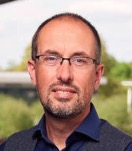 Matthew Hurles合作论文数Wellcome Sanger Institute;University of Cambridge29Richard Redon合作论文数27
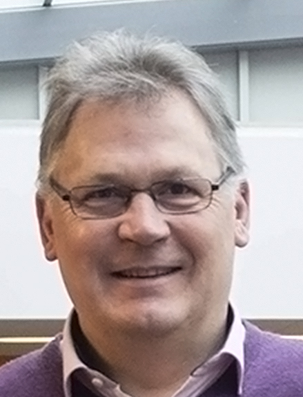 Ian Dunham合作论文数Open Targets25
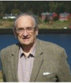 Malcolm A. Ferguson-Smith合作论文数Department of Veterinary Medicine, University of Cambridge23
 Tomas Fitzgerald合作论文数Wellcome Trust Sanger Institute23Susan M. Gribble合作论文数22
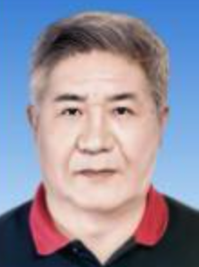 Fengtang Yang (杨凤堂)合作论文数School of Life Sciences and Medicine, Shandong University of Technology;Kunming Institute of Zoology, Chinese Academy of Sciences18Bee Ling Ng合作论文数18E. Prigmore合作论文数17
 Charles Lee合作论文数The Jackson Laboratory16Elena Prigmore合作论文数16
 Richard Durbin合作论文数Wellcome Sanger Institute;Department of Genetics, University of Cambridge15
 Helen V. Firth合作论文数Wellcome Sanger Institute;School of Clinical Medicine, University of Cambridge15Sarah Hunt合作论文数14S. Gribble合作论文数14
 Tim Hubbard合作论文数Wellcome Trust Sanger Institute;Genomics England;King's Health Partners;Department of Medical & Molecular Genetics, School of Basic & Medical Biosciences, Faculty of Life Sciences & Medicine, King's College London14Diana Rajan合作论文数14D C Burford合作论文数13
 Thomas Dan Andrews合作论文数John Curtin School of Medical Research, College of Health and Medicine, The Australian National University13Cordelia Langford合作论文数13David Beare合作论文数13
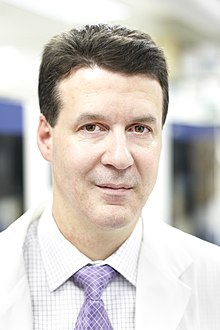 Stephen Scherer合作论文数Department of Molecular Genetics, Temerty Faculty of Medicine, University of Toronto;The Centre for Applied Genomics, The Hospital for Sick Children;Institute of Medical Science, University of Toronto;The McLaughlin Centre, Faculty of Medicine, University of Toronto13
 Stephan Beck合作论文数Research Department of Cancer Bio, University College London;Cancer Institute, University College London12Ruby Banerjee合作论文数12C. M. Clee合作论文数11cordelia langford合作论文数11
 Chris Tyler-Smith合作论文数Wellcome Sanger Institute11R. D. Bentley合作论文数10C Shaw-Smith合作论文数10
 Matthew Breen合作论文数10N Corby合作论文数10Kathryn Woodfine合作论文数10L Willatt合作论文数9G K Laird合作论文数9P Dhami合作论文数9J Bailey合作论文数9a mcmurray合作论文数9Adrienne Hunt合作论文数9Alan Coulson合作论文数9Jane Loveland合作论文数Wellcome Trust Sanger Institute;Wellcome Trust Genome Campus;Wellcome Trust Genome Campus, Wellcome Trust Sanger Institute9John Crolla合作论文数National Genetics Reference Laboratory (Wessex), Salisbury District Hospital9
 Robert Plumb合作论文数Imperial College London9
 Michael R. Speicher合作论文数Diagnostic and Research Institute of Human Genetics, Medical University of Graz9Helen Beasley合作论文数Wellcome Trust Sanger Institute;Wellcome Trust Genome Campus;Wellcome Trust Genome Campus, Wellcome Trust Sanger Institute9J E Collins合作论文数9
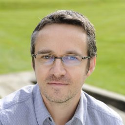 Alan Tracey合作论文数Wellcome Trust Sanger Institute;Wellcome Trust Genome Campus;Wellcome Trust Genome Campus, Wellcome Trust Sanger Institute9Laurens Wilming合作论文数9
 Lars Feuk合作论文数Department of Immunology, Genetics and Pathology, Uppsala University9




