We present a deep-learning approach for solving the problem of 2π phase ambiguities in two-dimensional quantitative phase maps of biological cells, using a multi-layer encoder-decoder residual convolutional neural network. We test the trained network, PhUn-Net, on various types of biological cells, captured with various interferometric setups, as well as on simulated phantoms. These tests demonstrate the robustness and generality of the network, even for cells of different morphologies or different illumination conditions than PhUn-Net has been trained on. In this paper, for the first time, we make the trained network publicly available in a global format, such that it can be easily deployed on every platform, to yield fast and robust phase unwrapping, not requiring prior knowledge or complex implementation. By this, we expect our phase unwrapping approach to be widely used, substituting conventional and more time-consuming phase unwrapping algorithms.
We report a cross-talk free simultaneous three-wavelength digital holographic microscopy setup for spectroscopic imaging of biological cells during flow. The feasibility of the proposed measurement technique is demonstrated on erythrocytes, due to their unique morphology and dependency of hemoglobin (Hb) molecule absorption on wavelength. From the spectroscopic quantitative phase profiles of cells acquired during flow in a microfluidic device, we decoupled the refractive index and the physical thickness. We then used our quantitative phase imaging results to dynamically calculate the mean cell volume (MCV), mean corpuscular Hb concentration (MCHC), mean corpuscular Hb content (MCH) and sphericity index.
Many medical and biological protocols for analyzing individual biological cells involve morphological evaluation based on cell staining, designed to enhance imaging contrast and enable clinicians and biologists to differentiate between various cell organelles. However, cell staining is not always allowed in certain medical procedures. In other cases, staining may be time-consuming or expensive to implement. Staining protocolsmay be operator-sensitive, and hence may lead to varying analytical results, as well as cause artificial imaging artifacts or false heterogeneity. We present a deep-learning approach, called HoloStain, which converts images of isolated biological cells acquired without staining by holographic microscopy to their virtually stained images. We demonstrate this approach for human sperm cells, as there is a well-established protocol and global standardization for characterizing the morphology of stained human sperm cells for fertility evaluation, but, on the other hand, staining might be cytotoxic and thus is not allowed during human in vitro fertilization (IVF). After a training process, the deep neural network can take images of unseen sperm cells retrieved from holograms acquired without staining and convert them to their stainlike images. We obtained a fivefold recall improvement in the analysis results, demonstrating the advantage of using virtual staining for sperm cell analysis. With the introduction of simple holographic imaging methods in clinical settings, the proposed method has a great potential to become a common practice in human IVF procedures, as well as to significantly simplify and radically change other cell analyses and techniques such as imaging flow cytometry.
We propose a new deep learning approach for medical imaging that copes with the problem of a small training set, the main bottleneck of deep learning, and apply it for classification of healthy and cancer cells acquired by quantitative phase imaging. The proposed method, called transferring of pre-trained generative adversarial network (TOP-GAN), is a hybridization between transfer learning and generative adversarial networks (GANs). Healthy cells and cancer cells of different metastatic potential have been imaged by low-coherence off-axis holography. After the acquisition, the optical path delay maps of the cells have been extracted and directly used as an input to the deep networks. In order to cope with the small number of classified images, we have used GANs to train a large number of unclassified images from another cell type (sperm cells). After this preliminary training, and after transforming the last layer of the network with new ones, we have designed an automatic classifier for the correct cell type (healthy/primary cancer/metastatic cancer) with 90-99% accuracy, although small training sets of down to several images have been used. These results are better in comparison to other classic methods that aim at coping with the same problem of a small training set. We believe that our approach makes the combination of holographic microscopy and deep learning networks more accessible to the medical field by enabling a rapid, automatic and accurate classification in stain-free imaging flow cytometry. Furthermore, our approach is expected to be applicable to many other medical image classification tasks, suffering from a small training set.
We present our latest advances in multi-wavelength label-free imaging using quantitative phase imaging. Basic QPI designs usually incorporate a single, coherent or partially coherent light source. The introduction of off-axis geometry allows for calculation of phase information from a single-exposure interferogram. For multi-wavelength imaging, it has been previously required to take sequential interferograms with different wavelengths, effectively reducing imaging throughput, or to use color cameras that have cross-talk between the color channels and artefacts caused by Bayer pattern. Alternatively, it was suggested to use spatial interferometric multiplexing in order to provide multi-wavelength data, where multiple interferograms of different off-axis angles are acquired in a single exposure. We introduce an external module for label-free QPI capable of multi-color imaging using spatial interferometric multiplexing of multiple color channels, with self-referencing that significantly reduces mechanical vibration noise. This technique has various applications such as interferometric spectroscopy of live cells and multiple wavelength phase unwrapping.
We present an external interferometric setup that is able to simultaneously acquire three wavelengths of the same sample instance without scanning or multiple exposures. This setup projects onto the monochrome digital camera three off-axis holograms with rotated fringe orientations, each from a different wavelength channel, without overlap in the spatial-frequency domain, and thus allows the full reconstruction of the three complex wavefronts from the three wavelength channels. We use this new setup for three-wavelength phase unwrapping, allowing phase imaging of thicker objects than possible with a single wavelength, but without the increased level of noise. We demonstrate the proposed technique for micro-channel profiling and label-free cell imaging.
We present a new interferometric imaging approach that allows for multiple-depth imaging in a single acquisition, using off-axis low-coherence holographic multiplexing. This technique enables sectioned imaging of multiple slices within a thick sample, in a single image acquisition. Each slice has a distinct off-axis interference fringe orientation indicative of its axial location, and the camera acquires the multiplexed hologram containing the different slices at once. We demonstrate the proposed technique for amplitude and phase imaging of optically thick samples.
We review our new imaging technique for quantitatively measuring the integral (thickness-average) refractive index of the nuclei of live biological cells during flow, combining quantitative phase microscopy with simultaneous 2-D fluorescence microscopy for nucleus localization.
We explore different deep learning-based approaches to solve the problem of phase unwrapping in objects with high spatial gradients, which is applicable to many fields in medicine, biology and remote sensing. We simulate data with high spatial gradients to compare the quality of the solution and the runtime obtained when addressing this problem either as an inverse problem or as a semantic segmentation problem.
We present a new external off-axis holographic module that doubles the acquired complex wavefront field of view, based on using both holographic flipping and multiplexing. In contrast to previous designs, this design does not require spatial filtering (no pinhole or lenses) to create the reference beam externally. In addition, the overlap area between the fields of view, as well as the off-axis angle between the sample and reference beams, can be fully controlled. As we demonstrate experimentally, this approach is useful for quantitative phase microscopy of extended stationary and dynamic samples, such as cancer cells during rapid flow and beating cardiomyocytes.
We suggest a new multimodal imaging technique for quantitatively measuring the integral (thickness-average) refractive index of the nuclei of live biological cells in suspension. For this aim, we combined quantitative phase microscopy with simultaneous 2-D fluorescence microscopy. We used 2-D fluorescence microscopy to localize the nucleus inside the quantitative phase map of the cell, as well as for measuring the nucleus radii. As verified offline by both 3-D confocal fluorescence microscopy and 2-D fluorescence microscopy while rotating the cells during flow, the nucleus of cells in suspension that are not during division can be assumed to be an ellipsoid. The entire shape of a cell in suspension can be assumed to be a sphere. Then, the cell and nucleus 3-D shapes can be evaluated based on their in-plain radii available from the 2-D phase and fluorescent measurements, respectively. Finally, the nucleus integral refractive index profile is calculated. We demonstrate the new technique on cancer cells, obtaining nucleus refractive index values that are lower than those of the cytoplasm, coinciding with recent findings. We believe that the proposed technique has the potential to be used for flow cytometry, where full 3-D refractive index tomography is too slow to be implemented during flow.
We present highly dynamic photothermal interferometric phase microscopy for quantitative, selective contrast imaging of live cells during flow. Gold nanoparticles can be biofunctionalized to bind to specific cells, and stimulated for local temperature increase due to plasmon resonance, causing a rapid change of the optical phase. These phase changes can be recorded by interferometric phase microscopy and analyzed to form an image of the binding sites of the nanoparticles in the cells, gaining molecular specificity. Since the nanoparticle excitation frequency might overlap with the sample dynamics frequencies, photothermal phase imaging was performed on stationary or slowly dynamic samples. Furthermore, the computational analysis of the photothermal signals is time consuming. This makes photothermal imaging unsuitable for applications requiring dynamic imaging or real-time analysis, such as analyzing and sorting cells during fast flow. To overcome these drawbacks, we utilized an external interferometric module and developed new algorithms, based on discrete Fourier transform variants, enabling fast analysis of photothermal signals in highly dynamic live cells. Due to the self-interference module, the cells are imaged with and without excitation in video-rate, effectively increasing signal-to-noise ratio. Our approach holds potential for using photothermal cell imaging and depletion in flow cytometry.
Measurements of biological cells during flow are highly important for medical diagnosis based on cell sorting. In the case of cell imaging during flow, very rapid image acquisition capabilities are required to enable fast cell flow for analyzing a sufficient number of cells. We present a new flipping interferometry (FI) module for simplified off-axis close-to-common-path interferometric phase microscopy. This wide-field off-axis interferometric module provides rapid quantitative phase microscopy of biological cells during flow in a microfluidic channel, with potential of integration into cell sorting devices. Various experimental demonstrations are presented.
We present a dual-wavelength external holographic microscopy module for quantitative phase imaging of 3D structures with extended thickness range. This is done by simultaneous acquisition of two off-axis interferograms, each at a different wavelength, and generation of a synthetic wavelength, which is larger than the sample optical thickness, allowing two-wavelength unwrapping. The simultaneous acquisition is carried out by using optical multiplexing of the two interferograms onto the camera, where each of them has orthogonal off-axis interference fringe direction in relation to the other one. We used the system to quantitatively image a 7.96 μm step target and 30.5 μm circular copper pillars.
The vast majority of quantitative phase imaging techniques face the same major issue of phase unambiguity. The phase of light is 2π-periodic, and as such can only be extracted in a small range of typically 0.4 µm to 0.7µm in the visible range. As most of the samples in biology and metrology are much higher, phase data is wrapped within this small range. One of the more reliant solutions of phase unambiguity is phase unwrapping, where the it is unwrapped based on the surrounding data. Unwrapping, however, works best on continuous phase changes and fails when abrupt phase jumps appear, e.g steps greater than the unambiguous range. An alternative method to overcome the unambiguous phase problem is multiplexing two wavelengths to capture the same phase image. Processing together the two phase images captured by different wavelengths can produce a new image, corresponding to a phase image with a synthetic wavelength that larger than each of the original wavelengths. This effectively enlarges the unambiguous range, making costly unwrapping algorithms redundant. We present a new dual-wavelength interferometry setup that can capture and multiplex two different phase images in an external module, portable to existing microscopy systems. The module is based on a self-interference multiplexing technique. As such, it is very flexible and can work with either transmission or reflection based microscopes. It can be used for either enlarging the unambiguous range or other dual-wavelength phase imaging applications.
We present a new holographic concept, named six-pack holography (6PH), in which we compress six off-axis holograms into a single multiplexed off-axis hologram without loss of magnification or resolution. The multiplexed hologram contains straight off-axis fringes with six different orientations, and can be generated optically or digitally. We show that since the six different complex wavefronts do not overlap in the spatial frequency domain, they can be fully reconstructed. 6PH allows more than 50% improvement in the spatial bandwidth consumption when compared to the best multiplexing method proposed so far. We expect the 6PH concept to be useful for a variety of applications, such as field-of-view multiplexing, wavelength multiplexing, temporal multiplexing, multiplexing for super-resolution imaging, and others.
We demonstrate photothermal quantitative phase imaging of living cells with functionalized nanoparticles in a cost-efficient setup and quantify the impact of nanoparticle excitation with low modulation frequencies on cell morphology and viability.
We correct a typo that repeated itself in several equations. Our previous results and conclusions are unchanged.
We explored photothermal quantitative phase imaging (PTQPI) of living cells with functionalized nanoparticles (NPs) utilizing a cost-efficient setup based on a cell culture microscope. The excitation light was modulated by a mechanical chopper wheel with low frequencies. Quantitative phase imaging (QPI) was performed with Michelson interferometer-based off-axis digital holographic microscopy and a standard industrial camera. We present results from PTQPI observations on breast cancer cells that were incubated with functionalized gold NPs binding to the epidermal growth factor receptor. Moreover, QPI was used to quantify the impact of the NPs and the low frequency light excitation on cell morphology and viability.
We present a portable, off-axis interferometric module for quantitative phase microscopy of live cells, positioned at the exit port of a coherently illuminated inverted microscope. The module creates on the digital camera an interference pattern between the image of the sample and its flipped version. The proposed simplified module is based on a retro-reflector modification in an external Michelson interferometer. The module does not contain any lenses, pinholes, or gratings and its alignment is straightforward. Still, it allows full control of the off-axis angle and does not suffer from ghost images. As experimentally demonstrated, the module is useful for quantitative phase microscopy of live cells rapidly flowing in a micro-channel.
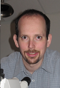 Natan Tzvi Shaked合作论文数Department of Biomedical Engineering, Tel Aviv University24
 Gili Dardikman合作论文数Fac Engn, Tel Aviv Univ6
 Itay Barnea合作论文数Faculty of Life Sciences, Tel Aviv University6
 Darina Roitshtain合作论文数Tel Aviv Univ, Fac Engn, Dept Biomed Engn, IL-69978 Tel Aviv, Israel5
 Simcha Kalman Mirsky合作论文数Dept Biomed Engn, Tel Aviv Univ4
 Yoav Nygate合作论文数4Moran Rubin合作论文数3Omry Blum合作论文数Department of Biomedical Engineering, Faculty of Engineering, and3
 Björn Kemper合作论文数Laboratory of Biophysics, University of Muenster3Barham Javidi合作论文数2Michael Isbach合作论文数2
 Dr. Gyanendra Singh合作论文数Instrument Design Development Centre, Indian Institute of Technology Delhi2
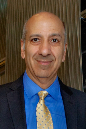 Bahram Javidi合作论文数Multidimensional Optical Sensing and Imaging Systems Laboratory, College of Engineering, University of Connecticut;Electrical & Computer Engineering Department, University of Connecticut2Steffi Ketelhut合作论文数2
 Burkhard Greve合作论文数2
 Jurgen Schnekenburger合作论文数Department of Medicine, University of Muenster2Omer Stein合作论文数1Noa Rotman-Nativ合作论文数1Lauren Wolbromsky合作论文数Faculty of Engineering, Tel Aviv University1Lidor Karako合作论文数Fac Engn, Tel Aviv Univ1
 Israel Gannot合作论文数Department of Electrical and Computer Engineering, Whiting School of Engineering, Johns Hopkins University;The Malone Center for Engineering in Healthcare, Johns Hopkins University;Opticul Diagnostics1Eravuchira Pinkie J合作论文数Departament d'Enginyeria Electrònica, Elèctrica i Automàtica, Universitat Rovira i Virgili1Habaza Mor合作论文数1Peled Anna合作论文数1
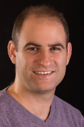 Handelman Amir合作论文数Iby and Aladar Fleischman Faculty of Engineering, Tel Aviv University1
 Mattan Levi合作论文数In Vitro Fertilization and Andrology Laboratories, Meir Medical Center;Department of Biomedical Engineering, The Iby And Aladar Fleischman Faculty of Engineering, Tel Aviv University;Faculty of Medical and Health Sciences, Tel Aviv University;QART Medical1
 Boris Apter合作论文数1
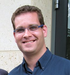 Idan Steinberg合作论文数1
 Avishay Eyal合作论文数Faculty of Engineering;Tel Aviv University;Faculty of Engineering, Tel Aviv University1
 Raja Giryes合作论文数Department of EE - Systems, School of Electrical Engineering, Faculty of Engineering, Tel Aviv University1
 Rafi Korenstein合作论文数Max-Planck-Institut für Ernährungsphysiologie, Rheinlanddamm 201, 4600 Dortmund, FRG1
 Gil Rosenman合作论文数School of Electrical Engineering, Tel Aviv University1
 Miki Haifler合作论文数1




